Biomarker for predicting preeclampsia and application thereof

A preeclampsia and biological technology applied in the biological field to achieve the effect of inhibiting migration and invasion

Active Publication Date: 2021-09-17
DALIAN MEDICAL UNIVERSITY
View PDF4 Cites 1 Cited by
  • Summary
  • Abstract
  • Description
  • Claims
  • Application Information

AI Technical Summary

Problems solved by technology

Several predictive markers have been studied, but none have been selected as predictive or diagnostic markers for PE according to the WHO definition

Method used


Image

  • Biomarker for predicting preeclampsia and application thereof
  • Biomarker for predicting preeclampsia and application thereof
  • Biomarker for predicting preeclampsia and application thereof

Examples


Embodiment 1

[0023] Materials and methods

[0024] Clinical samples (placental tissue) from women with symptoms of preeclampsia during pregnancy and healthy women with normal pregnancy, embedded in paraffin.

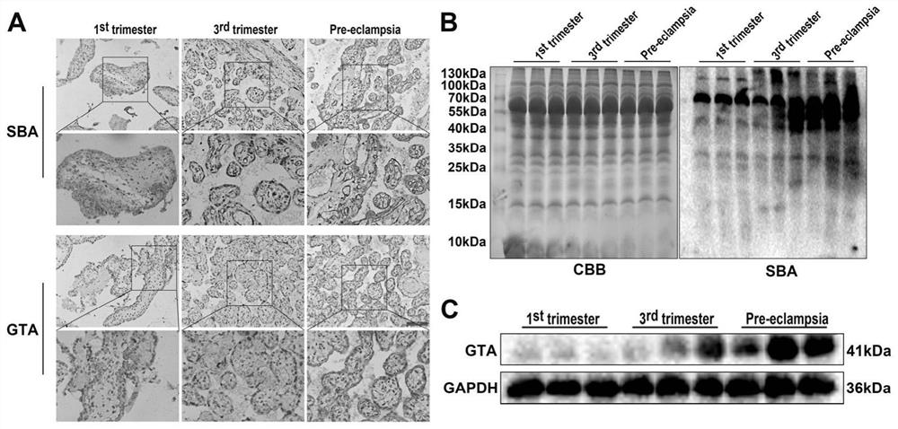
[0025] The expression of Galα1, 3GalNAc and GTA in the placental tissues of normal pregnant women and preeclampsia patients was detected.

[0026] 1. Detection of the localization and expression of Galα1,3GalNAc and GTA in placental tissue by immunohistochemical method

[0027] (1) Dewaxing: Put the prepared paraffin sections into the following solutions in turn: soak in xylene I for 10 minutes, then soak in xylene II for 10 minutes, soak in 100% ethanol I for 5 minutes, soak in 100% ethanol II for 5 minutes, and soak in 95% ethanol Soak for 5 minutes, soak in 85% ethanol for 5 minutes, soak in 70% ethanol for 5 minutes; then rinse with slow water for 10 minutes, do not directly wash the slices, soak in PBS 3 times, 5 minutes each time.

[0028] (2) Antigen retrieval: Put the slice...

Embodiment 2

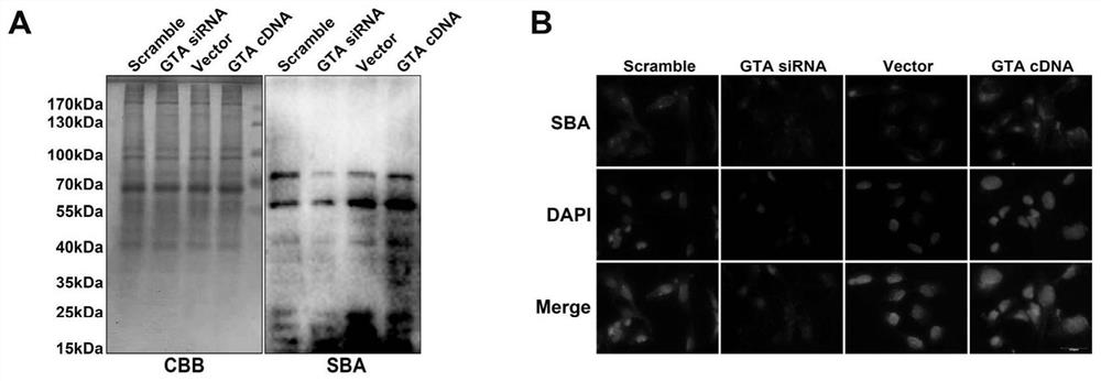
[0057] Example 2: Detection of Galα1,3GalNAc expression in trophoblast cells by Western blot and cell immunofluorescence

[0058] Climb slides: Human chorionic trophoblast cells (HTR8 / SVneo) were digested with trypsin, centrifuged at 800 rpm for 4 minutes, resuspended in fresh medium, and put the cell suspension into a petri dish with slides to allow the cells to grow in Climb on the sheet.

[0059] Collection: remove the culture medium in the culture dish, wash with PBS 3 times, 3 minutes each time.

[0060] Fixation: add 4% paraformaldehyde to the petri dish obliquely, and fix it for 20 minutes.

[0061] Paraformaldehyde was discarded, washed 3 times with PBS, 3 min each time.

[0062] Sealing: Select slides and place them in a wet box, add immunostaining blocking solution to seal for 1 hour.

[0063] Primary antibody incubation: clip the slices from the blocking solution, blot dry with filter paper, and incubate with primary antibody at 4°C overnight (rabbit anti-human G...

Embodiment 3

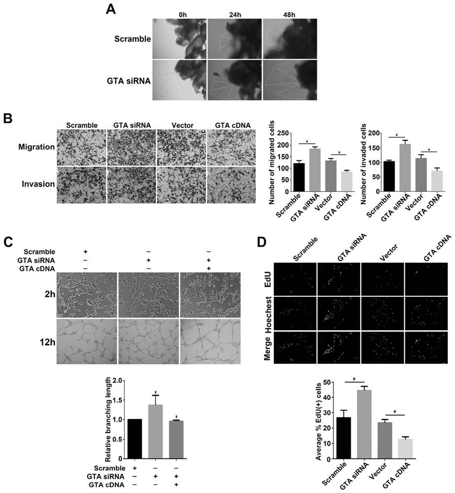
[0069] Embodiment 3: early pregnancy villi explant culture

[0070] (1) Laying gel: melt matrigel in a refrigerator at 4°C, dilute matrigel with serum-free medium DMEM / F-12 at a ratio of 1:2, gently blow and mix, add 50 μL to each well of a 96-well plate matrigel, try not to generate air bubbles, and place in a 37°C incubator.

[0071] (2) The villous tissue in the first trimester of pregnancy was obtained from healthy women undergoing artificial abortion in the obstetrics and gynecology operating room of the affiliated hospital for non-medical reasons, and the blood clots were rinsed with pre-cooled sterile PBS immediately.

[0072] (3) Put the villous tissue into a large dish containing PBS in a sterile operating table, and cut into 2-5mm fragments.

[0073] (4) Carefully transfer to the matrigel of the 96-well plate so that it is anchored in the center of the matrigel.

[0074] (5) After anchoring for 4-6 hours, add complete medium, observe the morphology under an inverte...


PUM


Abstract

The invention discloses a biological marker for predicting preeclampsia and application of the biological marker, and belongs to the technical field of biology. It is found for the first time that the expression of Gal alpha1, 3Gal NAc (glycosidic bond oligosaccharide) in placenta tissue of preeclampsia is remarkably improved compared with that of normal pregnancy tissue, Gal alpha1, 3Gal NAc can be catalytically synthesized by alpha1, 3N-acetylgalactosyl aminotransferase (GTA), Gal alpha1, 3Gal NAc and GTA serve as biological markers for diagnosing preeclampsia, and meanwhile it is proved that the Gal alpha1, 3Gal NAc and GTA have an effect on migration and invasion of trophoblastic cells, placental angiogenesis and proliferation capacity. According to the invention, prediction and diagnosis of preeclampsia are facilitated, an embryo implantation mechanism is deeply understood, and a new theoretical basis is provided for clinical treatment of preeclampsia with sugar as a target spot.

Description

technical field [0001] The invention belongs to the field of biotechnology, and in particular relates to a biological marker for predicting preeclampsia and its application. Background technique [0002] Preeclampsia is a systemic syndrome characterized by symptoms such as hypertension and proteinuria at ≥20 weeks of pregnancy; the basic pathological change of preeclampsia is systemic small artery spasm, involving multiple organs, often causing fetal Adverse outcomes such as intrauterine growth restriction, placental abruption, and even miscarriage. The main causes of preeclampsia include: activation of endothelial cells, intravascular inflammation, oxidative stress, abnormal trophoblast cell invasion, and impaired spiral artery remodeling; currently, the pathogenesis of preeclampsia is generally believed to be a combination of multiple factors As a result of the role of trophoblast cells, the abnormal invasion of trophoblast cells is considered to be the central link of it...

Claims


Application Information

Patent Timeline
Patent Type & Authority Applications(China)
IPC IPC(8): G01N33/573G01N33/53
CPCG01N33/573G01N33/5308G01N2800/368
Inventor 刘帅燕秋李雅琪陈天红崔馨元
Owner DALIAN MEDICAL UNIVERSITY
Why Eureka
Try Eureka