Method for diagnosing cancer using cfdna

A cancer cell, prostate cancer technology, applied in the field of detecting a biomarker gene specifically expressed or overexpressed in cancer by using the device, can solve the problem of spending a lot of time, etc.

Pending Publication Date: 2021-11-19
GENOPSY CO LTD
View PDF6 Cites 1 Cited by
  • Summary
  • Abstract
  • Description
  • Claims
  • Application Information

AI Technical Summary

Problems solved by technology

However, there are still limitations to using polymerase chain reaction to confirm which cfDNA is present
[0010] In order to amplify cfDNA by performing polymerase chain reaction, not only several kinds of primer sets are required, but also time-consuming and laborious steps are required

Method used


Image

  • Method for diagnosing cancer using cfdna
  • Method for diagnosing cancer using cfdna
  • Method for diagnosing cancer using cfdna

Examples


preparation example 1

[0584] Preparation example 1. Preparation of nanowires whose surface is treated with cationic polymer

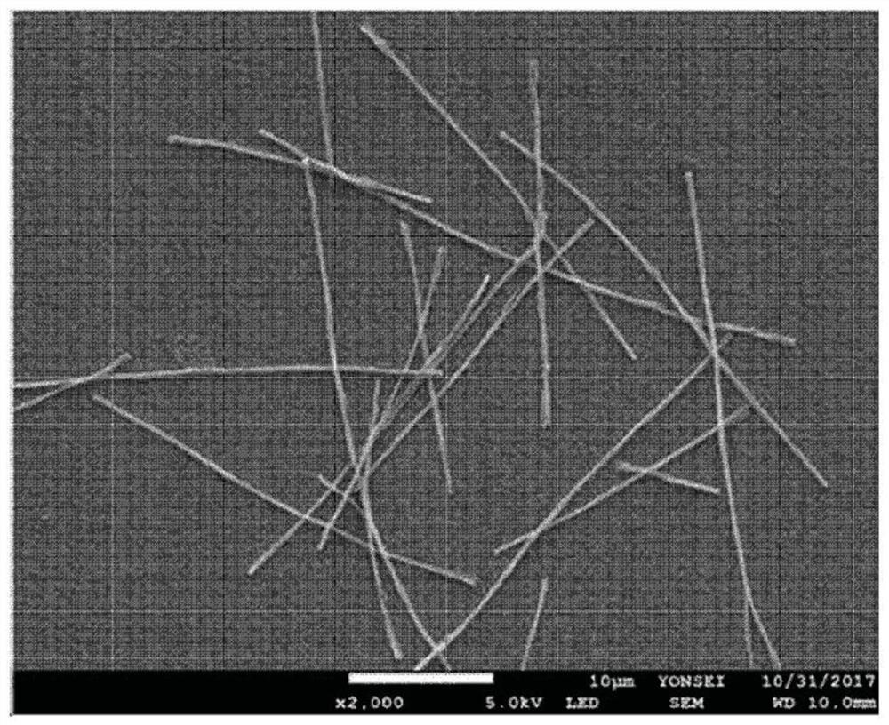
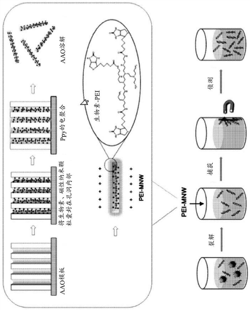
[0585] Such as Figure 1a As shown, nanowires with cationic polymer polyethyleneimine (PEI) conjugated to the nanowire surface were prepared. One surface of anodized aluminum oxide (AAO) was coated with a Q150T modular coating system (Quorum Technologies, UK) at 5 × 10 -3 A gold (Au) layer (thickness about 150 nm) was applied at mbar and 50 mA for 600 seconds. All electrochemical experiments were performed on AAO templates coated with gold (Au) using a potentiostat / galvanostat (BioLogic SP-150) equipped with a platinum wire counter electrode and an Ag / AgCl (3.0M NaCl type) reference electrode. .

[0586] To prepare cationic polymer-treated nanowires (PEI / Ppy NWs) together with 0.01 M poly(4-styrenesulfonic acid) and 0.01 M pyrrole solution containing 1 mg / ml biotin, chronoamperometry was performed at 1.0 V ( vs. Ag / AgCl) was applied to the pores of the AAO template for 7 ...

preparation example 2

[0589] Preparation example 2. Preparation of nanowires whose surface is treated with cationic polymer

[0590] According to substantially the same method as in Preparation Example 1 above, a nanostructure (PL / Ppy NW) having polylysine but not polyethyleneimine conjugated to the surface of the nanostructure was obtained.

preparation example 3

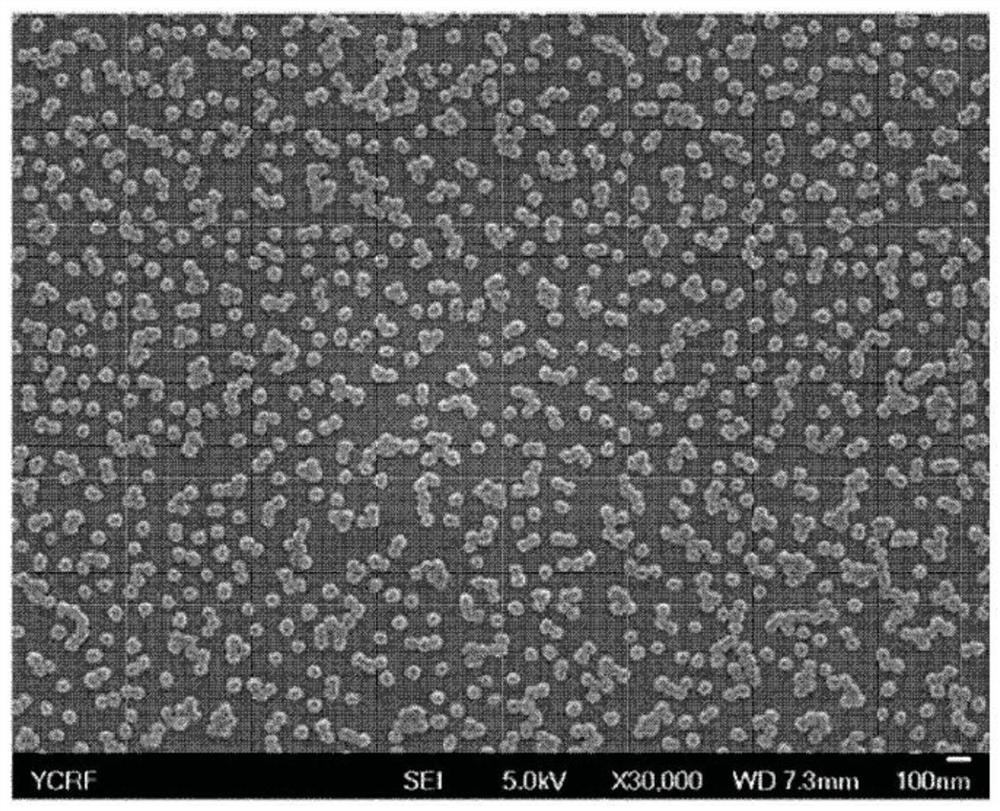
[0591] Preparation example 3. Preparation of poly(pyrrole) nanoparticles labeled with HRP and streptavidin

[0592] To prepare nanoparticles conjugated with HRP and streptavidin, 0.5 g of polyvinylpyrrolidone (PVP) was added to 12.5 mL of tertiary distilled water, stirred for 30 minutes, then 65 μL of pyrrole was added, and stirred for another 10 minutes. Afterwards, add 0.5 mL of FeCl at a concentration of 0.75 g / mL 3 solution and reacted for 3 hours. Afterwards, 20 mL of hyaluronic acid aqueous solution (400 mg / 20 mL) was added and stirred for 3 hours to prepare poly(pyrrole)-hyaluronic acid nanoparticles (Ppy-HA-NPs).

[0593] MWCO: Dialysis was performed for two days in triple distilled water using a 50,000 pore size membrane. Centrifuge at 1,200 rpm for 3 minutes to remove large particle aggregates, then lyophilize. Add 200 μg of Ppy-HA-NPs prepared above to 1 mL of triple distilled water, then add 100 mM EDC / 50 mM NHS solution and react for 45 minutes to activate the ...


PUM

PropertyMeasurementUnit
sizeaaaaaaaaaa
sizeaaaaaaaaaa

Abstract

A diagnosis method according to the present invention relates to a technique for concentrating and separating small cfDNA from a liquid specimen such as urine, cerebrospinal fluid, plasma, blood, pleural fluid, or body fluid, and then detecting biomarkers, overexpressed in a specific cancer, with extreme sensitivity and without a PCR. A detection method according to one example of the present invention does not require a PCR amplification reaction, and thus can significantly reduce the time it takes to diagnose cancer. In addition, the method enables immediate on-site analysis, and can be used as point-of-care testing (POCT) that can simultaneously search a large number of genes in a short time.

Description

technical field [0001] The present invention relates to a method for diagnosing cancer using free DNA (cell-free DNA) having a double helix structure, more particularly, the present invention relates to a method for detecting specifically expressed or A method of overexpressing a biomarker gene, and a device using the same. Background technique [0002] Recently, the importance of early diagnosis of cancer diseases has been greatly emphasized all over the world. Therefore, studies on early diagnosis methods of cancer are gradually increasing. However, until now, methods for diagnosing cancer have been through invasive methods such as collecting tissue samples and endoscopy. Specifically, a biopsy is performed by taking a portion of an area of ​​suspected disease and looking at it under a microscope. Therefore, in order to collect tissue samples using needles, punches, endoscopes or laparoscopes, the body must be incised, causing considerable discomfort to the patient and ...

Claims


Application Information

Patent Timeline
Patent Type & Authority Applications(China)
IPC IPC(8): C12Q1/6886C12Q1/6816C12N15/11
CPCC12Q1/6886C12Q1/6816C12Q2600/118C12Q2600/158C12Q2527/101C12Q2521/537C12Q2527/125C12Q2527/113C12Q1/6806C12Q2527/107C12Q2563/155C12Q1/6813C12Q2563/125C12Q2563/131
Inventor 曺荣南
Owner GENOPSY CO LTD
Why Eureka
Try Eureka