Method for determining titer of a swine transmissible gastroenteritis virus N protein antibody

A technology of antibody titer and determination method, which is applied in the direction of antiviral immunoglobulin, virus/bacteriophage, virus, etc., can solve the problems of high cost, low specificity, and various epitopes, and achieve the effect of rapid detection

Pending Publication Date: 2021-12-03
SHANDONG VOCATIONAL ANIMAL SCI & VETERINARY COLLEGE
View PDF0 Cites 0 Cited by
  • Summary
  • Abstract
  • Description
  • Claims
  • Application Information

AI Technical Summary

Problems solved by technology

At present, TGE detection kits on the market are prepared based on polyclonal antibodies and monoclonal antibodies. There are problems such as many epitopes and low specificity

Method used


Image

  • Method for determining titer of a swine transmissible gastroenteritis virus N protein antibody
  • Method for determining titer of a swine transmissible gastroenteritis virus N protein antibody
  • Method for determining titer of a swine transmissible gastroenteritis virus N protein antibody

Examples


Embodiment 1

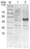
[0048] Example 1 Expression and purification of porcine transmissible gastroenteritis virus N protein

[0049] The recombinant vector was transformed into BL21(DE3), and 1.5 μL of the plasmid was added to 100 μL of competent cells. The EP tube was placed on ice for 30 minutes, and then heat-shocked in a water bath at 42°C for 90 seconds. After adding 1 mL of no-antibody LB into the EP tube Incubate on a shaker at 37°C for 1 hour, spread 150 μL of bacterial solution on the plate with a spreader and place it in a constant temperature incubator overnight. Pick a single colony with a regular shape on the plate, add 30mL Amp+LB liquid medium to culture, 210r / min 37°C, when the OD600nm value of the bacterial solution is 0.6-0.8, add a final concentration of 1mM / L IPTG inducer, and induce at 37°C After 6h, the recombinant N protein was successfully expressed by SDS-PAGE analysis ( figure 1 ).

Embodiment 2

[0050] Example 2 Construction of phage display library and panning of Nanobodies

[0051] 2.1 Immunization of experimental animals

[0052] 2.1.1 N protein immunization of Bactrian camels

[0053] Mix 1 mL of recombinant N protein (1 mg / mL) purified by affinity chromatography with an equal volume of Freund's complete adjuvant, emulsify completely to form a water-in-oil state, and inject it into the camel's neck. The injection method is subcutaneous injection. Freund's incomplete adjuvant was used for the second to fifth immunizations, and the interval between each immunization was two weeks. After the fifth immunization, 200 mL of peripheral blood was collected through the jugular vein.

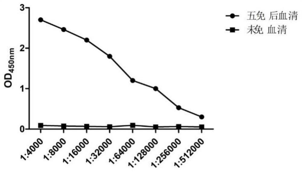
[0054] 2.1.2 N protein antibody titer determination

[0055] The serum was separated from the collected blood to detect the antibody titer against the N protein, and the serum before immunization was used as a control. Through indirect ELISA detection, the antibody titer against the N prote...

Embodiment 3

[0072] The establishment of embodiment 3 competition ELISA method

[0073] 3.1 Determine the concentration and dilution of biotin-labeled nanobody and optimal coating antigen by checkerboard titration

[0074] Use biotin-labeled Nb9 at a concentration of 1 mg / ml as the primary antibody, and HRP-labeled streptavidin as the secondary antibody, and find the concentration of the antibody with an OD value around 1.0. The concentration of Nb9 was 10 μg / mL-1 ng / mL, and 96-well plates were coated with N protein at different dilutions (0.5, 1, 2, 4 μg / mL), each well was blocked with 200 μL of blocking solution, and incubated at room temperature for 1 h. After washing 3 times, 100 μL of biotinylated Nb9 of different concentrations was added to each well of the enzyme-labeled plate, incubated for 1 h, after washing 3 times, 100 μL of horseradish peroxidase-labeled streptavidin was added and incubated for 30 min. After washing 3 times, add 100 μL of 3,3′,5,5′-tetramethylbenzidine, and re...


PUM


Abstract

The invention relates to a method for determining the titer of a swine transmissible gastroenteritis virus N protein antibody. The method comprises the following steps of: S1, performing an immune test on an experimental animal, comprising: determination of the titer of an N protein immune bactrian camel and an N protein antibody; S2, constructing a VHH bacteriophage library, comprising: amplification of a VHH gene, construction of a display vector, construction of a nano antibody library, and determination of the capacity of the library; and S3, elutriating an N specific nano antibody, comprising: library rescue and recombinant bacteriophage titer determination, N protein specific recombinant bacteriophage elutriation, protein specific recombinant bacteriophage enrichment and crude extract preparation, and ELISA detection of an N protein recombinant nano antibody.

Description

technical field [0001] The invention relates to the technical field of biological detection. In particular, it relates to a titer determination method for N protein antibody of porcine transmissible gastroenteritis virus. Background technique [0002] Porcine transmissible gastroenteritis (TGE) is an acute contact infectious disease caused by porcine transmissible gastroenteritis virus. Clinically, it mainly manifests as dehydration, severe diarrhea, vomiting, and atrophy of intestinal villi. Serious harmful effects can easily cause the death of lactating piglets and bring huge economic losses to my country's pig industry. The complete TGE virus contains four structural proteins: the nucleocapsid protein N (referred to as N protein) that wraps the genomic RNA, the membrane-bound protein M in the lipid envelope, the glycoprotein S that forms the virus protrusion, and the small membrane protein sM. Among them, the N protein is the nucleocapsid protein of porcine transmissibl...

Claims


Application Information

Patent Timeline
IPC IPC(8): C07K16/10C07K16/00C07K14/165C12N15/50C12N15/70G01N33/569
CPCC07K16/10C07K16/005C07K14/005C12N15/70G01N33/56983C07K2317/22C07K2317/33C07K2317/569C07K2317/35C12N2770/20022G01N2333/165
Inventor 王加才朱光彭晓蓓王云洲李申华
Owner SHANDONG VOCATIONAL ANIMAL SCI & VETERINARY COLLEGE
Why Eureka
Try Eureka