Reagent for modifying extracellular vesicles and preparation method thereof

A cell and vesicle technology, applied in the field of reagents and preparations for modifying extracellular vesicles, can solve problems such as affecting the biological function of extracellular vesicles, hindering the process of visualization research of extracellular vesicles, and low labeling efficiency, and achieves The effect of good visualization, efficient and controllable retouching, efficient and controllable labeling

Active Publication Date: 2022-03-25
天津外泌体科技有限公司
View PDF5 Cites 1 Cited by
  • Summary
  • Abstract
  • Description
  • Claims
  • Application Information

AI Technical Summary

Problems solved by technology

Therefore, this method affects the normal biological function of extracellular vesicles
However, the method of using conventional membrane dyes to label the lipid membrane of extracellular vesicles has the disadvantages of easy quenching of dyes, low labeling efficiency, and change

Method used


Image

  • Reagent for modifying extracellular vesicles and preparation method thereof
  • Reagent for modifying extracellular vesicles and preparation method thereof
  • Reagent for modifying extracellular vesicles and preparation method thereof

Examples


Embodiment 1

[0074] Milk-derived extracellular vesicles were obtained by separating and purifying fresh milk through acid precipitation (pH 4.0), ultracentrifugation, and column chromatography, and the particle size distribution of milk-derived extracellular vesicles was characterized by nanoflow detection technology , observing the morphology of extracellular vesicles by transmission electron microscopy, detecting the marker proteins of extracellular vesicles by Western blot, and detecting the purity of extracellular vesicles by ultra-high liquid phase-size-exclusion chromatography. The results will be as follows: image 3 E. Image 6 C. Figure 7 with Figure 8 Shown in B.

Embodiment 2

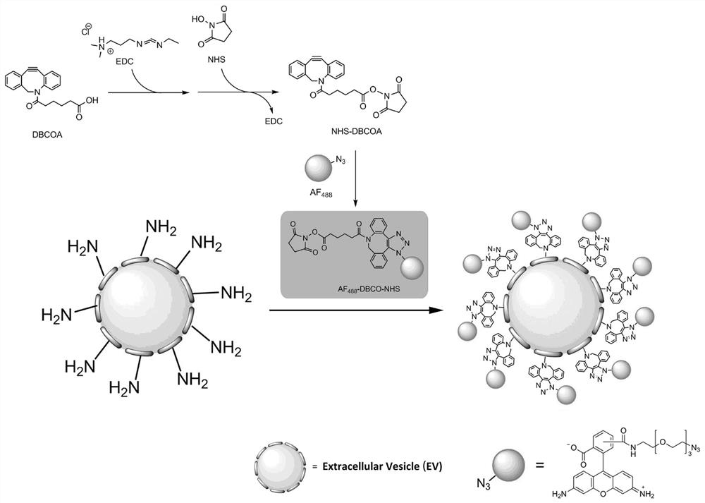
[0075] Embodiment 2: Preparation of fluorescent labeling reagent

[0076] EDC·HCl (5.8 mg, 30 μmol, 1.0 eq.) was mixed with NHS (3.5 mg, 30 μmol, 1.0 eq.), and DMSO (832 μL) was added to prepare 36.1 mM activated crosslinker. Add diphenylcyclooctyne adipic acid (10 mg, 30 μmol, 1.0 eq.) into the activated cross-linking agent, mix well at room temperature, and activate the carboxyl group to obtain diphenylcycloctyne succinimide ester.

[0077] The fluorescent probe azide fluorine 488 (1 mg, 1.74 μmol) was dissolved in DMSO (205 μL) to prepare 8.5 mM azide fluorine 488 fluorescent dye. Add 168 μL azide fluorine 488 fluorescent dye (1.428 μmol) to diphenylcyclooctyne succinimide ester, and mix thoroughly at room temperature to prepare a dichromate that is coupled with a fluorescent probe azide fluorine 488. Fluorescent labeling reagent for phenylcycloctyne succinimide ester (1 mL), store at 2-8 ℃.

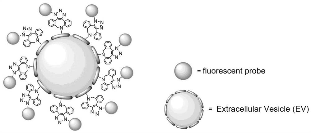
[0078] Mix 5 μL of fluorescent labeling reagent with 150 μL of milk-derived ext...

Embodiment 3

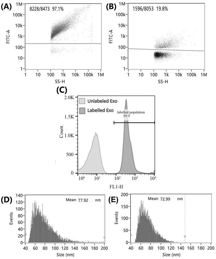
[0088] Example 3: Characterization of Fluorescence Labeling Rate and Particle Size Distribution of Extracellular Vesicles Using Nanoflow Detection Technology

[0089] The extracellular vesicles labeled with fluorescent molecules in Example 2 and Comparative Example were diluted 1000 times with PBS, and 250 ± 5 nm monodisperse nano-fluorescent silicon spheres were selected for optical calibration and counting standards of the nano-flow detector. Select A group of nano-silicon spheres with particle sizes of 68 ± 2 nm, 91 ± 3 nm, 113 ± 3 nm, and 155 ± 3 nm were used as particle size standards for the nano flow detection instrument, and the nano flow detection technology (Nano flow cytometry, NanoFCM) respectively detect the fluorescent labeling rate of at least 5000 extracellular vesicles in Example 2 and the comparative example.

[0090] Detection conditions: 488 nm laser, the detection channels are scattering channel and FITC fluorescence channel, the attenuation coefficient is...


PUM

PropertyMeasurementUnit
The average particle sizeaaaaaaaaaa

Abstract

The invention discloses a reagent for modifying extracellular vesicles and a preparation method thereof, and the reagent for modifying the extracellular vesicles, which has a modifier functional group and an active ester group at the same time, is formed by carrying out click chemical coupling on an azido-modified modifier and carboxyl-activated dibenzene ring octynoic acid. The method has the beneficial effects that the morphology and structural composition of the extracellular vesicles are not changed, the biological functions of the extracellular vesicles are not influenced, and the extracellular vesicles can be stably, efficiently and controllably modified; for example, when the modifier is a fluorescent probe, the extracellular vesicles can be efficiently and controllably modified, a good visualization effect of the extracellular vesicles is achieved, and a new strategy can be provided for developing an extracellular vesicle in-vivo tracing method and an extracellular vesicle tracing drug.

Description

technical field [0001] The invention belongs to the field of biotechnology, and in particular relates to a reagent for modifying extracellular vesicles and a preparation method. Background technique [0002] Extracellular vesicles (Extracellular Vesicles, EVs) are a type of vesicle-like bodies secreted and released by a variety of living cells, with a diameter in the range of 30-1000 nm and a lipid bilayer structure. Extracellular vesicles are widely found in cell culture supernatants and various body fluids of most animals (such as milk, blood, urine and lymph, etc.), and the cell-specific proteins and nucleic acids contained in them can be transmitted to other cells as signal molecules. Cells thus change the functions of other cells, and the advantages of stable lipid bilayer structure encapsulation and targetable modification have made extracellular vesicles become a research hotspot in the field of targeted drug delivery systems in recent years. At present, more and mor...

Claims


Application Information

Patent Timeline
IPC IPC(8): A61K47/54A61K49/00A61K31/704A61K31/7008A61K47/51A61P35/00
CPCA61K47/545A61K49/0052A61K47/51A61K49/005A61K49/0019A61K49/0021A61K49/0041A61K31/704A61K31/7008A61P35/00A61K49/0056
Inventor 葛啸虎王达董凤伟陆路马德美王淼程国钢白云飞韩春乐陈宁
Owner 天津外泌体科技有限公司
Why Eureka
Try Eureka