Chitosan-based biological tissue adhesive and its preparation method and application
A biological tissue and adhesive technology, used in applications, surgical adhesives, drug delivery, etc., can solve the problems of product cytotoxicity and weak adhesion, and achieve strong wet tissue adhesion, good mechanical properties, The effect of excellent hemostatic efficiency
- Summary
- Abstract
- Description
- Claims
- Application Information
AI Technical Summary
Problems solved by technology
Method used
Image
Examples
Embodiment 1
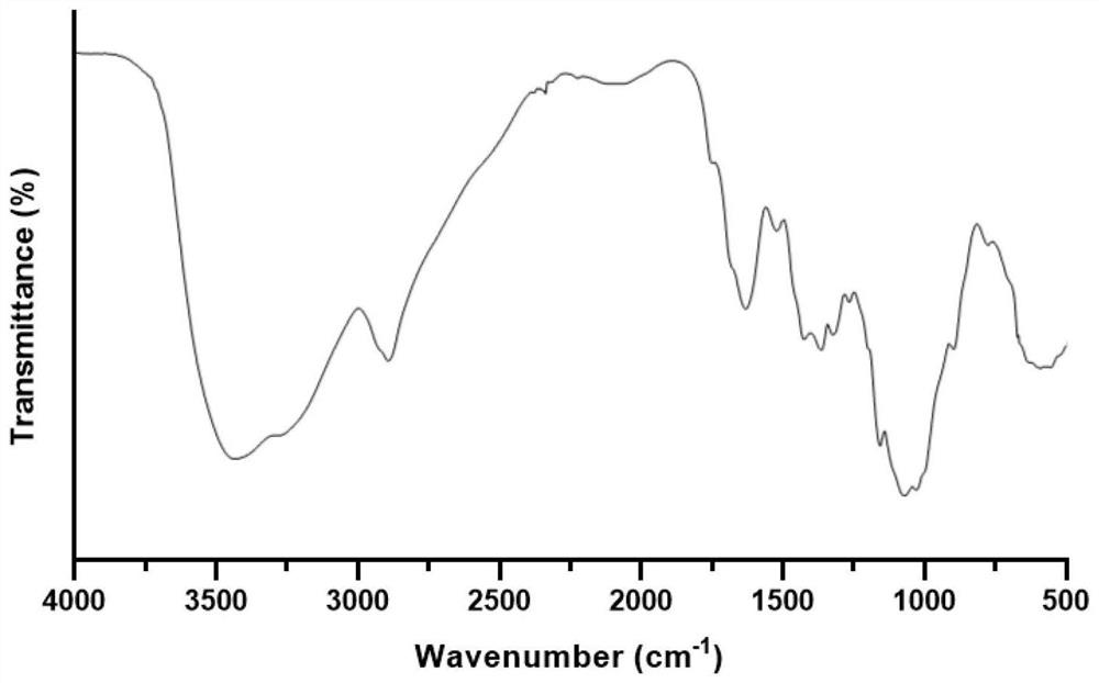
[0039] Example 1 Preparation of TEMPO oxidized chitosan
[0040] Under light-proof conditions, accurately weigh 0.2 g of TEMPO, dissolve it in deionized water, and continue stirring at 200 rpm until the dissolution is uniform. Then 200 mL of sodium hypochlorite solution (available chlorine concentration 4%), the main oxidant, was slowly added, and the pH was adjusted to 7.8 using 1 M hydrochloric acid.
[0041] 12 g of powdered chitosan was added to the above TEMPO oxidation system, and the reaction was continued and slowly stirred for 2 hours at room temperature. During the reaction, 0.5M NaOH solution was used to maintain the pH value of the reaction system stable. Anhydrous ethanol (1:10, v / v) was added to stop the oxidation reaction, and the pH was adjusted to 7.0, and then the oxidation product was precipitated after standing for 30 min. The pellets were washed 6 times by repeated centrifugation using graded ethanol solution to remove unreacted reagents or impurities. T...
Embodiment 2
[0044] Example 2 Preparation of biological tissue adhesive
[0045]Disperse 2 g of TEMPO oxidized chitosan and 30 g of organic polymer monomer acrylic acid prepared in Example 1 in 100 ml of deionized water, continue stirring at room temperature at 300 rpm until a homogeneous solution is obtained, and then add 2.0 mL of 0.35M The free radical initiator ammonium persulfate solution, the hydrogel prepolymerization solution was injected into a closed glass reaction mold with a thickness of 100 μm using a disposable sterile syringe, and the hydrogel was prepared overnight at 60 °C through a free radical initiating polymerization reaction. glue. After the hydrogel was formed, the hydrogel was immersed in a 75% ethanol solution to remove unreacted impurities, and then it was completely dried. Finally, the tissue adhesive patch and desiccant were placed in a sealed bag and stored at -20°C for later use.
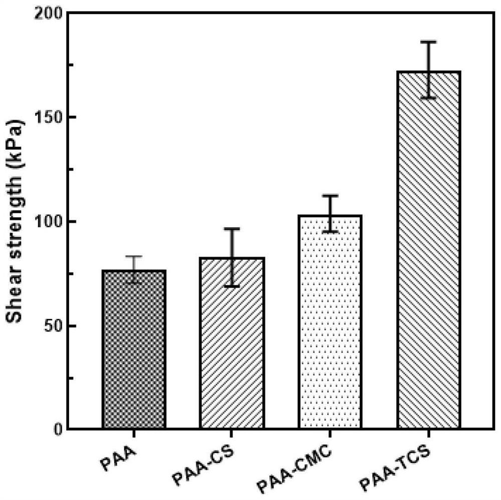
[0046] Using a microcomputer-controlled electronic universal testing machine ...
Embodiment 3
[0047] Example 3 Application of biological tissue adhesive
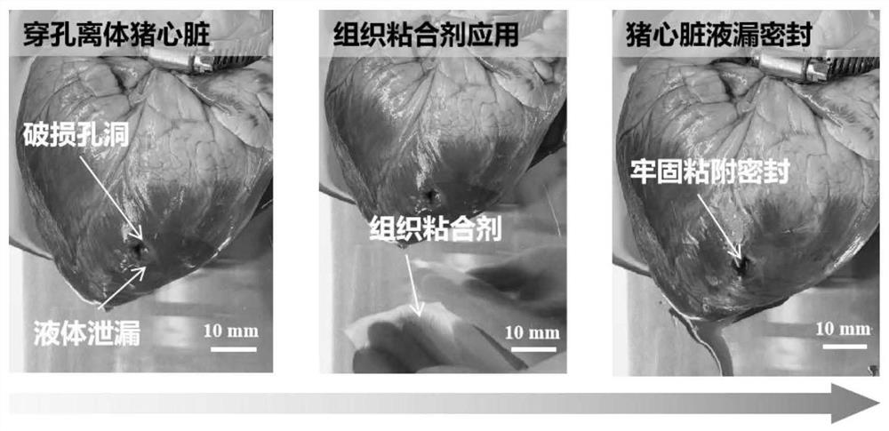
[0048] Using an isolated porcine organ model, the adhesive hemostatic properties of tissue adhesives under wet and dynamic conditions were evaluated. In vitro adhesion hemostasis experiments were performed using fresh porcine hearts as models. A hole with a diameter of 10-20 mm was punctured in the pig heart, and then a perfusion tube with a liquid flow rate of 20 mL / min was inserted into the pig heart to simulate massive hemorrhage. Use tissue adhesive (prepared in Example 2) to attach to the damaged organ and seal the incision after pressing for 5 s, and monitor the liquid leakage and sealing of the tissue adhesive.
[0049] The experimental results are as image 3 As shown, the tissue adhesive can be directly applied to the damaged part of the organ, and after being pressed with mild pressure (about 1 kPa) for 5 s, it can achieve tight adhesion and form a liquid seal, and no leakage of liquid at the damaged part...
PUM
 Abstract
Description
Claims
Application Information
 - R&D
- Intellectual Property
- Life Sciences
- Materials
- Tech Scout
- Unparalleled Data Quality
- Higher Quality Content
- 60% Fewer Hallucinations
Browse by: Latest US Patents, China's latest patents, Technical Efficacy Thesaurus, Application Domain, Technology Topic, Popular Technical Reports.
© 2025 PatSnap. All rights reserved.Legal|Privacy policy|Modern Slavery Act Transparency Statement|Sitemap|About US| Contact US: help@patsnap.com


