3D printing bone defect repair stent and preparation method thereof

A technology of 3D printing and bone printing, which is applied in the direction of processing and manufacturing, manufacturing auxiliary devices, prostheses, etc., can solve the problems of poor mechanical strength and low immunogenicity, and achieve regular scaffold structure, good uniformity, and easy mixing. Effect

Pending Publication Date: 2022-07-15
STOMATOLOGY AFFILIATED STOMATOLOGY HOSPITAL OF GUANGZHOU MEDICAL UNIV
View PDF7 Cites 0 Cited by
  • Summary
  • Abstract
  • Description
  • Claims
  • Application Information

AI Technical Summary

Problems solved by technology

Gelatin / sodium alginate hydrogel is a mixture of natural polymer materials, which has the advantages of good biocompatibility, tissue absorbability, low immunogenicity, etc., especially for its combination with highly biologically active inorganic powders Carry out 3D printing molding, but its mechanical strength is relatively poor

Method used


Image

  • 3D printing bone defect repair stent and preparation method thereof
  • 3D printing bone defect repair stent and preparation method thereof
  • 3D printing bone defect repair stent and preparation method thereof

Examples


Embodiment 1

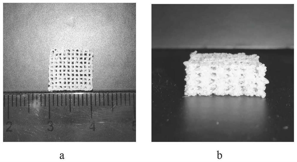
[0038] A 3D-printed bone defect repair scaffold is a multi-layer interconnected porous network structure; the first layer includes a plurality of mutually parallel lines, and the second layer is a plurality of mutually parallel lines vertically connected to the upper surface of the first layer of lines Lines, the third layer is a plurality of mutually parallel lines vertically connected to the upper surface of the lines of the second layer, and so on; the diameter of each layer of lines in the 3D printed bone defect repair scaffold is about 500 μm, and adjacent lines The distance between them is about 700μm;

[0039] The preparation method of the 3D printed bone defect repair scaffold includes the following steps:

[0040] S1. dissolve gelatin, sodium alginate and 58S bioglass in water to obtain a solution, wherein the mass / volume concentration of each component in the solution is 16% gelatin, 6.5% sodium alginate, and 8.5% 58S bioglass;

[0041] S2. Stir the solution evenly ...

Embodiment 2

[0044] A 3D-printed bone defect repair scaffold is a multi-layer interconnected porous network structure; the first layer includes a plurality of mutually parallel lines, and the second layer is a plurality of mutually parallel lines vertically connected to the upper surface of the first layer of lines Lines, the third layer is a plurality of mutually parallel lines vertically connected to the upper surface of the lines of the second layer, and so on; the diameter of each layer of lines in the 3D printed bone defect repair scaffold is about 400 μm, and adjacent lines The distance between them is about 600μm;

[0045] The preparation method of the 3D printed bone defect repair scaffold includes the following steps:

[0046] S1. dissolve gelatin, sodium alginate and 58S bioglass in water to obtain a solution, wherein the mass / volume concentration of each component in the solution is 16% gelatin, 6.5% sodium alginate, and 8.5% 58S bioglass;

[0047] S2. Stir the solution evenly ...

Embodiment 3

[0050] A 3D-printed bone defect repair scaffold is a multi-layer interconnected porous network structure; the first layer includes a plurality of mutually parallel lines, and the second layer is a plurality of mutually parallel lines vertically connected to the upper surface of the first layer of lines Lines, the third layer is a plurality of mutually parallel lines vertically connected to the upper surface of the lines of the second layer, and so on; the diameter of each layer of lines in the 3D printed bone defect repair scaffold is about 400 μm, and adjacent lines The distance between them is about 500 μm;

[0051] The preparation method of the 3D printed bone defect repair scaffold includes the following steps:

[0052] S1. dissolve gelatin, sodium alginate and 58S bioglass in water to obtain a solution, wherein the mass / volume concentration of each component in the solution is 16% gelatin, 6.5% sodium alginate, and 8.5% 58S bioglass;

[0053] S2. Stir the solution evenly...


PUM

PropertyMeasurementUnit
diameteraaaaaaaaaa
porosityaaaaaaaaaa
porosityaaaaaaaaaa

Abstract

A preparation method of the 3D printing bone defect repair scaffold comprises the following steps that S1, gelatin, sodium alginate and 58S bioglass are dissolved in water to obtain a solution, and the concentration of mass / volume of each component in the solution is that the concentration of the gelatin is 16%, the concentration of the sodium alginate is 6.5%, and the concentration of the 58S bioglass is 8.5%; s2, the solution is stirred to be uniform, 3D printing slurry is obtained, and then 3D printing is conducted; a needle with the aperture of 0.41 mm is adopted for 3D printing, and printing is conducted according to the printing speed of 6 mm / s under the conditions that the air pressure is 0.38 Mpa and the temperature is 29 DEG C; s3, after printing is completed, a semi-finished stent product is obtained, the semi-finished stent product is subjected to chemical crosslinking with a calcium chloride solution for 8-15 min, and then the semi-finished stent product is soaked in a glutaraldehyde solution to be subjected to chemical crosslinking for 1.5-2.5 h; and finally, cleaning and freeze-drying. The 3D printing bone defect repair stent prepared by the invention is applied to bone defect repair, is good in degradability, has the average Young modulus of 290kPa, is good in elasticity, can effectively promote the formation of bone tissues and vascular tissues, and is good in osteogenesis effect.

Description

technical field [0001] The invention belongs to the technical field of bone tissue engineering repair and reconstruction, and mainly relates to a 3D printing bone defect repair bracket and a preparation method thereof. Background technique [0002] With the increase of bone tissue damage caused by trauma such as aging, joint degenerative diseases, and traffic accidents, more and more attention has been paid to the repair of bone defects. Autologous bone transplantation is the "gold standard" for defect repair, but the source of autologous bone is limited, and the supply is often in short supply. Allogeneic bone transplantation has the risk of infectious diseases, while artificial bone transplantation lacks osteoinductive activity and has poor osteogenesis efficiency. Organizing nascent tissues with similar structures. Therefore, the research on new regenerative bone defect repair materials with high biological activity and efficient osteogenesis has become a difficult and h...

Claims


Application Information

Patent Timeline
Patent Type & Authority Applications(China)
IPC IPC(8): A61L27/40A61L27/22A61L27/20A61L27/10A61L27/02A61L27/12A61L27/50A61L27/54A61L27/56A61L27/58B33Y10/00B33Y70/10B33Y80/00C08L89/00C08L5/04C08K3/40C08J9/00C08J9/36B29C64/106B29C64/393B33Y50/02
CPCA61L27/222A61L27/20A61L27/10A61L27/025A61L27/12A61L27/50A61L27/54A61L27/56A61L27/58B33Y10/00B33Y70/10B33Y80/00C08J9/0061C08J9/0066C08J9/36B29C64/106B29C64/393B33Y50/02A61L2430/02A61L2300/412C08J2389/00C08J2405/04C08L5/04B33Y40/20A61L27/446A61F2/30771A61F2002/2835A61F2002/3092A61F2002/30985
Inventor 江千舟涂欣冉郭吕华谭国忠陈荣丰张阳
Owner STOMATOLOGY AFFILIATED STOMATOLOGY HOSPITAL OF GUANGZHOU MEDICAL UNIV
Why Eureka
Try Eureka