

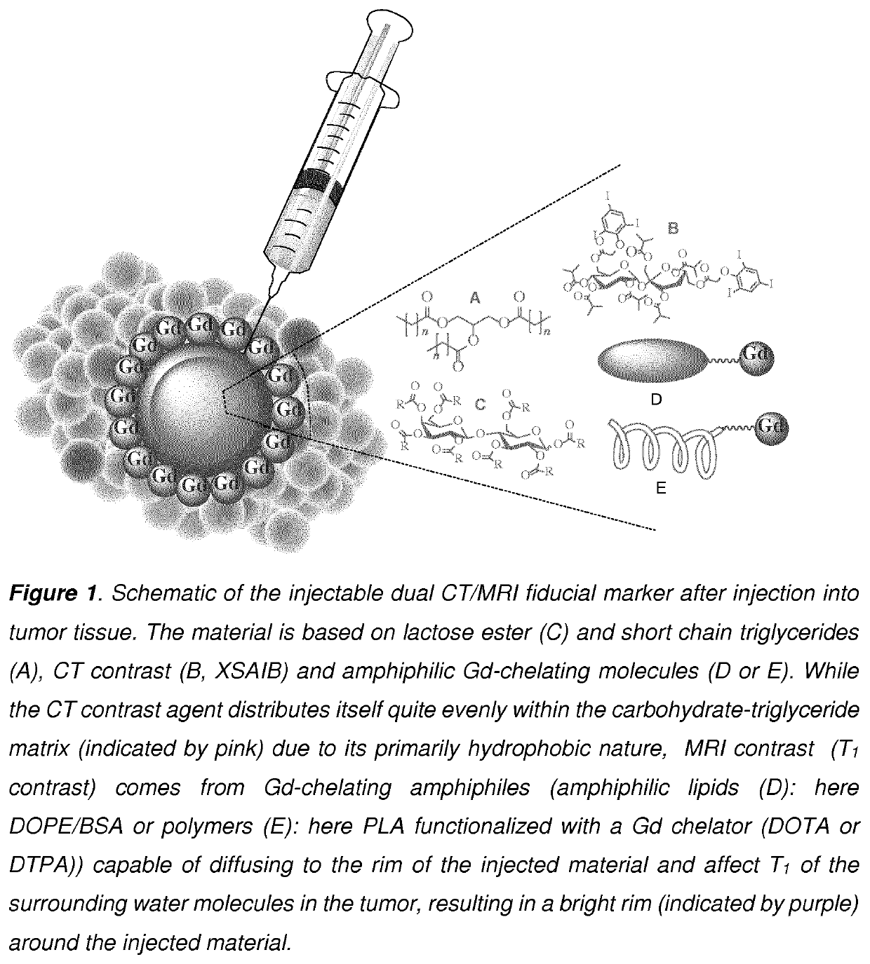
Development of injectable fiducial markers for image guided radiotherapy with dual MRI and CT visibility
a technology of image guided radiotherapy and injectable fiducial markers, which is applied in the direction of general/multifunctional contrast agents, medical preparations, non-active ingredients, etc., can solve the problems of difficult visualization in tumor tissue, ct-based target delineation of soft tissue tumors tends to only improve the accuracy of tumor delineation for radiotherapy treatment, and the mri is tsub>1/sub>-weighted, and the signal is not strong enough
- Summary
- Abstract
- Description
- Claims
- Application Information
AI Technical Summary
Benefits of technology
Problems solved by technology
Method used
Image

Examples
example 1
of Carbohydrate Esters
[0098]General experimental conditions: All reactions were carried out under inert atmosphere (N2). Water sensitive liquids and solutions were transferred via syringe. Water used for washing of the isolated products was in all cases MilliQ water. Organic solutions were concentrated by rotary evaporation at 30-60° C. at 200-0 mbar. Thin layer chromatography (TLC) was carried out using aluminum sheets pre-coated with silica 60F (Merck 5554). The TLC plates were inspected under UV light or developed using a cerium ammonium sulphate solution (1% cerium (IV) sulphate and 2.5% hexa-ammonium molybdate in a 10% sulfuric acid solution).
[0099]Reagents: DOTA-NHS was purchased from Macrocyclics. All other chemicals were purchased from Sigma Aldrich and were used as received. Dry pyridine was obtained by drying over molecular sieves (4 Å) for 2-3 days prior to use.
[0100]Instrumentation: Nuclear Magnetic Resonance (NMR) was conducted on a Bruker Ascend™ 400 MHz—operating at 4...
example 2
of Fluorescently Labeled PLA
[0112]PLA-RhB
[0113]
[0114]PLA-NH2 (Mn˜2500) (260 mg, 0.1 mmol) was suspended in dry DCM (˜5 mL). Then, a pre-mixed mixture of Rhodamine-B (105, 0.2 mmol), EDC-HCl (80 mg, 0.4 mmol) and DMAP (106 mg, 0.9 mmol) dissolved in 5 mL dry DCM was added, and the reaction was continued at r.t. for 2 days, where Kaiser test was negative, indicating full functionalization. The solvents were removed in vacuo, and the crude mixture was dissolved in DMSO and purified by dialysis (Mw cutoff: 1000 da) against MQ water for 14 days. Yield: 299 mg (97%). 1H NMR (400 MHz, DMSO-d6) δ 8.31 (d (br), J=7.7 Hz, 1H), 7.95 (br. t, J=7.4 Hz, 1H), 7.87 (br. t, J=7.6 Hz, 1H), 7.57-7.44 (m, 2H), 7.05-7.00 (m, 1H), 6.39-6.24 (m, ˜5H), 5.20 (q, J=7.1 Hz, ˜33H), 3.65 (dd (br), J=13.8, 6.9 Hz, 4H), 3.31 (br. q, J=7.1 Hz, 8H), 3.12-2.94 (m, 2H), 1.47 (d, J=7.1 Hz, ˜99H), 1.08 (t, J=6.9 Hz, 12H).
example 3
of Gd-Chelators
[0115]DOPE-DOTA
[0116]
[0117]Diacylphosphatidylethanolamine (DOPE) (12 mg, 0.016 mmol) was suspended in dry dichloromethane (3 mL), followed by addition of DOTA-NHS (˜18 mg, 0.036 mmol) and triethyl amine (50 μL). The reaction was continued for 2.5 days at r.t., where after kaisertest (negative) and MALDI-TOF of the reaction mixture indicated completion. The solvent was removed in vacuo, and the crude mixture was dissolved in MeOH:H2O 40:60 and purified by preparative HPLC (Xterra C8 column, MeCN / H2O / TFA system. Gradient: 50→100% MeCN in 10 minutes). Yield: 13.5 mg, 74%. 1H NMR (400 MHz, Chloroform-d) δ 7.93 (s, 1H), 5.39-5.28 (m, 5H), 5.21 (dddd (br), J=5.6, 3.0 Hz, 1H), 4.32 (dd, J=12.0, 3.1 Hz, 1H), 4.09 (dd, J=12.1, 6.8 Hz, 1H), 3.95-3.78 (m, 4H), 3.49 (dd, J=14.6, 7.3 Hz, 8H) 3.09 (dd, J=14.6, 7.3 Hz, ˜15H), 2.63 (s, 4H), 2.25 (dt, J=10.0, 7.5 Hz, 4H), 1.97 (q, J=6.4 Hz, 8H), 1.40-1.13 (m, ˜40H), 0.84 (t, 6H). MALDI-TOF MS: Calc [M+H]+: Calc 1131.45, Found: 1131.5....
PUM
| Property | Measurement | Unit |
|---|---|---|
| viscosity | aaaaa | aaaaa |
| viscosity | aaaaa | aaaaa |
| concentrations | aaaaa | aaaaa |
Abstract
Description
Claims
Application Information