Automated hybridization/imaging device for fluorescent multiplex DNA sequencing

a hybridization/imaging device and fluorescent multiplex technology, applied in the field of automatic hybridization/imaging device for fluorescent multiplex dna sequencing, can solve the problems of hazardous and unstable acquisition of sequence data, inability to achieve high-resolution direct imaging of radioactive signals, and inability to achieve straight-forward removal. , to achieve the effect of easy removal

Inactive Publication Date: 2002-01-31
WEISS ROBERT B +6
View PDF1 Cites 73 Cited by
  • Summary
  • Abstract
  • Description
  • Claims
  • Application Information

AI Technical Summary

Benefits of technology

[0022] Another aspect of the invention is providing a fluorogenic substrate that is converted into a fluorescent product by an enzyme, wherein enzyme turnover produces many copies of the fluorescent product, the fluorescent product produces a clear pattern of hybridization with the support-bound nucleic acid, and the fluorescent product is easily removed for subsequent rounds of hybridization. Illustrative of suitable fluorogenic substrates is the benzothiazole derivative, 2'-(2-benzothiazolyl)-6'-hydroxybenzothiazole phosphate (BBTP)-Alkaline phosphatase catalyzes the conversion of BBTP into the fluorescent product BBT. BBT does not diffuse on nylon membranes thus providing a sharp fluorescent image of membrane-bound DNA when illuminated with a wavelength of light that excites fluorescence. BBT is easily removed from the membrane by washing in detergent so that subsequent hybridizations can be performed on the same membrane.

Problems solved by technology

The savings made in sequencing reactions and electrophoresis by multiplex sequencing are offset to some extent, however, by new steps that are unnecessary in conventional sequencing protocols.
A remaining problem is the acquisition of sequence data in electronic form.
Although radioactive probes can detect minute g quantities of DNA, they are hazardous and unstable, and high-resolution direct imaging of radioactive signals is not straight-forward.
Although calorimetric detection of sequence ladders has been achieved, P. Richterich et al, 7 Bio / Techniques 52 (1989), the inability to remove the colored product from the membrane precludes its use for sequential probing.
Low fluorescence membranes, such as amine derivatized polypropylene (e, g., U.S. Pat. No. 5,112,736), are known, however such low flourescence membranes are restricted by a limit of detection about 100-fold too high for multiplex sequencing and the membranes are more fragile than nylon membranes.
However the light output from chemiluminescence is quite low.
Due to the low level of light emitted, a sensitive, low-noise detector, such as a cryogenically cooled CCD, is required for imaging, and a long exposure time is needed.
A fully automated system based on chemiluminescence could be constructed, but it would be expensive and slow.
When compared to other methods of visualization, however, such as autoradiography using isotope labels and X-ray film, the most obvious limitation of CCD imaging lies in the dimensions of the sensor arrays most commonly used in analytical applications.
Their limited size rules out the recording of high-resolution electropherograms on a single frame.
However, this procedure is time consuming and labor intensive, and the quality of the resulting composite image is compromised by discontinuities.
CCD arrays consisting of 2048 elements square are commercially available, although at prices that are often prohibitive for analytical applications.
The task of converting relative band positions into nucleotide sequence is conceptually simple, however, the 1-3% error rate of human readers indicates that reading is more complex in practice.

Method used


Image

  • Automated hybridization/imaging device for fluorescent multiplex DNA sequencing
  • Automated hybridization/imaging device for fluorescent multiplex DNA sequencing
  • Automated hybridization/imaging device for fluorescent multiplex DNA sequencing

Examples


example 2

[0042] The membrane from Example 1 was placed in an automated imaging hybridization chamber, to be described momentarily, where it was probed with a biotinylated oligonucleotide complementary to positions 80 to 110 of the ladder, and then treated with a streptavidin-alkaline phosphatase conjugate. Complete cycle time was 4.5 hours. The rotation of the inner cylinder within the hybridization chamber device was set to sweep a bead of fluid across the convex surface of the membrane, by movement of the membrane through the fluid, at approximately 20 second intervals. Hybridization volumes were 50 ml total, and wash volumes were 100 ml each. Unbound probe was removed by 8 washes with phosphate buffered saline (PBS) containing 5% SDS. Then a 1 / 5000 dilution of streptavidin-alkaline phosphatase (Boehringer Mannheim, 1000 U / ml) was applied in a total volume of 40 ml of PBS, 5% SDS. The enzyme conjugate was allowed to bind for 45 minutes. Then, unbound conjugate was removed by 1 wash with PB...

example 3

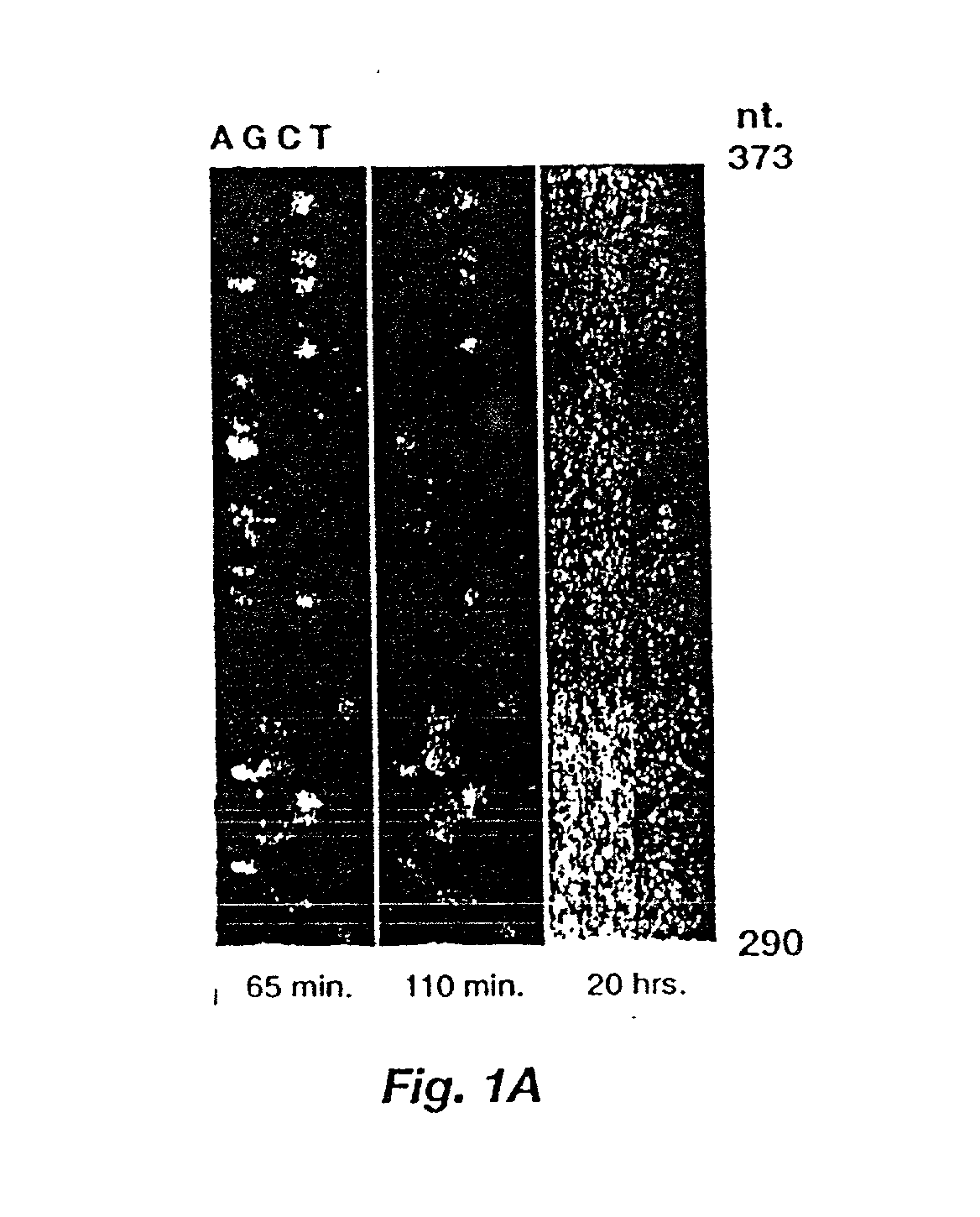
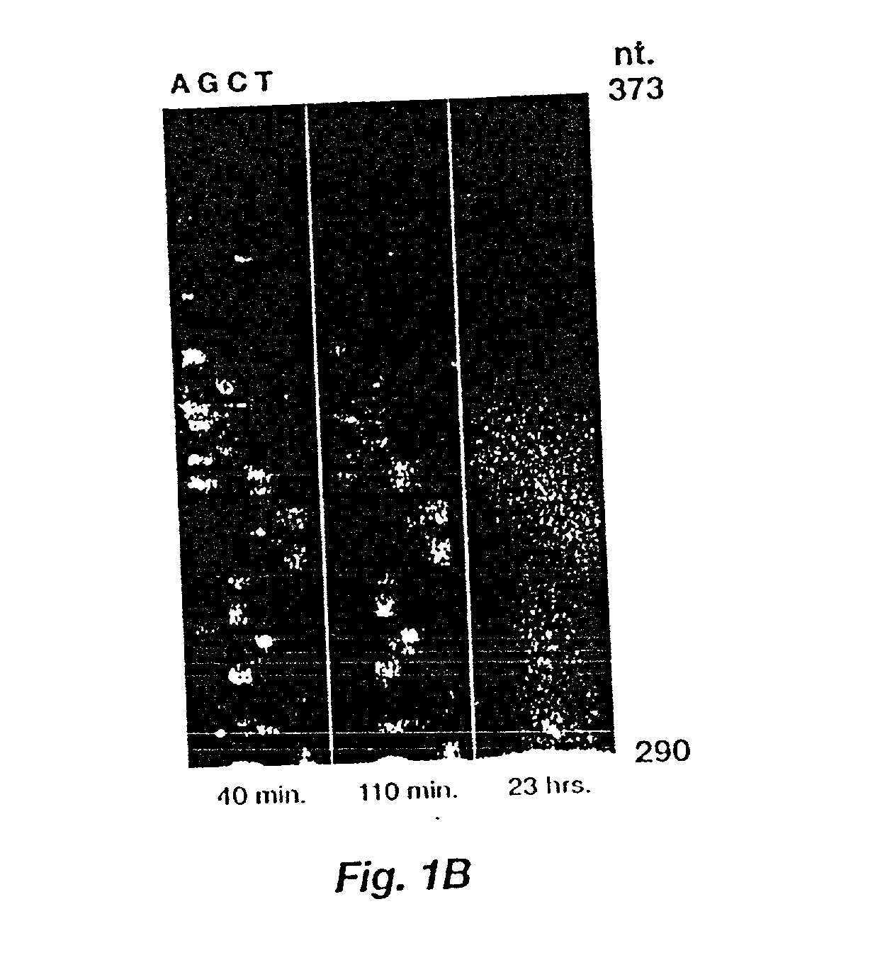
[0043] The membrane from Example 2 was cut so that each of the three substrates could be applied to a sequencing ladder. The enzymatic reaction was started by addition of 1 ml of a fluorogenic alkaline phosphatase substrate for every 300 cm.sup.2 of membrane. The stock solutions were: MUFP-50 .mu.g / ml in 0.1 M diethanolamine, pH 10; 5MFP-50 .mu.g / ml in 0.1 M diethanolamine, pH 10; and BBTP-600 .mu.g / ml in 2.4 mM diethanolamine, pH 10. The substrate solution was applied as an even coat on the membrane.

[0044] In this example, the membranes were imaged outside the chamber, thus membranes were then placed on glass plates and wrapped with transparent plastic film. Fluorescence emission was excited by a 458 nm line of an argon ion laser (Lexel model 96) for 5MFP and BBTP or a long wave UV mercury lamp for MUFP. Laser light was passed through a lens to widen the beam distribution and through a 450 nm, 40 nm bandwidth bandpass filter (Melles Griot). Images were obtained by a cryogenically c...

example 4

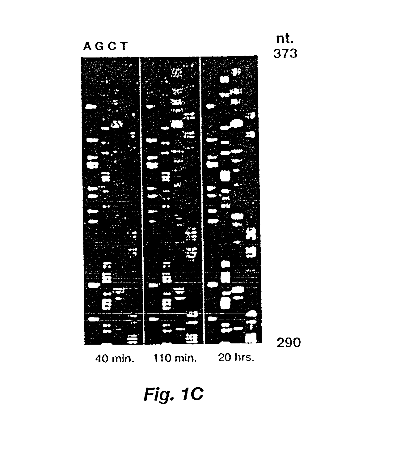
[0062] A full length image of the BBTP membrane, 40 minutes after addition of substrate, shown in FIG. 1C was acquired with a CCD camera operating in TDI mode and was processed using the automatic gel reading software of serial no. 07 / 978,915 modified to accept a positive fluorescent image rather than a negative one as is usual with imaging on X-ray film. FIG. 5 shows a portion of the unprocessed one-dimensional traces, obtained by averaging horizontally across several pixels at the lane centers. The BBTP membrane was up-sampled by a factor of two to produce a 300 dots-per-inch image expected by the gel reader. Lanes were found manually using a Macintosh program, and the data shown were produced by averaging the value of 15 pixels per row around the lane centers. The individual traces have not been processed in FIG. 5, but the traces have been slightly shifted relative to one another to obtain better alignment.

[0063] The automated reader yielded reasonably accurate sequence as far a...


PUM

PropertyMeasurementUnit
pHaaaaaaaaaa
pHaaaaaaaaaa
timeaaaaaaaaaa

Abstract

A method is disclosed for automated multiplex sequencing of DNA with an integrated automated imaging hybridization chamber system. This system comprises an hybridization chamber device for mounting a membrane containing size-fractionated multiplex sequencing reaction products, apparatus for fluid delivery to the chamber device, imaging apparatus for light delivery to the membrane and image recording of fluorescence emanating from the membrane while in the chamber device, and programmable controller apparatus for controlling operation of the system. The multiplex reaction products are hybridized with a probe, then an enzyme (such as alkaline phosphatase) is bound to a binding moiety on the probe, and a fluorogenic substrate (such as a benzothiazole derivative) is introduced into the chamber device by the fluid delivery apparatus. The enzyme converts the fluorogenic substrate into a fluorescent product which, when illuminated in the chamber device with a beam of light from the imaging apparatus, excites fluorescence of the fluorescent product to produce a pattern of hybridization. The pattern of hybridization is imaged by a CCD camera component of the imaging apparatus to obtain a series of digital signals. These signals are converted by the controller apparatus into a string of nucleotides corresponding to the nucleotide sequence an automated sequence reader. The method and apparatus are also applicable to other membrane-based applications such as colony and plaque hybridization and Southern, Northern, and Western blots.

Description

[0002] This invention relates generally to the field of nucleic acid hybridization on membranes. More particularly, this invention relates to a method for automated multiplex sequencing of DNA.[0003] Large scale nucleotide sequencing initiatives, such as a project to sequence the human genome, have created a need for increased efficiency and productivity. J. Watson, 248 Science 44 (1990). Automation of the various steps involved in sequencing is one area in which gains in efficiency and productivity are being made.[0004] Multiplex sequencing, one scheme for reducing the number of sequencing reactions and electrophoresis steps, involves the processing of a mixture of sequencing templates followed by sequential hybridization with selected probes. G. Church & S. Kieffer-Higgins, 240 Science 185 (1988); U.S. Pat. No. 4,942,124. In this method, many sequencing templates, each carrying a short known sequence or tag, are processed together. A single DNA preparation yields a mixture of temp...

Claims


Application Information

Patent Timeline
Patent Type & Authority Applications(United States)
IPC IPC(8): B01L7/00C12Q1/68C12Q1/6869G01N21/64G01N27/447
CPCB01L7/52C12Q1/6869G01N27/44721
Inventor WEISS, ROBERT B.KIMBALL, ALVIN W.GESTELAND, RAYMOND F.FERGUSON, F. MARKDUNN, DIANE M.DI SERA, LEONARD J.CHERRY, JOSHUA L.
Owner WEISS ROBERT B
Why Eureka
Try Eureka