
Treating or preventing the early stages of degeneration of articular cartilage or subchondral bone in mammals using carprofen and derivatives

Inactive Publication Date: 2003-01-09
EVANS NIGEL A +4
View PDF2 Cites 3 Cited by
  • Summary
  • Abstract
  • Description
  • Claims
  • Application Information

AI Technical Summary

Problems solved by technology

Thus, upregulation of IL-1 may have long-term adverse effects on cartilage maintenance.
However, the specific contribution of cytokine action to the pathophysiology of OA is not well understood.
Cats, dogs and horses among mammals are especially vulnerable to inflammatory diseases and processes such as rheumatoid arthritis, osteoarthritis, traumatic or degenerative joint disease, use of impaired joints, actual or incipient hip dysplasia, and osteochondrosis.
Treating mammals with anti-inflammatory agents is especially troublesome in two regards.
First, the pathologic changes in cartilage and subchondral bone in the joints of mammals most prevalently accompanies osteoarthritis, which is a multifactorial and variably expressed disease which is still not fully understood, making decisions about appropriate therapy often difficult.
More recently, however, this paradigm has been questioned.
Second, long-term application of most NSAIDs, especially those more established in use, may actually exacerbate the progress of osteoarthritis.
The loss of subchondral bone increases the mechanical strain on the overlying articular cartilage, leading to its degeneration.
The subsequent thickening of the subchondral plate negatively affects intrinsic repair mechanisms and thereby contributes to the progression of cartilage breakdown.
This stage of hypertrophic repair of the articular cartilage may persist for some time, but the repair cartilage tissue which is formed lacks the resiliency and resistance to mechanical stress possessed by normal hyaline cartilage.
Eventually, proteoglycan production subsides and the chondrocytes are no longer able to maintain their extracellular matrix.
This end stage results in full-thickness loss of articular cartilage.
However, when a comparison is made between a group of mammals with synovitis and a group of mammals without synovitis, changes in articular cartilage from the two groups are indistinguishable.
The makeup of the repair cartilage is deficient however, due to altered composition and distribution of the glycosaminoglycan component and a change in its capacity to aggregate with the hyaluronic acid component.
Particles released during these pathologic changes may also lead to inflammatory changes in the synovial membrane.
However, there is a significant disparity between IL-1 and IRAP potency, with approximately 130-fold more IRAP being required to abolish the effects of IL-1, as measured in chondrocytes and cartilage explants.
Any imbalance between IL-1 and IRAP will further exacerbate the degeneration of articular cartilage.
Further still, changes in subchondral bone occur before gross alterations in the articular cartilage become apparent because cytokines responsible for initiating and maintaining the inflammatory process gain access to the lower layers of cartilage through microcracks across the calcified zone The metabolism of the chondrocytes involved is adversely affected, and in addition the chondrocytes in the middle zone of the articular cartilage produce many cytokines, including those responsible for initiating and maintaining the inflammatory process.
The increased levels of stromelysin may occur for only a fairly short period of time, but where the damage to the joint transcends the tidemark zone of the articular cartilage, and reaches into the subchondral bone, there is a substantial likelihood of subsequent articular cartilage degeneration, usually preceded by a stiffening of the subchondral bone.
These alterations include increased stiffening of the subchondral bone, accompanied by loss of shock-absorbing capacity.
These subchondral bone changes are caused by inappropriate repair of trabecular microfractures which result, in turn, from excessive loading of the joint.
These alterations in subchondral bone density are not only evidence of an imbalance in the bone remodeling process, but also are a key ingredient in eventual focal cartilage loss.
Further, site-related differences in osteoblast metabolism occur which lead to the production of different cartilage-degrading molecules.
These changes in osteoblast metabolites in turn lead to corresponding changes in chondrocyte metabolism, rendering them more susceptible to cytokine-induced activity of the types above-described.
On the other hand, increased levels of viscosity in synovial fluids pose problems in immunoassay systems which must be addressed by the artisan.
However, said chondroprotective compound may provide less than such optimal results and still be within the scope of the present invention.
The more the disease has progressed, the more difficult it will become to arrest or reverse the disease process.
Said pathologic changes include changes in the composition, form and density of the articular cartilage from that present before the onset of said disease process, which result in a degradation of the beneficial properties of said articular cartilage including strength, resilience, elasticity, conformational integrity and stability, viability, and the ability to successfully resist various kinds of mechanical stress, especially the ability to absorb mechanical shocks.
Occasionally supersaturated solutions may be utilized, but these present stability problems that make them impractical for use on an everyday basis.
However, the use of depots and implants as well as delayed-, sustained-, and controlled-release formulations has tended to blur these distinctions.
However, this generalization does not take into account such important variables as the specific type of articular cartilage or subchondral bone degeneration or destruction involved, the specific therapeutic agent involved and its pharmacokinetics, and the specific patient (mammal) involved.
One of the most significant of these is heartworm, which is a very damaging and often fatal parasitic affliction of cats and dogs.
This follows from the fact that invasive surgery on the joint of a mammal, especially a dog, inevitably degrades the ability of that joint to bear its accustomed load as efficiently as before surgery.

Method used


Image

  • Treating or preventing the early stages of degeneration of articular cartilage or subchondral bone in mammals using carprofen and derivatives
  • Treating or preventing the early stages of degeneration of articular cartilage or subchondral bone in mammals using carprofen and derivatives
  • Treating or preventing the early stages of degeneration of articular cartilage or subchondral bone in mammals using carprofen and derivatives

Examples


example 1

[0144] Three groups of skeletally mature crossbred mammals, weighing 20 to 25 kg each, are used in a study whose objective is to demonstrate subchondral bone changes which are a marker for the early stages of articular cartilage degeneration in mammals, and which, accordingly, may be used to identify mammals which are appropriate candidates for pharmacologic intervention.

[0145] In Group I, osteoarthritis is induced in these mammals (n=4) using the surgical procedure described in Pelletier, J-P , Martel-Pelletier, J.; Altman, R. D.; Ghandur-Mnaymneh, L.; Howell, D S.; Woessner, J. F , Jr; "Collagenolytic Activity and Collagen Matrix Breakdown of the Articular Cartilage in the Pond-Nuki Mammal Model of Osteoarthritis", Arthritis Rheum, 26, 1983, 866-874; wherein said mammals are anesthetized with intravenous injection of sodium pentobarbital (25 mg / kg), and the anterior cruciate ligament of the right knee is sectioned by a stab incision. After surgery the mammals are housed and allowe...


PUM

PropertyMeasurementUnit
Frequencyaaaaaaaaaa
Frequencyaaaaaaaaaa
Ratioaaaaaaaaaa

Abstract

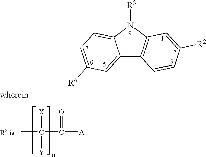
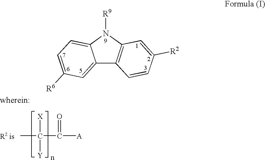
Treating or preventing the early stages of degeneration of articular cartilage or subchondral bone in the affected joint of a mammal is accomplished by administering a chondroprotective compound of Formula (I): where A is hydroxy, (C1-C4)alkoxy, amino, hydroxy-amino, mono-(C1-C2)alkylamino, di-(C1-C2)alkylamino; X and Y are independently H or (C1-C2)alkyl; and n is 1 or 2; R6 is halogen, (C1- C3)alkyl, trifluoromethyl, or nitro; R9 is H; (C1-C2)alkyl; phenyl or phenyl-(C1- C2)alkyl, where phenyl is optionally mono-substituted by fluoro or chloro; -C(=O)-R, where R is (C1-C2)alkyl or phenyl, optionally mono-substituted by fluoro or chloro; or -C(=O)-O-R1, where R1 is (C1- C2)alkyl. This treatment ameliorates, diminishes, actively treats, reverses or prevents any injury, damage or loss of articular cartilage or subchondral bone subsequent to said early stage of said degeneration. Whether or not a mammal needs such treatment is determined by whether or not it exhibits a statistically significant deviation from normal standard values in synovial fluid or membrane from the affected joint, with respect to at least five of the following substances: increased interleukin-1 beta (IL-1beta), increased tumor necrosis factor alpha (TNFalpha); increased ratio of IL-1beta to IL-1 receptor antagonist protein (IRAP); increased expression of p55 TNF receptors (p55 TNF-R), increased interleukin-6 (IL-6); increased leukemia inhibitory factor (LIF); decreased insulin-like growth factor-1 (IGF-1); decreased transforming growth factor beta (TGFbeta); decreased platelet-derived growth factor (PDGF); decreased basic fibroblast growth factor (b-FGF); increased keratan sulfate; increased stromelysin; increased ratio of stromelysin to tissue inhibitor of metalloproteases (TIMP); increased osteocalcin; increased alkaline phosphatase; increased cAMP responsive to hormone challenge; increased urokinase plasminogen activator (uPA); increased cartilage oligomeric matrix protein; and increased collagenase.

Description

[0001] The present invention relates to the use of carprofen in mammals as a means of treating and preventing cartilage and subchondral bone injury and loss in the inflamed joints of such mammals. Such damage to the cartilage and subchondral bone is a natural sequelae of the process of osteoarthritis and its aftermath when it occurs in the mammal. The ability of carprofen to achieve this unexpected result is referred to as "chondroprotection".[0002] Carprofen has been used heretofore as a COX-2 selective non-steroidal anti-inflammatory drug (NSAID) whose activity was based at least in part on the potent and selective inhibition of the inducible cyclooxygenase II (COX-2) isoenzyme. Such activity does not, however, exclude the possibility that carprofen, like other NSAIDs, possesses inhibitory activity with respect to the enzymes involved in the lipoxygenase pathway, or that it is active against the suppression, recruitment and migration of inflammatory cells and the release of enzyme...

Claims


Application Information

Patent Timeline
IPC IPC(8): A61K31/00A61K31/403A61K31/40A61K31/405A61P19/00A61P19/02A61P19/08A61P43/00C07D209/88
CPCA61K31/405A61P19/00A61P19/02A61P19/08A61P43/00A61K31/40
Inventor EVANS, NIGEL A.KILROY, CAROLYN R.LUNDY, KRISTIN M.PELLETIER, JEAN-PIERRERICKETTS, ANTHONY P.
Owner EVANS NIGEL A
Learn More