Microcapsules containing biomedical materials

a biomedical material and microcapsule technology, applied in the field of microcapsules, can solve the problems of insufficient stability of capsules, deformation, and limitation of the application of this approach to treatment, and achieve the effect of low cos

Inactive Publication Date: 2005-06-02
MCMASTER UNIV
View PDF8 Cites 41 Cited by
  • Summary
  • Abstract
  • Description
  • Claims
  • Application Information

AI Technical Summary

Benefits of technology

[0023] The process of the invention is simple, low cost, requires no complex steps or chemical syntheses and has the benefit that the biomedical materia...

Problems solved by technology

However, it suffers from a major drawback in that the capsules are insufficiently stable and degrade with time.
The rate of this degradation and failure, which depends on the nature of the host organism, severely limits the application of this approach to the treatment. of human patients.
The capsules have an inherent lack of strength such that when subject to an osmotic shock they disintegrate.
However, they are inconvenient for the following reasons.
Reagents such as acrylic anhydride are expensive as their preparation and isolation are difficult.
These reagents cannot be conveniently stored for long periods.
In addition, the capsules produced by such methods have relatively rough surfaces and are smaller in diameter and thus more dense than capsules made using the previously kno...

Method used


Image

  • Microcapsules containing biomedical materials
  • Microcapsules containing biomedical materials
  • Microcapsules containing biomedical materials

Examples


example 1

Detailed Procedure with Acrylic Acid using Irgacure as the Photoinitiator with Long Wavelength UV Light

[0068] A solution containing 100 μl of 0.2% Irgacure 2959 in saline, 30 μl of 1.39 M Acrylic Acid in saline and 50 μl of 0.834M N-vinylpyrrolidone in saline were added to 2 ml of calcium microcapsule in a 60 mm cell culture dish. After a gentle shaking, the microcapsules were immediately enclosed to UV light (wavelength of approximately 350 nm) for varying periods at 4° C. Afterwards, the capsules were washed with fresh 1.1% CaCl2 to remove unreacted reagents. The capsules were then treated with poly-L-lysine and alginate in the standard manner. Sterile techniques were used throughout the whole procedure.

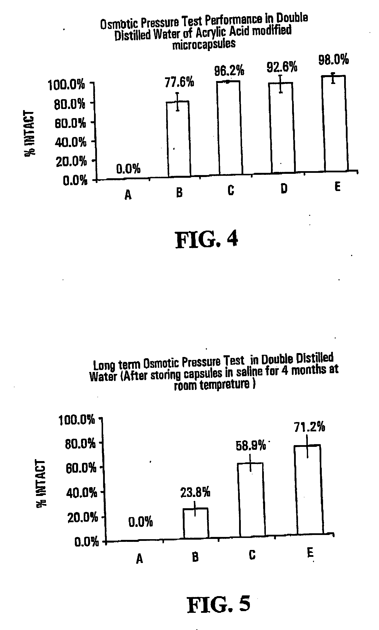
[0069]FIG. 4 shows results of osmotic pressure tests in double distilled water on capsules of the invention and “standard” capsules, i.e., capsules that had not been subjected to photopolymerization with an ethylenically unsaturated monomer. The osmotic pressure test measures th...

example 2

Detailed Procedure with Acrylic Acid using Ethyl Eosin as the Photoinitiator

[0078] The capsules as obtained in Example 1 were suspended in 10 ml of an ethyl eosin solution (see above for formulation) for 2 min to allow uptake of the dye, then washed three times with fresh 1.1% CaCl2 to remove non-absorbed dye. The microcapsules were transferred from the CaCl3 solution to a 0.9% NaCl solution for photomoaification.

[0079] A solution was prepared by admixing 100 μl of 4% w / v of triethanolamine in physiological saline, 30 μL of 1.39M acrylic acid in physiological saline and 25 μl of 0.832M N-vinylpyrrolidone in physiological saline. The solution was added to 2 ml of these microcapsules contained in a 60 mm cell culture dish. After a gentle shaking, the microcapsules were immediately exposed to visible light (wavelength greater than 400 nm) for a defined period at 4° C. After the irradiation, the capsules were washed with fresh 1.1% CaCl2 solution to remove unreacted reagents. The cap...

example 3

Detailed Procedure with Sodium Acrylate using Ethyl Eosin as the Photoinitiator with Visible Wavelength Light

[0088] A procedure similar to Example 2 was used, with acrylic acid are the ethylenically unsaturated monomer and with ethyl eosin as an initiator. The same molar amount of sodium acrylate (30 μl of a 1.39M solution) and varying concentrations of N-vinylpyrrolidone were added to 2 ml of the suspended capsules. (The amount of sodium acrylate used corresponds to a 10% modification.)

[0089]FIGS. 7 and 8 show results of osmotic pressure tests conducted on capsules formed with varying amounts of sodium acrylate. The tests were carried out upon formation of the capsules, and after storage in saline for Tofthu at room temperature, respectively. Details are as follows: [0090] A—Standard alginate-poly-L-Lysine-alginate microcapsules [0091] B—10% w / w sodium acrylate (SA) (30 μl 1.35M) to Alginate [0092] C—20% w / w sodium acrylate (SA) 60 μl 1.39M to Alginate [0093] D—50% w / w sodium ac...


PUM

PropertyMeasurementUnit
Wavelengthaaaaaaaaaa
Strengthaaaaaaaaaa
Molar ratioaaaaaaaaaa

Abstract

Biomedical materials are encapsulated in ionically crosslinked polymer capsules, preferably alginate microcapsules. The alginate capsules are then subjected, in a liquid vehicle, to an ethylenically unsaturated monomer and an initiator, to induce polymerization of the unsaturated monomer and therey enhance the strength of the capsule wall. The microcapsules can be after-treated with, for example, polysine and alginate to reduce their tendency to elicit an immune response if implanted in an animal. The invention extends to the microcapsules and also to a method of treating or preventing medical conditions in an animal particularly a human, by implanting microcapsule containing biomedical material in the animal.

Description

FIELD OF THE INVENTION [0001] The present invention relates to microcapsules. More specifically, the present invention relates to the formation of microcapsules that can be implanted in an animal and / or human. The microcapsules may contain biomedical material for example, cells, especially recombinant cells for gene therapy, proteins and / or drugs for long term delivery. BACKGROUND OF THE INVENTION [0002] It is known that microcapsules can be prepared from alginate cross-linked with Ca2+. These capsules are well suited for the incorporation of living cells, and allow the diffusion of nutrients into and expressed proteins out of the capsules. Particularly if the microcapsules are coated first with poly-L-lysine ad then subsequently with alginate, they show little image response when implanted within a mammalian host. They have been used as a convenient means of supplying a hormone to a human or non-human animal lacking the ability to produce such a material. This classical method of e...

Claims


Application Information

Patent Timeline
IPC IPC(8): A61K9/50A61K9/52B32B5/16B65B1/00
CPCA61K9/0024A61K9/5026Y10T428/2984A61K9/5073A61K9/5089A61K9/5036
Inventor CHILDS, RONALD F.SHEN, FENGWANG, SANJU
Owner MCMASTER UNIV
Why Eureka
Try Eureka