
Nuclear magnetic resonance-docking of compounds

Inactive Publication Date: 2005-10-20
TRIAD THERAPEUTICS
View PDF3 Cites 4 Cited by
  • Summary
  • Abstract
  • Description
  • Claims
  • Application Information

AI Technical Summary

Benefits of technology

[0021] An advantage of the invention is that a structure model of a test ligand bound to the macromolecule can be obtained at sufficient resolution to assist in structure-based design of a biologically active agent or drug without the requirement for a complete determination of the structure of the macromolecule-test ligand complex. In particular, by comparing the spectra arising from different complexes, structural constraints for the bound ligand can be obtained without the need to characterize atoms of the macromolecule that do not interact with the ligand. For example, where the spectroscopic method is nuclear magnetic resonance (NMR) spectroscopy, selective observation of magnetic signals arising from ligand-macromolecule interactions allows a structure model of the ligand to be obtained more rapidly than by conventional NMR methods which typically require that resonances be assigned for non-binding site atoms of the macromolecule. Moreover, the methods of the invention can be used with larger macromolecules compared to conventional NMR methods because selective observation of magnetic signals arising from ligand-macromolecule interactions reduces problems associated with resonance overlap.
[0022] The invention further provides a method for determining a structure model for a macromolecule bound to a ligand. In the method, structural constraints derived from spectroscopically observed interactions of the macromolecule and ligand are used to guide molecular modeling or to evaluate the results of a molecular modeling simulation. An advantage of the method is that by combining binding site-focused spectroscopic measurements with molecular modeling, an accurate structure model of the macromolecule can be obtained more rapidly and efficiently than with conventional spectroscopic methods. Definitions
[0023] As used herein, the term “structure model” is intended to mean a representation of the relative locations of atoms of a molecule. A representation included in the term can be defined by a coordinate system that is preferably in 3 dimensions, however, manipulation or computation of a model can be performed in 2 dimensions or even 4 or more dimensions in cases where such methods are desired. The location of atoms in a molecule can be described, for example, according to bond angles, bond distances, relative locations of electron density, probable occupancy of atoms at points in space relative to each other, probable occupancy of electrons at points in space relative to each other or combinations thereof. A representation included in the term can contain information for all atoms of a particular molecule or a subset of atoms thereof. Examples of representations included in the term that contain a subset of atoms are those commonly used for polypeptide structures such as ribbon diagrams, and the like, which show the coordinates of the polypeptide backbone while omitting coordinates for all or a portion of the side chain moieties of the polypeptide. Representations for other macromolecules and small molecules included in the term can similarly contain all or a subset of atoms.
[0024] A structure model can include a representation that is determined from empirical data derived from, for example, X-ray crystallography or nuclear magnetic resonance spectroscopy. A representation included in the term can also be derived from a theoretical calculation including, for example, comparison to a known structure such as in homology modeling or ab initio molecular modeling. A representation of a structure model can include, for example, an electron density map, atomic coordinates, x-ray structure model, ball and stick model, density map, space filling model, surface map, Connolly surface, Van der Waals surface or CPK model.
[0025] As used herein, the term “binding site-localized” is intended to mean an atom of a macromolecule or bound ligand that is proximal to one or more atoms of a second ligand in a complex containing the macromolecule and second ligand or a complex containing the macromolecule and both ligands. Proximal atoms included in the term are those that are within a distance sufficient to cause a chemical interaction such as a hydrogen bond, van der Waals interaction or ionic interaction or to cause a magnetic interaction detectable by a nuclear magnetic resonance spectroscopy measurement used in the methods of the invention. Examples of magnetic effects included in the term are a relaxation effect which can be detected for atoms that are about 10 Å apart or closer, the Nuclear Overhauser Effect which can be detected for atoms that are about 6 < apart or closer or chemical shift due to shielding or de-shielding which can be detected for atoms that are about 10 Å or closer. Atoms that are about 5 Å apart or closer, 4 Å apart or closer, 3 Å apart or closer, 2 Å apart or closer or 1 Å apart or closer are also proximal atoms that are included in the term.
[0026] As used herein, the term “macromolecule” is intended to mean a polymeric molecule or complex of polymeric molecules that are associated in solution, including biological and synthetic polymers. Proteins and other polypeptides are particularly useful biological polymers. Other useful biological polymers include polysaccharides and polynucleotides. Polynucleotides are also referred to herein as nucleic acids. Synthetic polymers include plastics and mimetics of biological polymers such as protein-nucleic acids.

Problems solved by technology

However, for many drug targets the throughput of available screens is prohibitively low.
Furthermore, even in cases where high throughput detection is available, limitations on available resources for obtaining a library with sufficient size or diversity, or for obtaining a sufficient quantity of the drug target to support a large screen, can be prohibitive.
For many drug targets of interest, three-dimensional structure models are not presently available.
Although methods for structure determination are evolving, it is currently difficult, costly and time consuming to determine the structure of a macromolecule drug target at sufficient resolution to render structure-based drug design practical.
It can often be even more difficult to produce a macromolecule-ligand complex in a condition allowing determination of the bound conformation of the ligand.
The typically long time period required to obtain structure information useful for developing drug candidates is particularly limiting with regard to exploiting the growing number of potential drug targets identified by genomics research.

Method used


Image

  • Nuclear magnetic resonance-docking of compounds
  • Nuclear magnetic resonance-docking of compounds
  • Nuclear magnetic resonance-docking of compounds

Examples


example i

Docking of a Furoic Acid-Based Inhibitor into the Binding Site of DHPR

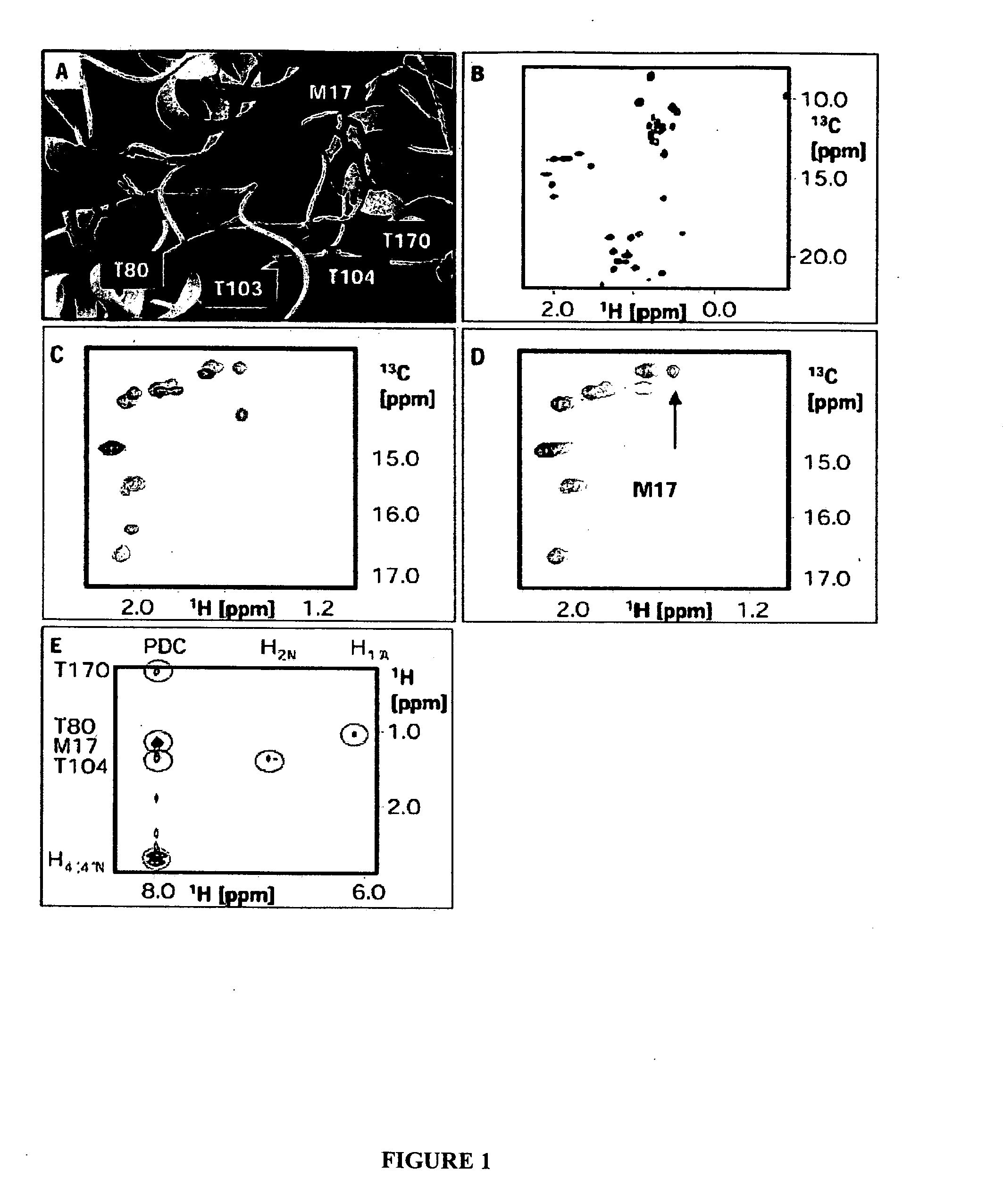
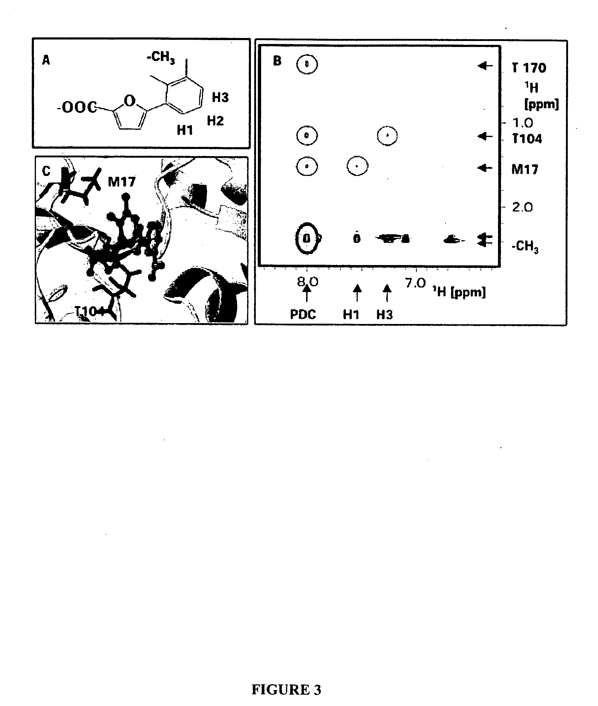
[0126] This Example demonstrates determination of a three dimensional model of a furoic acid-based inhibitor bound to the NADH binding site of E. coli Dihydrodipicolinate reductase (DHPR). In particular, this example describes, expression and purification of isotopically labeled DHPR; NMR measurements of a DHPR-NADH complex to assign DHPR binding site residues that interact with NADH; NOE measurements of a DHPR-inhibitor complex to determine distances between the binding site residues and the inhibitor; and docking of the inhibitor to a previously determined structure model of DHPR based on distance constraints derived from the NOE measurements.

A. Expression of Isotopically Labeled DHPR

[0127]E. coli DHPR was selectively labeled with 13Cε / 1H Met, 13Cδ / 1H Ile and 13C / 1H Thr and uniformly labeled with 2H. The resulting labeled protein is referred to as MIT-DHPR. This labeling scheme was chosen based on analysis o...

example ii

Overlay of a Furoic Acid-Based Inhibitor onto DHPR-Bound NADH

[0146] This Example describes determination of a three dimensional model of a furoic acid-based inhibitor (TTM2000—29—85) by comparison to the structure of NADH when bound to E. coli Dihydrodipicolinate reductase (DHPR). In particular, this example describes comparing cross-peaks for a 2D NOESY spectrum of a DHPR-NADH complex with cross-peaks for a 2D NOESY spectra of a DHPR-TTM2000—29—85 complex and overlaying a structure model of TTM2000—29—85 and NADH based on distance constraints derived from the NOE measurements. As described below, neither assignment of DHPR-derived peaks to particular binding site residues nor a structural model of DHPR is necessary to determine structural properties of the inhibitor by ligand overlay.

[0147] DHPR is expressed, isotopically labeled and purified and NMR measurements are obtained as described in Example 1.

[0148] Binding site cross-peaks are identified from NOESY spectra for the tern...

example iii

Validation of a Binding Site Homology Model for 1-Deoxy-D-Xylulose-5-Phosphate Reductoisomerase

[0151] This example demonstrates generation of a homology model for 1-Deoxy-D-xylulose 5-phosphate reductoisomerase (DOXPR) based on sequence analysis. Validation of the model using nuclear magnetic resonance spectroscopy is also demonstrated.

[0152] 1-Deoxy-D-xylulose 5-phosphate reductoisomerase (DOXPR) is an enzyme involved in isoprenoid biosynthesis, catalyzing the formation of 2-C-methyl-D-erythritol from 1-deoxy-D-xylulose 5-phosphate (Takahashi et al., Proc. Natl. Acad. Sci. USA 95:9879-9884 (1998)). The deoxyxylulose pathway, found in some bacteria, algae, plants and protozoa, is an alternate to the ubiquitous mevalonate pathway for isoprenoid biosynthesis (Eisenreich et al., Trends Plant Sci. 6:78-84 (2001)). Because a three dimensional model of the DOXPR structure was not available and to aid in the design of inhibitors of DOXPR, a model for the NADPH-binding, N-terminal domain ...


PUM


Abstract

The invention provides a method for determining a structure model for a test ligand bound to a macromolecule binding site. Structural constraints for the test ligand are derived from spectroscopic signals arising from interactions between the test ligand and macromolecule. The structure constraints are used as constraints in docking a structure model of the ligand to a structure model of the macromolecule, or as constraints in overlaying a structure model of the test ligand on the known structure for a reference ligand that binds to the macromolecule. The invention further provides a method for determining a structure model for a macromolecule bound to a ligand. Structural constraints derived from spectroscopically observed interactions of the macromolecule and a reference ligand are used to guide molecular modeling or to evaluate the results of a molecular modeling simulation of the macromolecule.

Description

[0001] This application is based on, and claims the benefit of, U.S. Provisional Application No. 60 / 294,675, filed May 30, 2001, which is incorporated herein by reference.BACKGROUND OF THE INVENTION [0002] The present invention relates generally to interactions between macromolecules and ligands and more specifically to Nuclear Magnetic Resonance (NMR) methods for determining structure-related properties of a ligand when bound to a macromolecule. [0003] Structure determination plays a central role in chemistry and biology due to the correlation between the structure of a molecule and its function. Although a full understanding of this correlation is not yet established, one can gain insight into the function of a molecule from its deduced structure. Thus, the structure can provide a strong basis for directing the development of molecules having a desired function. Conversely, the eventual disclosure of a structure for a well studied molecule can have a significant effect in convergi...

Claims


Application Information

Patent Timeline
IPC IPC(8): G01N24/08G01R33/46G01R33/465
CPCG01N24/08Y10T436/24G01R33/465G01R33/4625
Inventor SEM, DANIEL S.PELLECCHIA, MAURIZIO
Owner TRIAD THERAPEUTICS
Features
  • Generate Ideas
  • Intellectual Property
  • Life Sciences
  • Materials
  • Tech Scout
  • Unparalleled Data Quality
  • Higher Quality Content
  • 60% Fewer Hallucinations
Learn More