

Method for detecting analytes based on evanescent illumination and scatter-based detection of nanoparticle probe complexes
a nanoparticle probe and probe complex technology, applied in the field of specific binding partner interactions, evanescent waveguides and light scattering, can solve the problems of unable to achieve sufficient sensitivity of methods, and low detection limit of 10 fmol targetsup>9 /sup>9 /sup>, etc., to achieve the effect of higher sensitivity
- Summary
- Abstract
- Description
- Claims
- Application Information
AI Technical Summary
Benefits of technology
Problems solved by technology
Method used
Image

Examples
example 1
Preparation of Nanoparticle-Oligonucleotide Conjugate Probes
[0108] In this Example, a representative nanoparticle-oligonucleotide conjugate detection probe was prepared for the use in the detection of APC, Factor V Leiden gene, or mecA gene targets.
(a) Preparation of 15 nm Diameter Gold Nanoparticles
[0109] Gold colloids (˜15 nm diameter) were prepared by reduction of HAuCl4 with citrate as described in Frens, 1973, Nature Phys. Sci., 241:20-22 and Grabar, 1995, Anal. Chem. 67:735. Briefly, all glassware was cleaned in aqua regia (3 parts HCl, 1 part HNO3), rinsed with Nanopure H2O, then oven dried prior to use. HAuCl4 and sodium citrate were purchased from Aldrich Chemical Company. Aqueous HAuCl4 (1 mM, 500 mL) was brought to reflux while stirring. Then, 38.8 mM sodium citrate (50 mL) was added quickly. The solution color changed from pale yellow to burgundy, and refluxing was continued for 15 min. After cooling to room temperature, the red solution was filtered through a Micron...
example 2
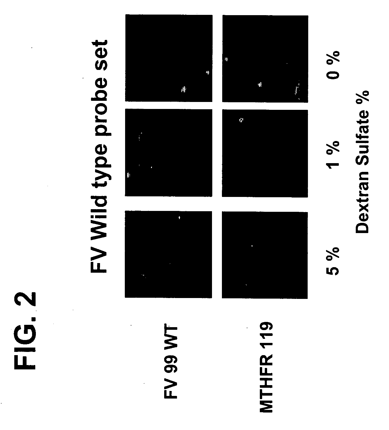
The Use of Dextran Sulfate as a Hybridization Facilitator for Detection of Double Stranded Nucleic Targets with DNA-Modified Gold Nanoparticle Probes
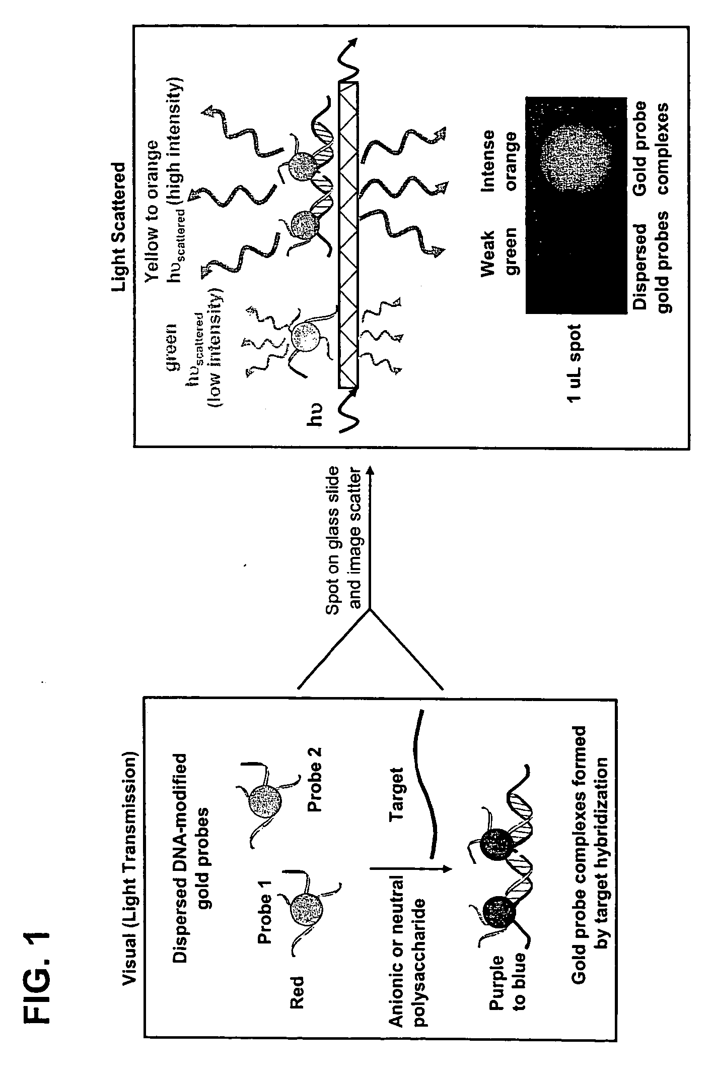
[0118] Metallic nanoparticles (30-120 nm diameter) have the potential to be used in homogeneous scatter-based detection of specific nucleic acid sequences in PCR amplicons or even genomic DNA samples, which has never been demonstrated. Light scattered from gold or silver particle increases with the sixth power of the radius, and a specific color of scattered light is emitted based on the particle composition, size, and shape.36,37 For example, 30-60 nm diameter gold particles scatter green colored light based on the surface plasmon resonance frequency, and light scattered from gold particles in this size range can be detected at picomolar—femtomolar particle concentrations using detection instrumentation that monitors non-evanescent light scattering.36,37 In homogeneous reactions, the color of absorbed or scattered light changes if the...
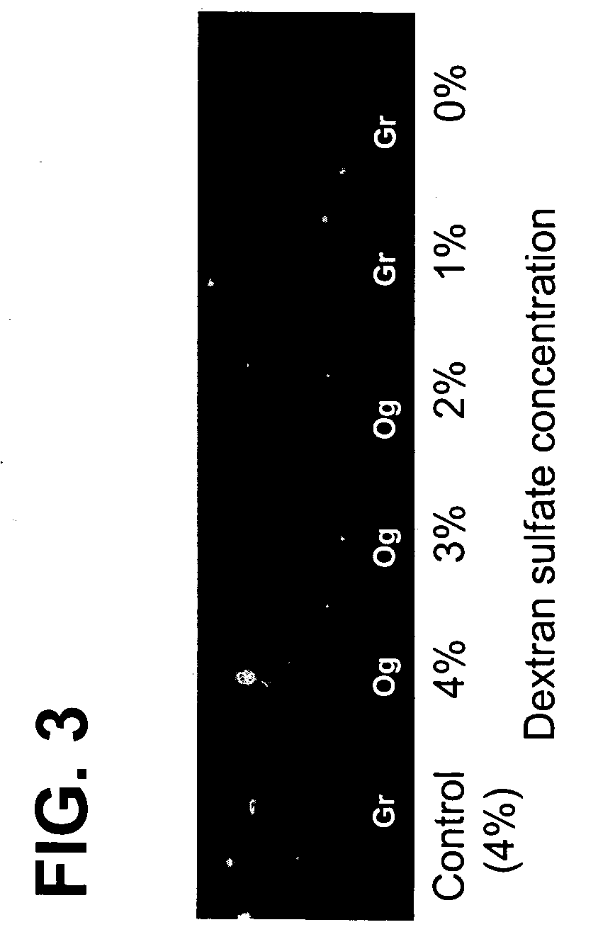
example 3
The Use of Dextran Sulfate as a Hybridization Facilitator for Detection of Double Stranded Nucleic Targets with DNA-Modified Gold Nanoparticle Probes
[0121] An experiment similar to that described in Example 2 was performed using a mecA gene sequence in place of the Factor V Leiden gene. A pair of 40 nm diameter gold probes (SEQ ID NO: 11 and 12) designed to bind to a mecA gene PCR amplicon (281 bp, SEQ ID NO: 14) was used in initial testing. A ˜6 nM mecA 281 base-pair PCR fragment (5 μL, fragment length and approximate concentration determined with an Agilent Bioanalyzer) was mixed with 6 μL of 40 nm diameter gold probe (1:1 ratio, 50 pM total probe), and 4 μL of hybridization buffer (buffer contains 20% formamide, 16% dextran sulfate, and 3.75 mM MgCl2). The solutions were heated to 95° C. for 30 seconds and incubated in a water bath at 40° C. for 15 min. A 1 μL aliquot of each sample was spotted and imaged wet. As shown in FIG. 3, gold probe samples containing more than 2% dextra...
PUM
| Property | Measurement | Unit |
|---|---|---|
| Color | aaaaa | aaaaa |
| Volume | aaaaa | aaaaa |
| Metallic bond | aaaaa | aaaaa |
Abstract
Description
Claims
Application Information