
Contrast agents for magnetic resonance imaging

a magnetic resonance imaging and contrast agent technology, applied in applications, nanomedicine, diagnostic recording/measuring, etc., can solve the problems of poor sensitivity when compared to other imaging techniques, induce anaphylactic reactions, and mr imaging, and achieve enhanced relaxation, high signal-to-noise ratio, and target ability

Inactive Publication Date: 2005-11-24
PRIAVOID GMBH +1
View PDF8 Cites 61 Cited by
  • Summary
  • Abstract
  • Description
  • Claims
  • Application Information

AI Technical Summary

Benefits of technology

[0009] The present invention meets these and other needs by contrast agents based on superparamagnetic iron oxide in a core-shell structure. The contrast agents provide enhanced relaxivity, high signal-to-noise ratios, and targeting abilities. In addition, the contrast agents possess resistance to agglomeration, controlled particle size, blood clearance rate, and biodistribution. A nanoparticle having a signal generating core and a stabilizing coating is also disclosed. Methods of making such contrast agents and nanoparticles are also disclosed.
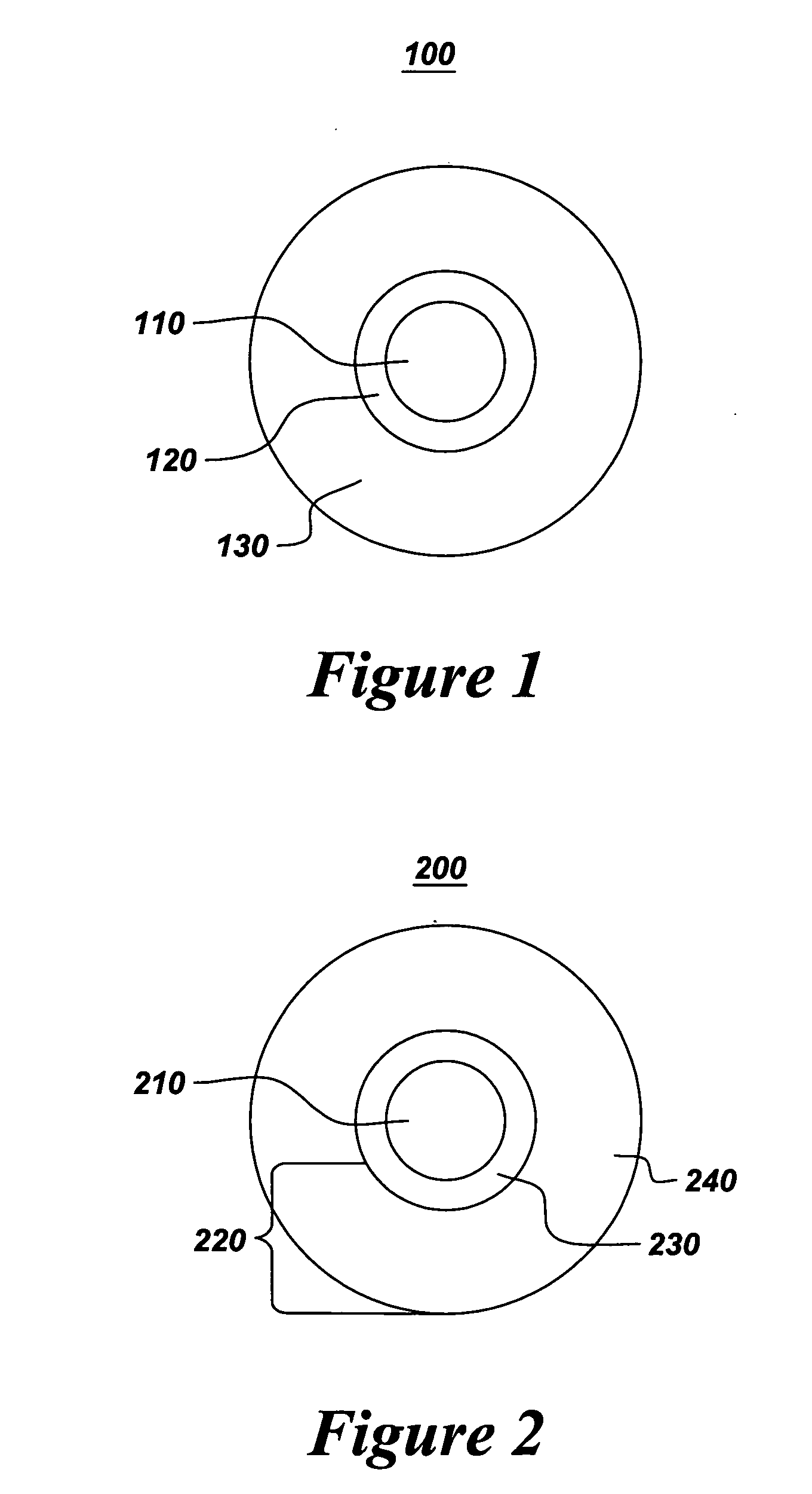
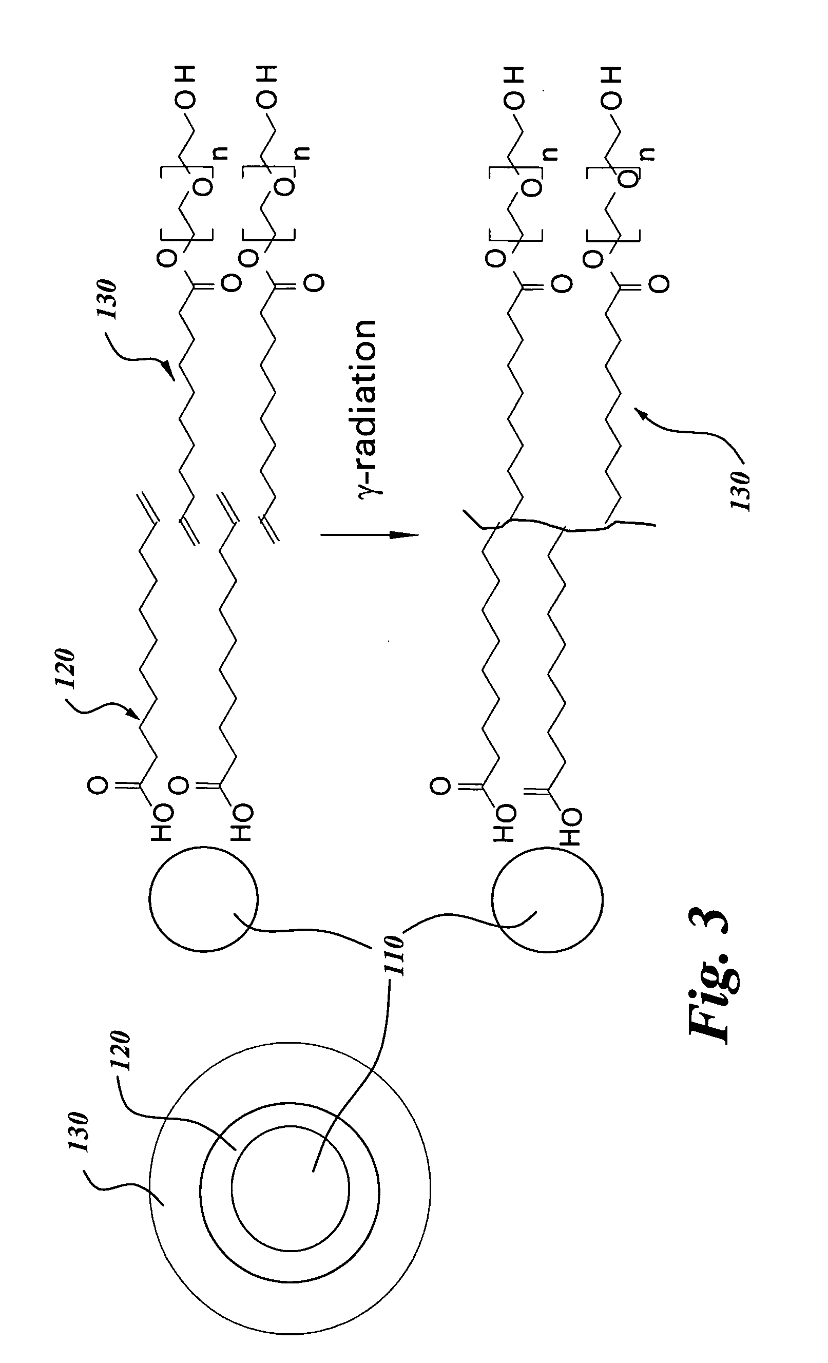
[0010] Accordingly, one aspect of the invention is to provide a contrast agent comprising a plurality of nanoparticles. Each of the plurality of nanoparticles comprises: a signal generating core having a diameter of up to 10 nm; at least one organic layer comprising at least one of a polymer, a monomer, and a surfactant; and a water soluble outer shell comprising at least one of a polymer, a monomer, and a ligand. The at least one organic layer is adsorbed upon and substantially surrounds the signal generating core, and stabilizes the signal generating core. The water soluble outer shell solubilizes each of the plurality of nanoparticles and provides biocompatibility for each of the plurality of nanoparticles.
[0012] A third aspect of the invention is to provide a contrast agent comprising a plurality of nanoparticles. Each of the plurality of nanoparticles comprises: a signal generating core having a diameter of up to 10 nm, wherein the signal generating core is superparamagnetic; and a stabilizing coating disposed on and substantially covering the signal generating core. The stabilizing coating comprises: an inner shell comprising at least one of a polymer, a monomer, and a surfactant, wherein inner shell is adsorbed upon and substantially surrounds the signal generating core, and wherein inner shell stabilizes the signal generating core; and a water soluble outer shell. The water soluble outer shell is disposed on an outer surface of the inner shell and substantially surrounds the inner shell. The water soluble outer shell comprises at least one of a second polymer, a second monomer, and a second ligand. The water soluble outer shell solubilizes each of the plurality of nanoparticles and provides biocompatibility for each of the plurality of nanoparticles.

Problems solved by technology

In in-vivo diagnostics, MR imaging provides good resolution characteristics, but has poor sensitivity when compared to other imaging techniques.
Dextran, however, may induce anaphylactic reactions.
In addition, nanoparticles obtained using current methods also have a low level of crystallinity, which significantly impacts the sensitivity of the contrast agent.
Moreover, nanoparticles tend to agglomerate, due to weak and reversible adsorption of the coating material on the magnetic crystal surface and strong interparticle interactions.
Aggregation increases the size of the nanoparticle, resulting in rapid blood clearance as well as reducing targeting efficiency.
Contrast agents have inherent problems that limit targeting efficiency, such as large particle sizes, tendency to agglomerate, quick blood clearances, low efficiency of ligand attachment, and the accessibility of ligands to the biomarker targets.

Method used


Image

  • Contrast agents for magnetic resonance imaging
  • Contrast agents for magnetic resonance imaging
  • Contrast agents for magnetic resonance imaging

Examples


example 1

[0054] Iron oxide nanocrystals coated with surfactant and monomer, as described hereinabove, were synthesized according to the non-aqueous synthesis route referred to hereinabove. A mixture of trimethylamine-N-oxide, 10-undecenoic acid (or, alternatively, lauric acid or oleoic acid), and deoxygenated dioctyl ether, each individually dehydrated and deoxygenated, was added under an inert atmosphere to a 50 ml 2-neck Schlenk flask. The mixture was homogenized with vigorous stirring and heating to about 100° C. Iron carbonyl (Fe(CO)5) was then added to the reaction solution, which was at a temperature in a range from about 100° C. to about 105° C., resulting in instantaneous and aggressive reaction. The reaction mixture was then heated to a temperature in a range from about 120° C. to about 130° C. under nitrogen and maintained at temperature for about 1 hour while being vigorously stirred. Additional iron carbonyl (Fe(CO)5) was then added to the reaction mixture, and the temperature wa...

example 2

[0055] Monodisperse, bilayer surfactant- or monomer-coated magnetic nanoparticles were synthesized according to the aqueous route described hereinabove. In a typical preparation, NaNO3, FeCl2 (anhydrous) and FeCI2.6H2O were dissolved in deoxygenated Milli-Q water with vigorous stirring under nitrogen. The Fe2+ / Fe3+ molar ratio in the solution was 0.5. The solution was heated to 80° C. and then charged with rapid sequential injections of NH4OH solution and 10-undecenoic acid. Crystal growth proceeded for about 45 minutes at 80° C. with constant vigorous stirring, producing a stable colloidal suspension of nanoparticles, which was then cooled slowly to room temperature with stirring. The suspension was placed on a magnet for at least 1 hour, and then filtered to remove any insoluble material. The material obtained was found to comprise monodisperse spinel-structured mixed iron oxide (γ-Fe2O3)1-y(Fe3O4)y nanocrystals. The average particle size of the nanocrystals an about 8.5±1.2 nm, a...

example 3

[0056] In this example, the preparation of nanoparticles having an iron oxide core, an inner layer comprising a monomer, and a water soluble outer shell that includes at least one ligand comprising PEG and undecenoic acid (PEGylated ligands), all of which are disclosed hereinabove, is described. The PEGYlated ligands were first prepared using either PEGs, or alternatively, PEG monomethyl ethers with molecular weights between 300-5,000 g / mol as starting materials. In one instance, PEG (2,000 Da) was dissolved in dry methylene chloride. Trimethyl amine, and dimethylamino pyridine (DMAP) were added to the solution and stirred under nitrogen in an ice bath. 10-undecenoyl chloride diluted with dry methylene chloride was added dropwise to the chilled solution, and the reaction mixture was stirred for about two hours, first in an ice bath and then at room temperature. The reaction mixture was then filtered, diluted with methylene chloride, and washed three times with 0.1N HCl, 0.1N NaOH an...


PUM

PropertyMeasurementUnit
Temperatureaaaaaaaaaa

Abstract

A contrast agent for magnetic resonance imaging having a plurality of nanoparticles. Each of the nanoparticles has: a signal generating core having a diameter of up to 10 nm; at least one organic layer of at least one of a polymer, a monomer, and a surfactant; and a water soluble outer shell of at least one of a polymer, a monomer, and a ligand. The organic layer is adsorbed upon and substantially surrounds and stabilizes the signal generating core. The water soluble outer shell solubilizes and provides biocompatibility for each of the nanoparticles. The contrast agents provide enhanced relaxivity, high signal-to-noise ratios, and targeting abilities. In addition, the contrast agents possess resistance to agglomeration, controlled particle size, blood clearance rate, and biodistribution. Methods of making such contrast agents and nanoparticles are also disclosed.

Description

CROSS-REFERENCE TO RELATED APPLICATION [0001] This application claims the benefit of U.S. Provisional Application No. 60 / 572,726, filed May 18, 2004.BACKGROUND OF THE INVENTION [0002] The present invention relates generally to the field of magnetic resonance imaging (MRI) contrast agents comprising a plurality of magnetic nanoparticles. More particularly, the present invention relates to the design and synthesis of magnetic nanoparticles comprising a monocrystalline superparamagnetic core coated with an organic shell and decorated with targeting moieties. [0003] Diagnostic imaging procedures and contrast agents are used to study organs, tissues, and diseases in a body. MRI is most effective at providing images of tissues and organs that contain water, such as the brain, internal organs, glands, blood vessels, and joints. Magnetic resonance imaging is based on the magnetic properties of atoms. When focused radio wave pulses are broadcast towards aligned hydrogen atoms in a tissue of ...

Claims


Application Information

Patent Timeline
IPC IPC(8): A61K49/00A61K49/18
CPCA61K49/1839B82Y5/00A61K49/186
Inventor ACAR, HAVVA YAGCISYUD, FAISAL AHMEDGARAAS, RACHEL NICOLEBONITATEBUS, PETER JOHN JR.KULKARNI, AMIT MOHAN
Owner PRIAVOID GMBH
Learn More