Methods for in vitro expansion and transdifferentiation of human pancreatic acinar cells into insulin-producing cells

a technology of pancreatic acinar cells and in vitro expansion, which is applied in the direction of gastrins/cholecystokinins, drug compositions, metabolic disorders, etc., can solve the problems of not being able to obtain insulin-producing cells in good numbers, relative rarity of stem-like cells that can be harnessed, etc., to achieve superior cell attachment, increase the number of cells, and enhance culture efficiency

Inactive Publication Date: 2006-06-08
BECTON DICKINSON & CO
View PDF1 Cites 18 Cited by
  • Summary
  • Abstract
  • Description
  • Claims
  • Application Information

AI Technical Summary

Benefits of technology

[0013] The cell attachment surface for this cell culture system is any surface to which the cells can attach and expand, including both 2 dimensional (e.g. plates, flasks, roller bottles, petri dishes, wells etc.) and 3 dimensional (e.g. scaffold) environments. Preferably the surface comprises at least one type of ECM, or a peptide fragment thereof. Cells may, in some circumstances, detach from these surfaces and form self-supporting aggregates. Suitable fragments include peptides consisting of a sequence of three of more amino acid residues that are identical to any portion of the amino acid sequence of the ECM. Such fragments can be easily made and tested by means known to those of skill in the art. Most preferably the surface is a layer of collagen I. Many other surfaces known in the art are also suitable, such as Collagen VI, Collagen IV, Vitronectin, or Fibronectin. Collagen I is preferred due to ease and cost.
[0017] By the use of this attachment surface and medium, the expansion and transdifferentiation of primary pancreatic cells with the desired phenotype is simplified greatly.
[0019] The cell culture system enables superior attachment in vitro of primary pancreatic epithelial cells for adherent culture compared to prior methods, while creating a cellular environment that promotes expansion of the epithelial component of primary pancreatic cultures with concomitant transdifferentiation of the acinar cells present in the starting material into IP cells, while minimizing emergence of undesired fibroblasts. Advantages of this culture system are ease of construction, few components needed, and that all components are readily available and easily used in the required manner.
[0022] The collagen I surface provides superior cell attachment (thereby increasing the number of cells that adhere during initial culture and thus enhancing culture efficiency), while the collagen I and the combination of soluble active factors (e.g., HGF, TGFA and EGF) promote continued proliferation of cells over time, leading to an increase in cell number above what has been previously reported for primary pancreatic acinar cells. Furthermore, the expansion of the acinar cells is accompanied by a transdifferentiation in the majority of cells to an IP phenotype, which is potentially a therapeutically useful cell phenotype for the treatment of diseases such as diabetes. This likely occurs due to convergence of the intracellular signaling pathways associated with collagen I, HGF, TGFA and EGF, creating a synergistic response.

Problems solved by technology

The challenges presented by these approaches are related to maintenance of function of islets over long periods of culture, and of the relative rarity of stem-like cells that can be harnessed for insulin production from the bone marrow and pancreas.
Prior to the development of the present system, primary pancreatic acinar cells were expanded without differentiation into insulin-producing cells, either in serum-containing medium (undesirable both because of the risk and the uncertainty associated with the use of serum), or in complex serum free media formulations.
Furthermore, it has not been previously possible to obtain insulin-producing cells in good numbers using acinar cells as starting material.

Method used


Image

  • Methods for in vitro expansion and transdifferentiation of human pancreatic acinar cells into insulin-producing cells
  • Methods for in vitro expansion and transdifferentiation of human pancreatic acinar cells into insulin-producing cells
  • Methods for in vitro expansion and transdifferentiation of human pancreatic acinar cells into insulin-producing cells

Examples


example 1

Characterization of Cell Culture Conditions

A. Serum-Free Medium

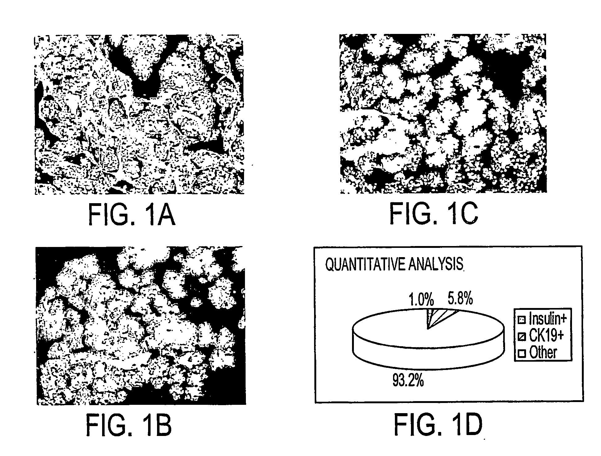
[0080] Freshly isolated primary human pancreatic cells were collected as a pellet from a COBE cell separator, fixed in formalin, paraffin-embedded, sectioned, and analyzed with antibodies to amylase, CK19, and Insulin. Images (FIGS. 1A and 1B) were collected on a Universal Imaging System (Universal Imaging Corporation) and analyzed with MetaMorph Software. This cell pellet (FIG. 1C) was comprised of 1.0% insulin+ cells (beta cells of the islet), 5.8% CK19+ cells (primary ductal cells), and 93.2% amylase+ and unlabeled (acinar cells and other cell types).

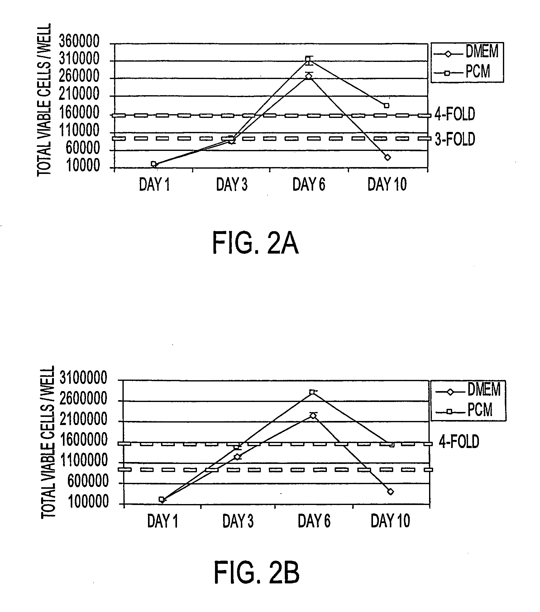
[0081] Primary human pancreatic cells were then seeded at 104 or 105 cells / cm2 onto tissue culture treated polystyrene in either DMEM commercial medium plus 10% fetal bovine serum or in PCM plus 10% fetal bovine serum. Replicate cultures were harvested at 3 day intervals via trypsinization and live cells (as determined by trypan blue exclusion) and enumerated on a hem...

example 2

Further Studies with ECM Surfaces and Various Media Components

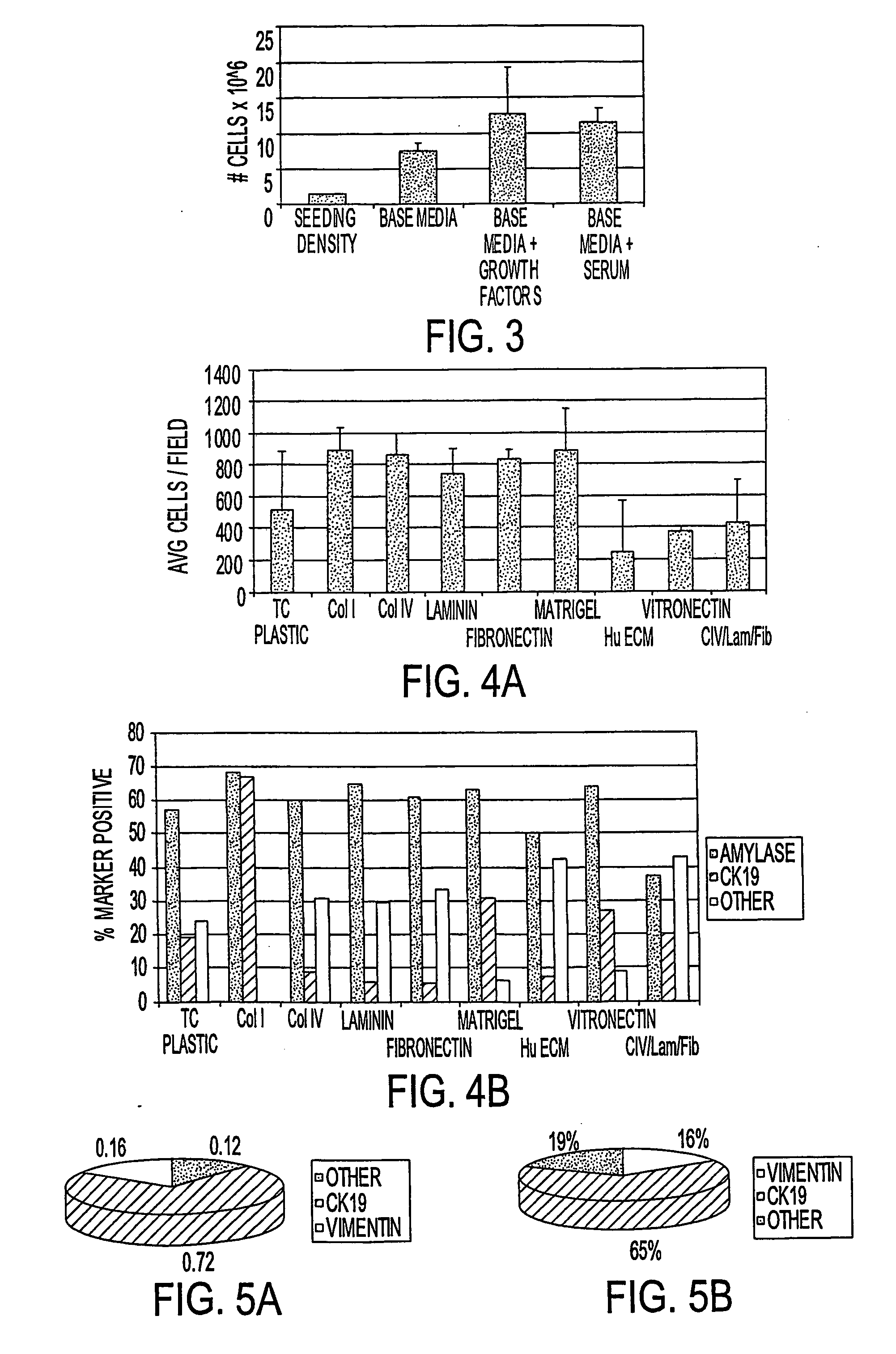
[0084] Primary pancreatic cells, composed of >90% non-islet / non-duct cells, were plated onto various coated surfaces at a density of 28,900 cells / well (105 cells / cm2). Unattached cells were washed off after 18 hours, and cultures were re-fed and allowed to grow for 8 days. Cultures were fixed in formalin (10%) and subjected to phenotypic analysis with antibodies to CK19 and Amylase. The results are shown in FIG. 4A-B. While Collagens I, IV, Laminin, Fibronectin, and Matrigel provide a suitable surface for cell attachment and expansion, maintenance of acinar (amylase+) phenotype along with the presence of an increased proportion of cells with a glandular epithelial phenotype (CK19+) was superior on Collagen I. More than 50% of cells analyzed expressed amylase and more than 50% of cells analyzed expressed CK19, suggesting that a subpopulation of cells in these experimental conditions express both markers.

[0085] Tissue cul...

example 3

Density of Cell Seeding

[0086] Primary pancreatic cells were seeded at (3) densities on tissue-culture treated polystyrene dishes (60 mm) and fed with PCM. Light microcopic observations were made daily. At the 24-hour timepoint, dishes were sacrificed and stained with trypan blue to assess viability. The results are shown in Table 3.

TABLE 3SeedingAfter 3 daysDensityAt 24 Hours:At 48 Hours:Growth:104 cells / cm2Most cells attached,Mitotic Figures presentEpithelialtrypan blue negative(light microscopy)monolayer(live)forming105 cells / cm2Most cells attached,Mitotic Figures presentEpithelialtrypan blue negative(light microscopy)monolayer(live)forming106 cells / cm2Some cells attached,Few Mitotic FiguresCells aremost are trypan bluepresent (lightdetached;positive (dead)microscopy)somefibroblastspresent


PUM

PropertyMeasurementUnit
concentrationaaaaaaaaaa

Abstract

This invention relates, e.g., to a method for expanding mammalian acinar cells, comprising culturing the cells in a cell culture system comprising a cell culture medium and a cell attachment surface, under conditions wherein the acinar cells undergo a 3-4 fold expansion together with transdifferentiation into a modified cell phenotype (IP cells) showing characteristics of acinar cells and liver cells. The invention also relates to a method for transforming these IP cells to insulin-producing cells in vitro, comprising culturing the cells in a novel, defined medium. Also disclosed are suitable culture media for performing these methods, isolated cells having the phenotype of IP cells and / or produced by these methods, and kits for performing the methods.

Description

[0001] This application claims the benefit of provisional application 60 / 384,000, filed May 28, 2002, which disclosure is incorporated by reference in its entirety herein.BACKGROUND OF THE INVENTION [0002] 1. Field of the Invention [0003] The invention relates to compositions and methods whereby, e.g. human pancreatic acinar cells are cultured under conditions that support expansion and transdifferentiation into glandular epithelial cells and subsequently into insulin-producing cells. [0004] 2. Background Information [0005] The potential benefits of taking insulin-producing cells from organ donors and transplanting them into insulin-dependent, Type I diabetic patients is clear. In the Edmonton clinical trials, many patients have lived free from the delivery of exogenous insulin for approximately 2 years after being transplanted with intact islets from organ donor sources. However, current technology requires two organ donor pancreata to generate a sufficient number of islets (about ...

Claims


Application Information

Patent Timeline
Patent Type & Authority Applications(United States)
IPC IPC(8): C12N5/08A61K38/18A61K35/12C12N5/00C12N5/02C12N5/071
CPCA61K35/12A61K2035/126C12N5/0031C12N5/0676C12N2500/25C12N2500/38C12N2500/46C12N2501/01C12N2501/105C12N2501/11C12N2501/113C12N2501/115C12N2501/117C12N2501/12C12N2501/135C12N2501/15C12N2501/16C12N2501/165C12N2501/235C12N2501/315C12N2501/335C12N2501/34C12N2501/345C12N2501/35C12N2501/37C12N2501/39C12N2501/392C12N2501/41C12N2501/83C12N2501/85C12N2501/998C12N2506/22C12N5/0037A61P3/08A61P43/00A61P3/10Y02A50/30
Inventor PRESNELL, SHARON C.HEIDARAN, MOHAMMADHAALAND, PERRYROBBINS, NEILSCHARP, DAVID WILLIAMLATTA, PAUL PRESLEYCOUTTS, MARGARETMCINTYRE, CATHERINE ANNE
Owner BECTON DICKINSON & CO
Why Eureka
Try Eureka