Adipose-derived stem cells for tissue regeneration and wound healing

a technology of stem cells and tissue, applied in the direction of skeletal/connective tissue cells, extracellular fluid disorder, peptide/protein ingredients, etc., can solve the problems of inability to fully heal wounds, injury or trauma site remains open, and death due to infection

Active Publication Date: 2006-07-06
PRIMEGEN BIOTECH LLC
View PDF16 Cites 62 Cited by
  • Summary
  • Abstract
  • Description
  • Claims
  • Application Information

AI Technical Summary

Benefits of technology

[0061] An advantage of the present invention is that wound healing with adipose-derived stem cells results in wound healing by regeneration of like tissues rather than scar formation.
[0062] In an embodiment of the present invention, the adipose-derived stem cell is differentiated prior to use in tissue regeneration. In a non-limiting example, for tissue regeneration of cardiac tissue after myocardial infarction, it is possible to differentiate the adipose-derived stem cells into a cardiogenic precursor cell or cardiomyocyte prior to transplantation of the cells to the treatment site.
[0063] The present inventive method also can involve the co-administration of bioactive agents with the ADSC cells. By “co-administration” is meant administration before, concurrently with, e.g., in combination with bioactive agents in the same formulation or in separate formulations, or after administration of a therapeutic composition as described above.
[0064] As used herein, the phrase, “bioactive agents” refers to any organic, inorganic, or living agent that is biologically active or relevant. For example, a bioactive agent can be a protein, a polypeptide, a polysaccharide (e.g. heparin), an oligosaccharide, a mono- or disaccharide, an organic compound, an organometallic compound, or an inorganic compound. It can include a living or senescent cell, bacterium, virus, or part thereof. It can include a biologically active molecule such as a hormone, a growth factor, a growth factor producing virus, a growth factor inhibitor, a growth factor receptor, an anti-inflammatory agent, an antimetabolite, an integrin blocker, or a complete or partial functional insense or antisense gene. It can also include a man-made particle or material, which carries a biologically relevant or active material. An example is a nanoparticle comprising a core with a drug and a coating on the core.
[0065] Bioactive agents also can include drugs such as chemical or biological compounds that can have a therapeutic effect on a biological organism. Non-limiting examples include, but are not limited to, growth factors, anti-rejection agents, anti-inflammatory agents, anti-infective agents (e.g., antibiotics and antiviral agents), analgesics and analgesic combinations, anti-asthmatic agents, anticonvulsants, antidepressants, anti-diabetic agents, anti-neoplastics, anticancer agents, anti-psychotics, antioxidants, immunosuppressive agents, vitamins and minerals, and agents used for cardiovascular diseases such as anti-restenosis and anti-coagulant compounds.
[0066] Bioactive agents also can include precursor materials that exhibit the relevant biological activity after being metabolized, broken-down (e.g. cleaving molecular components), or otherwise processed and modified within the body. These can include such precursor materials that might otherwise be considered relatively biologically inert or otherwise not effective for a particular result related to the medical condition to be treated prior to such modification.

Problems solved by technology

It is estimated that eight million people per year in the United States suffer from wounds caused by mechanical trauma, vascular insufficiencies or diabetes and if these wounds are left untreated, death due to infection can occur.
Occasionally these wounds never fully heal and the injury or trauma site may remain open for periods ranging from months to years.
These wounds require long-term medical treatment, which in addition to the devastating health implications can become costly to the patient and / or the health care system.
Without formation of new capillaries into the wound, required nutrients fail to reach the wound resulting in a chronically unhealed wound.
Although scar formation is critical to wound healing, excessive scar formation can have additional cosmetic and / or pathologic consequences, such as keloids and / or hypotrophic scars.

Method used


Image

  • Adipose-derived stem cells for tissue regeneration and wound healing
  • Adipose-derived stem cells for tissue regeneration and wound healing
  • Adipose-derived stem cells for tissue regeneration and wound healing

Examples


example 1

Generation and Culture of Adipose-Derived Stem Cells

[0098] This example demonstrates the isolation of adipose-derived stem cells. While the following example described the isolation of adipose-derived stem cells from mouse tissue, it would be possible for a person of ordinary skill in the art to use these techniques to isolate adipose-derived stem cells from other mammalian tissues.

[0099] Briefly, visceral fat encasing the stomach and intestines was removed and finely minced with sterile scissors. The dissected fat was then washed ten times with calcium / magnesium-free Dulbecco's phosphate-buffered saline containing 1 mM EDTA (DPBS(−) / EDTA) and centrifuged at 500 xg for 5 min after each wash step to remove floating adipocytes. The adipose tissue was then incubated in DPBS(−) / EDTA for ten minutes at room temperature and the adipose tissue was then mechanically disaggregated with surgical scissors or with a BD Medimachine (BD Biosciences, San Jose, Calif.). The disaggregated adipose ...

example 2

Identification of Adipose-Derived Stem Cell Side Population Cells

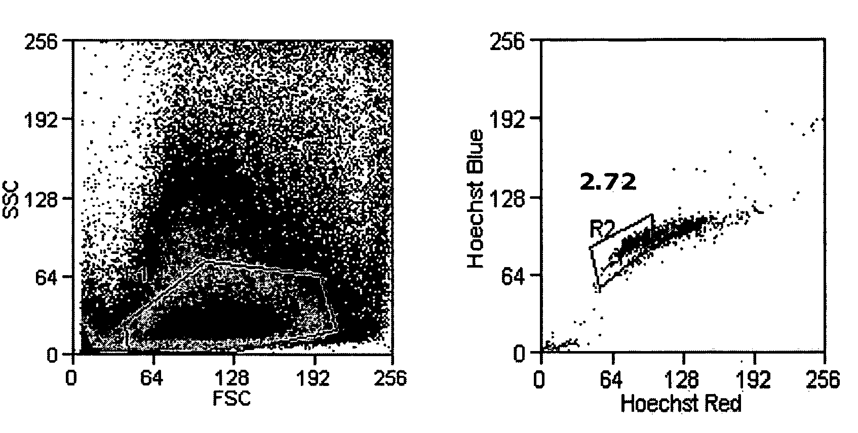
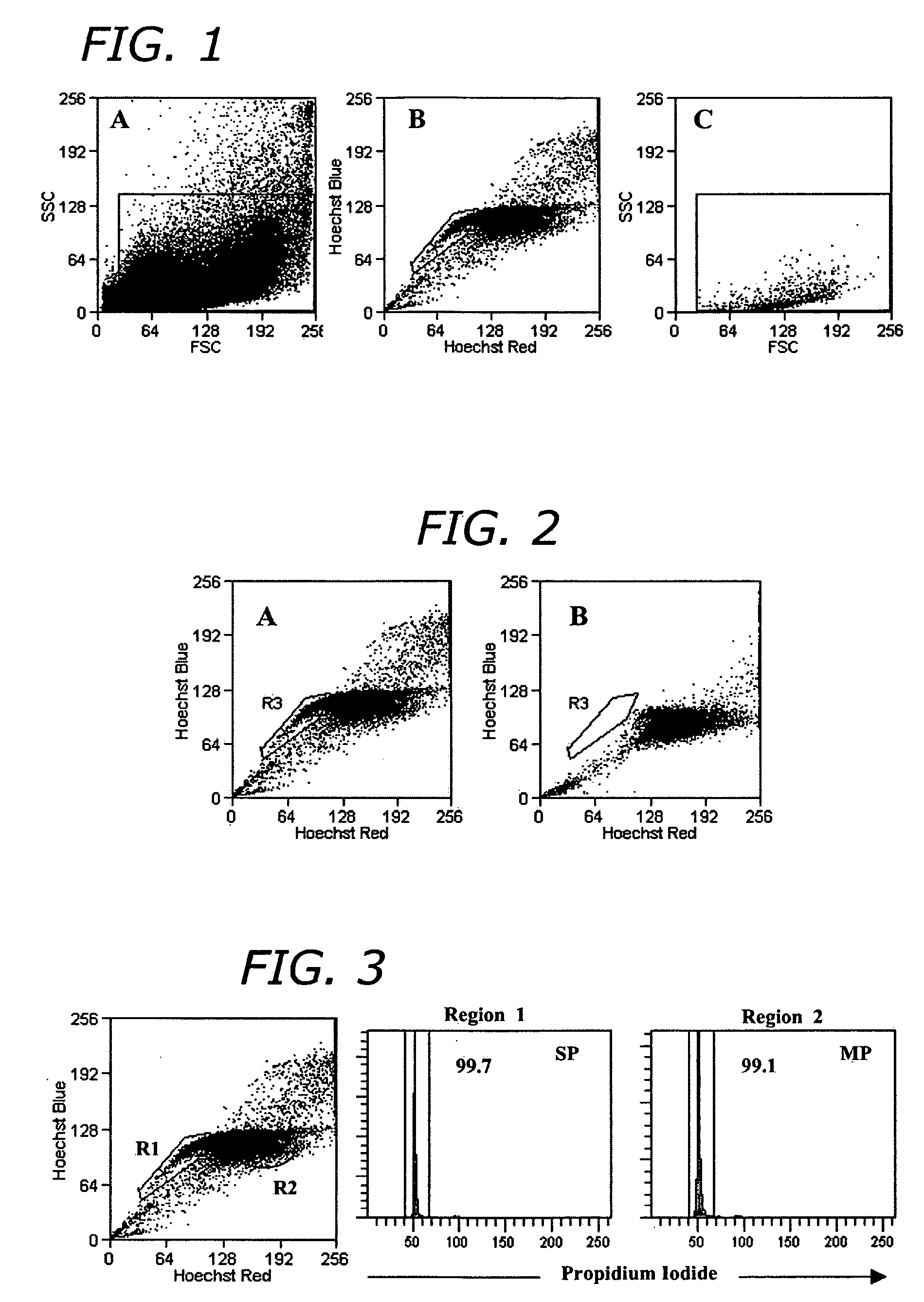
[0101] For adipose-derived stem cell side population cell (ADSC-SP) analysis, adipocytes were suspended in a concentration of 1×10⁶ cells / mL in DMEM with 10% FBS. The cells were incubated with Hoechst 33342 (Sigma) at a final concentration of 2.5 μg / mL. The cells were gently agitated every 20 min in a 37° C. water bath for a total of 90 min. After incubation, cells were pelleted by centrifugation and kept on ice until flow sorting. To demonstrate Hoechst efflux inhibition, cells were incubated with verapamil (Sigma) at a final concentration of 25 μg / mL in addition to Hoechst staining for the same incubation period.

[0102] Sorting was done on a Cytopeia InFlux Cell Sorter (Seattle, Wash.). Hoechst-stained cells were excited with a 355 nm 20 mW UV laser (Lightwave Electronics, Mountain View, Calif.) and Hoechst blue and red emission was separated with a 560 nm dichroic mirror and collected using a 460 / 50 and 670 / 40 band...

example 3

Differentiation of ADSC-SP Cells

[0109] After sorting, cells were washed once with DMEM supplemented with 10% FBS and cultured on a feeder layer of mouse embryonic fibroblast STO cells. The STO feeder cells were plated on dishes coated with 0.1% (wt / vol) gelatin, treated with mitomycin C (Sigma) at a concentration of 10 μg / ml for 2.5 h at 37° C., and washed three times with PBS. Sorted ADSC-SP cells were plated onto mitomycin C-treated STO feeder cells with a daily change of culture medium (FIG. 5).

[0110] To examine the functional capabilities of ADSC-SP cells, they were differentiated into different cell types. For osteogenesis, adipogenesis, and neurogenesis 50,000 cultured ADSC-SP cells at passage four were plated. For chondrogenesis, 80,000 cultured ADSC-SP cells at passage four were plated as a micromass.

[0111] Adipogenesis

[0112] Fifty thousand cells were grown in adipogenic induction and maintenance medium (Cambrex, Walkersville, Md.) according to manufacturer's specificati...


PUM

PropertyMeasurementUnit
concentrationaaaaaaaaaa

Abstract

Compositions and methods for promoting tissue regeneration, particularly skin regeneration, with adipose-derived stem cells are provided. Additionally methods and compositions for promoting tissue regeneration with adipose-derived stem cell side population cells are provided. The adipose-derived cells are administered in a tissue regenerating effect amount optionally with a bioactive agent. Additionally the adipose derived cells can be autologous or syngeneic.

Description

CROSS-REFERENCE TO RELATED APPLICATIONS [0001] The present application claims priority under 37 CFR §119(e) to U.S. Provisional Patent Application No. 60 / 641,034 filed Dec. 30, 2004.FIELD OF THE INVENTION [0002] The present invention provides compositions, including adipose-derived stem cells and adipose-derived stem cell side population cells, for promoting tissue regeneration, particularly skin regeneration. Additionally, the present invention provides methods for inducing tissue regeneration and wound healing. BACKGROUND OF THE INVENTION [0003] Stem cells have been shown to repopulate and repair tissues, organs and / or organ systems. Of interest for regenerative medicine is the use of adult or post-natal stem cells for cell-based therapies. One particular type of post-natal stem cell is an adipose-derived stem cell which is found in the connective tissues within adipose tissue. Adipose-derived stem cells have exhibited multi-potency in vitro by being able to differentiate into car...

Claims


Application Information

Patent Timeline
Patent Type & Authority Applications(United States)
IPC IPC(8): A61K35/12C12N5/08A61K35/28A61K35/36C12N5/0775
CPCA61K35/28C12N5/0667A61K38/18A61K33/00A61K35/36A61K2300/00A61P17/02A61P25/00A61P43/00A61P7/02A61P9/00
Inventor SAYRE, CHAUNCEY B.SILVA, FRANCISCO J.
Owner PRIMEGEN BIOTECH LLC
Why Eureka
Try Eureka