
Drug/gene eluting stent

a stent and gene technology, applied in the field of drug/gene eluting stents, can solve the problems of delayed endothelial regeneration, no established method, and high rate of “restenosis” or relapse of stenosis of once dilated cardiac arteries,

Inactive Publication Date: 2007-04-05
EGASHIRA KENSUKE
View PDF2 Cites 19 Cited by
  • Summary
  • Abstract
  • Description
  • Claims
  • Application Information

AI Technical Summary

Benefits of technology

[0022] The stent of the present invention having a layer containing the gene encoding a hybrid polypeptide on the surface will gradually release the anti-inflammatory factor or angiogenic factor by acidosis when it is indwelled in the arterial lesion and for example, and in addition, has an enhanced efficiency of the gene transfer into cells of the lesion. Thus, the stent directly and continuously exerts anti-inflammatory action and angiogenesis action for a long period in tissues under inflammatory or hypoxic conditions. The transfer efficiency is shown by an equation of number of genes introduced in cells of the lesion / amount of released genes×100.
[0024] The drug / gene eluting stent of the present invention has advantages that can directly deliver the genes encoding a hybrid polypeptide, particularly in uniform microcapsules of hybrid polypeptide, to the lesion and can reduce the given doses, and thus can improve safety and efficacy and further can maintain the efficacy for a long period.

Problems solved by technology

However, a high rate of “restenosis” or relapsed stenosis of the once dilated cardiac artery has become a medical problem.
Therefore, a new therapeutic approach inhibiting the restenosis has been expected, however, no established method has been found.
However, it has some drawbacks such as emergence of dead cases due to subacute stage thrombosis, hypersensitivities (such as pain, eczema and blood pressure fluctuation), persistent chronic inflammation and delayed endothelial regeneration / vascular reparation reaction, and no sufficient improvement has been obtained.
Furthermore, the above mentioned reference also disclosed an invention with similar purpose, but no satisfactory result was also obtained.

Method used


Image

  • Drug/gene eluting stent
  • Drug/gene eluting stent
  • Drug/gene eluting stent

Examples


example 1

Preparation of a Drug / Gene Eluting Stent

[0035] A drug / gene eluting stent with four layers structure was prepared by the following steps (1) to (4);

[0036] (1) A silane coupling agent was diluted in a methanol / water (volume ratio: 50 / 50), applied on an exterior surface of a stent with inner diameter of about 2.8 mm, and formed a primer layer after drying.

[0037] (2) A 2 mass % solution of a water absorptive polyether type thermoplastic polyurethane dissolved in tetrahydrofuran (THF) was coated on a primer layer and formed a drug layer base.

[0038] (3) The stent prepared by step (2) in an aqueous solution of a gene encoding a hybrid polypeptide at 10 mass % was dipped and thoroughly swelled. Thereafter, the stent was vacuum dried to remove water and formed a drug layer.

[0039] (4) A 2 mass % solution of a highly in vivo stable polycarbonate type thermoplastic polyurethane dissolved in tetrahydrofuran (THF) was coated on the drug layer to form a protective layer and the stent of the p...

example 2

Investigation of a Gene Expression and Inflammation Image in a Cholesterol Loaded Rabbit•Model

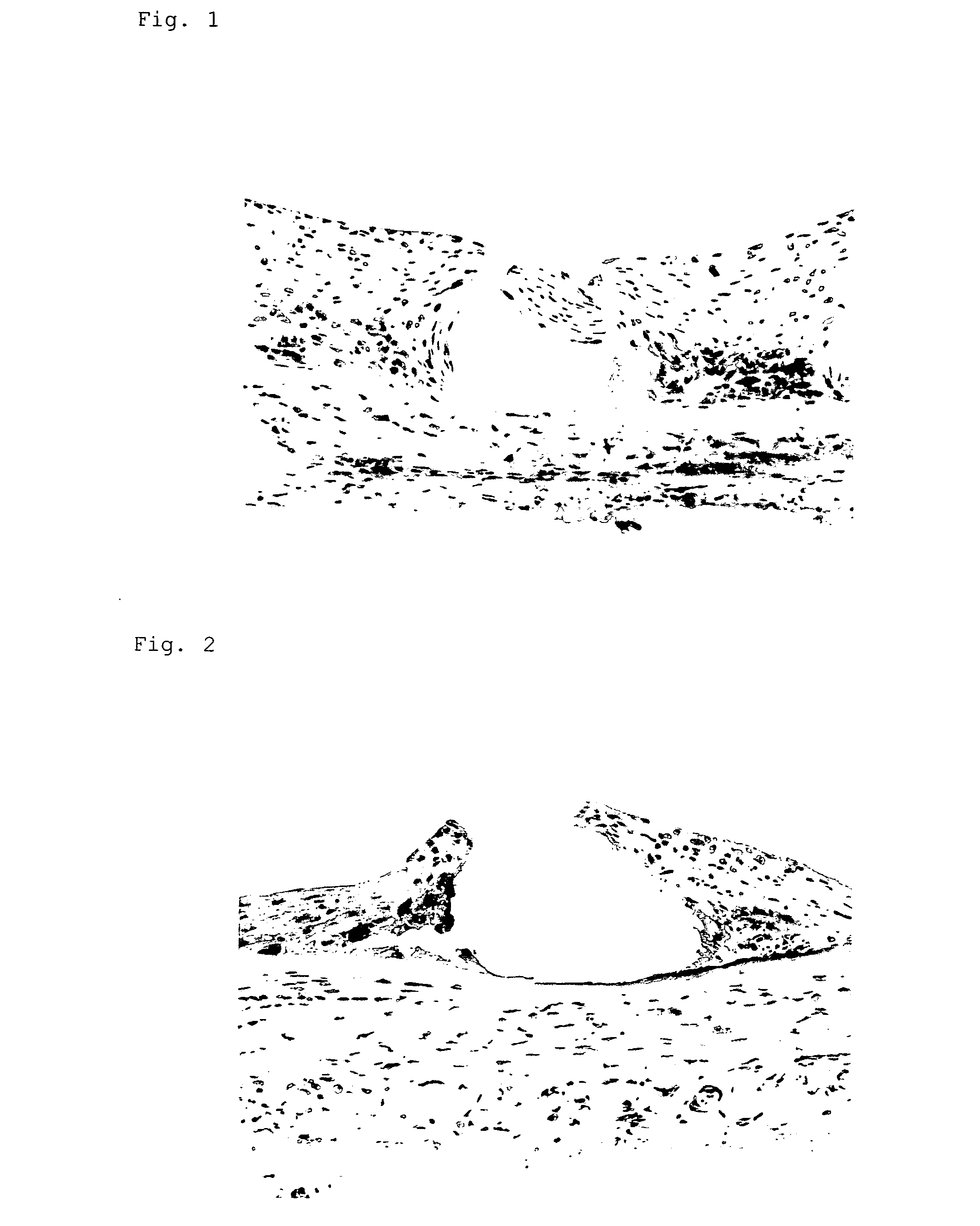
[0040] A stent (inner diameter of about 2.8 mm) coated with a gene encoded a marker gene, LacZ, was indwelled in an iliac artery of a hyperlipemic domestic rabbit loaded with 1% cholesterol for four weeks and investigated the gene expression after three days. Cells positive to gene expression were observed in 60-70% of intimal cells and 50% of medial and outer membrane cells. No positive cell was observed in a stent without gene coating. This was tenfold or over of the transfer efficiency compared to the result shown in Nat. Biotechnol., November 2000,; 18(11): 1181-4.

example 3

Investigation of an Inflammatory Image in Cholesterol Loaded Rabbit•Model

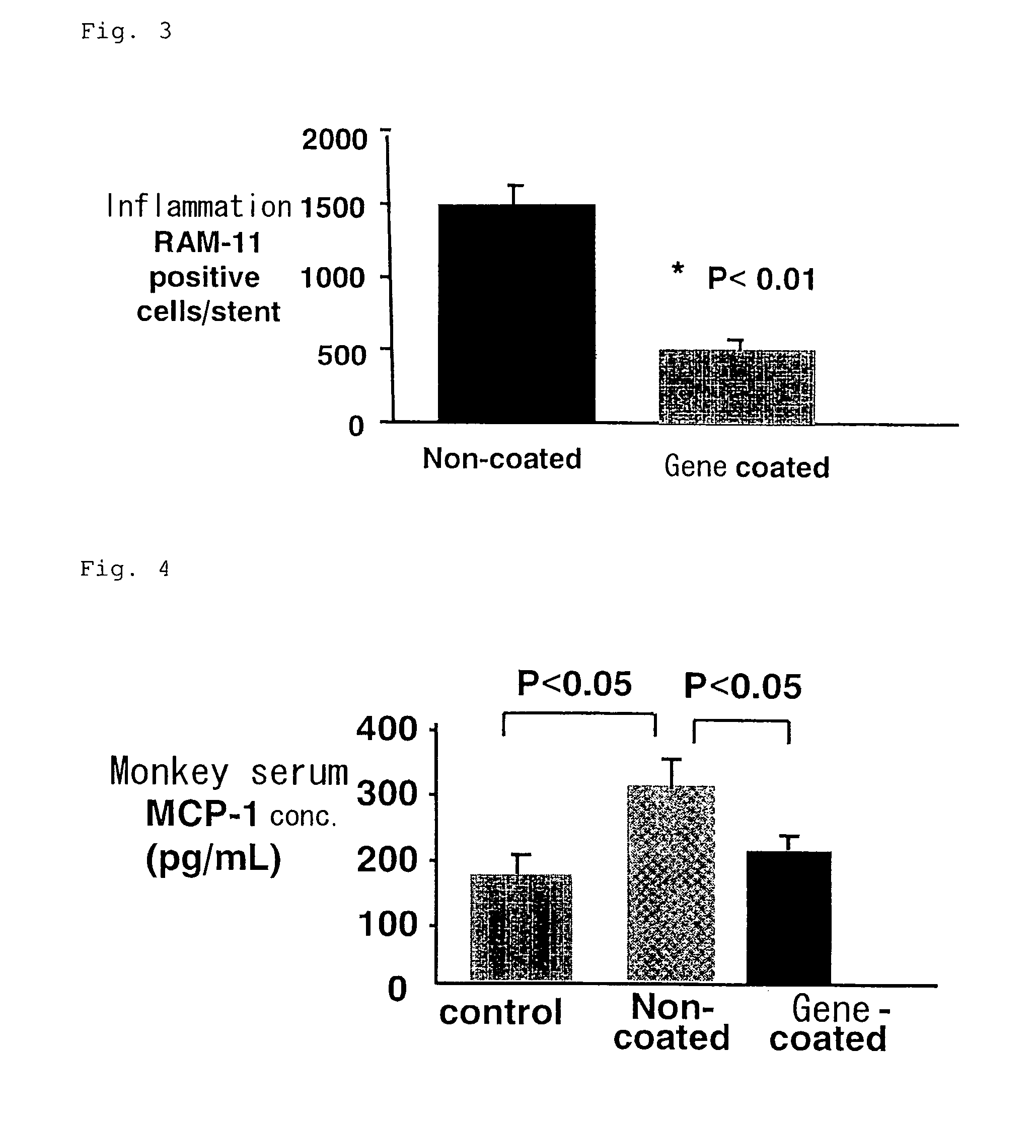
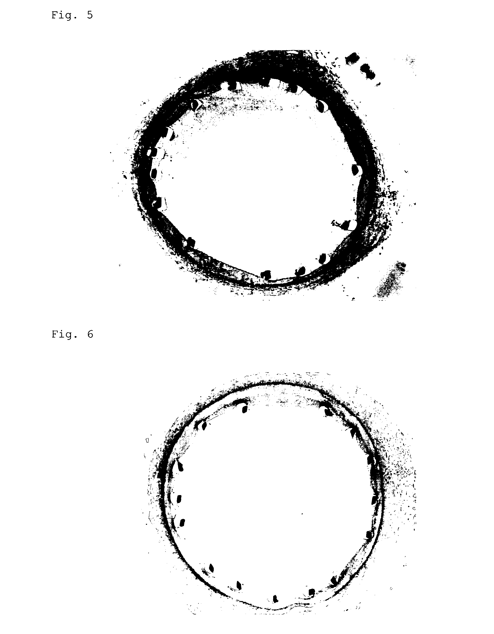
[0041] Metal stents (n=5) or gene-coated stents (n=5) (both have inner diameter of about 2.8 mm) were indwelled in an iliac artery of hyperlipemic domestic rabbit loaded with 1% cholesterol for 12 weeks. Determination of macrophage infiltration and blood level of monocyte migration factor, MCP-1, were performed after 10 days.

[0042] In addition, the gene-coated stent was prepared with the metal stent similar to that of the control, and a biodegradable base material containing a plasmid composed of a gene encoding FNCBD-7ND hybrid polypeptide in a similar manner with that of the preparation example.

[0043] Macrophage infiltration in arteries was searched using an antibody which specifically recognizes rabbit macrophage in histochemical investigation and a significant inhibition of the infiltration degree (p<0.01) was observed in the gene-coated stent group in comparison with that in the metal stent group.

[0044...


PUM

PropertyMeasurementUnit
Particle sizeaaaaaaaaaa

Abstract

The present invention provides a more safe and highly effective stent having functions such as anti-inflammatory action, antithrombotic action, maintenance of tissue restoration response and maintenance of endothelial regeneration. More specifically, the drug / gene eluting stent has a layer containing a gene encoding a hybrid polypeptide on the surface. The hybrid polypeptide is preferably a bound of fibronectin-derived collagen binding domain (FNCBD) polypeptide and an anti-inflammatory factor or an angiogenic factor. The uniform fine particle size capsules can directly deliver the gene encoding the hybrid polypeptide to the lesion and have benefits of reducing the given doses, and thus improving safety and efficacy and further maintaining the efficacy for a long period.

Description

TECHNICAL FIELD OF THE INVENTION [0001] The present invention relates to a drug / gene eluting stent. PRIOR ART [0002] The fundamental pathology of angina pectoris and cardiac infarction is a stenosis and thrombosis of cardiac artery due to arteriosclerosis, and coronary interventions (PCI, balloon dilation and stent dilation) dilating arteriosclerotic stenosis have been popularly used as a useful therapy in the world. [0003] However, a high rate of “restenosis” or relapsed stenosis of the once dilated cardiac artery has become a medical problem. The recurrent cardiac ischemia due to restenosis may highly causes relapsed angina pectoris and cardiac infarction, and not a few cases require re-operation of PCI or coronary artery bypass surgery. [0004] Therefore, a new therapeutic approach inhibiting the restenosis has been expected, however, no established method has been found. [0005] WO-A 02 / 47739, JP-A 2002-060400, JP-A 2002-284698, Artif. Organs, February 2003; 27(2): 147-54, and Nat...

Claims


Application Information

Patent Timeline
IPC IPC(8): A61K48/00A61F2/02A61F2/82A61K38/00A61K38/16A61K38/22A61K38/39A61K47/48A61L27/00A61L31/10A61L31/16A61P9/00A61P9/10
CPCA61K38/39A61K47/48992A61L31/10A61L31/16A61L2300/258A61L2300/62A61K2300/00A61K47/6957A61P9/00A61P9/10
Inventor EGASHIRA, KENSUKE
Owner EGASHIRA KENSUKE
Learn More