
Antibody complexes and methods for immunolabeling

an antibody complex and immunolabeling technology, applied in the field of immunolabeling complexes, can solve the problem that the time required for the labeling reagent to selectively bind to the target-binding antibody is typically very short, than 10 minutes

Inactive Publication Date: 2007-11-22
MOLECULAR PROBES
View PDF54 Cites 55 Cited by
  • Summary
  • Abstract
  • Description
  • Claims
  • Application Information

AI Technical Summary

Benefits of technology

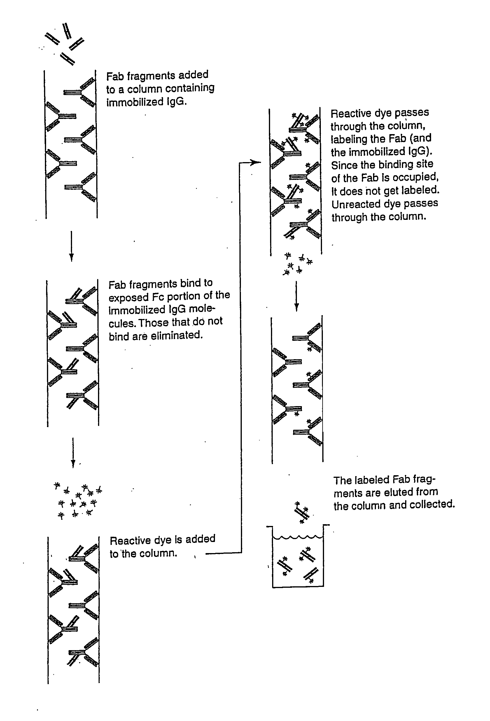
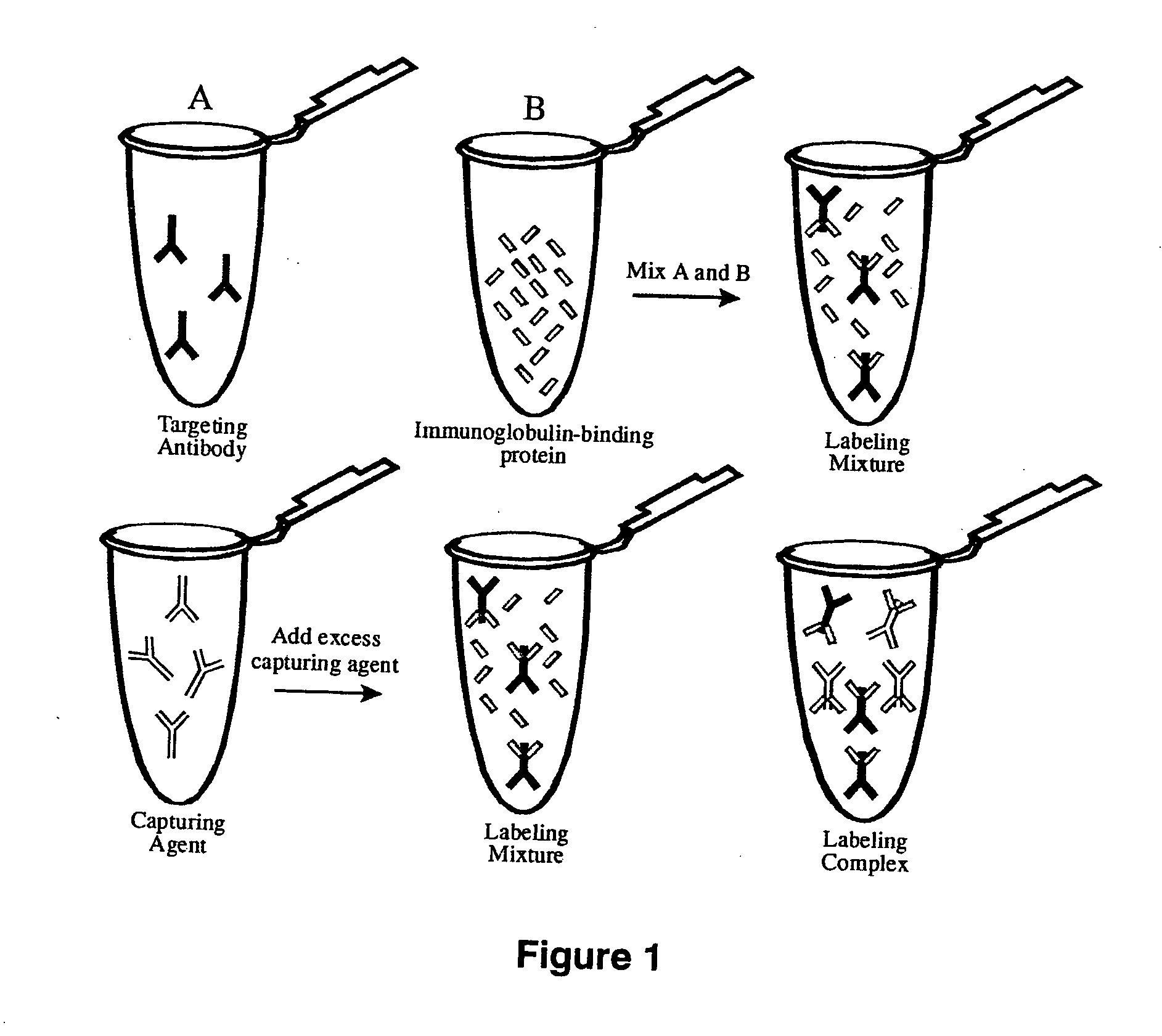
[0157] When preparing the immuno-labeled complex using purified target-binding antibody, stock solutions of both the labeling reagent and the target-binding antibody are typically near 1 mg / mL in an appropriate buffer, although more or less concentrated solutions are also suitable. Generally, the labeling reagent is mixed in a molar ratio of at least one to 50 moles of labeling reagent to one mole of the target-binding antibody to be complexed. More commonly a ratio of at least one to as many as 10 moles of labeling reagent per mole of target-binding antibody is combined. With an anti-Fc region Fab to a target-binding antibody, a molar ratio of approximately 2 to 10 is typical, more typically 3 to 5 (particularly for complexes in which the labeling reagent has been labeled while immobilized on an affinity matrix). The ease of formation of the complex permits rapid optimization of the complex and assessment of the effect of variation in experimental parameters. A particularly unique advantage of the invention is that the stoichiometry of the complex is easily adjusted to provide complexes with different ratios of labeling reagent to target-binding antibody, and thus there is control over the ultimate detectability of the target in the sample. Complexes that have been labeled with the same dye but at different molar ratios can be separately detected by the differences in their intensities.
[0158] Complex formation appears to occur almost within the mixing time of the solutions (<1 minute) but the reaction typically is allowed to proceed for at least 5 minutes and can be longer before combining the immuno-labeled complex with the sample. Although complex formation can be reversed by addition of an unlabeled antibody that contains the same binding region, reversibility is very slow; furthermore, following binding of the immuno-labeled complex to a target in a sample, the sample can be “fixed” using aldehyde-based fixatives by methods that are commonly practiced by those skilled in the art of immunolabeling.
[0159] The labeling process optionally further comprises the addition of a capture component to remove excess labeling reagent. For applications in which immunolabeling complexes of multiple primary antibodies from the same species (e.g. mouse monoclonal antibodies) or cross-reacting species (e.g. mouse and human antibodies) are to be used simultaneously or sequentially, it is necessary to quench or otherwise remove any excess labeling reagent by use of a capture component or by other means to avoid inappropriate labeling of the sample. The most effective capturing components to capture excess labeling reagent are those that contain the binding site of the labeling reagent but are themselves not labeled, preferably an antibody or antibody fragment. Capture components may be free in solution or immobilized on a matrix, such as agarose, cellulose, or a natural or synthetic polymer, to facilitate separation of the excess capture component from the immuno-labeled complex. The capture component is optionally attached to a microsphere or magnetic particle. However, separation of excess labeling reagent is not essential for successful utilization of the invention, particularly when using a single target-binding antibody.
[0160] The steps of the labeling process for the target-binding antibodies can be repeated to form discrete immuno-labeled complex subsets that can be used individually or pooled in an assay to detect individual or multiple targets. As used herein the term immuno-labeled complex subsets refers to subsets that are distinguished from each other i) a ratio of label to labeling reagent, or ii) a physical property of the label, or iii) a ratio of labeling reagent to the target-binding antibody, or iv) by the target-binding antibody, or a combination thereof. For example a panel of subsets may comprise a target-binding antibody that is bound by a labeling reagent comprising a subset of different ratios of the same label on the labeling reagent resulting in a discrete subset of immuno-labeled complexes. This subset of immuno-labeled complexes can be used individually wherein a target is identified by the intensity of the detectable label or used in combination with another subset of immunocomplexes that differ in the target-binding antibody to identify multiple targets. C. Ligand-Detection Reagent
[0161] The immuno-labeled complexes can be modified by the addition of a ligand analog of the present invention to form a ligand-detection reagent. The present ligand-detection reagents comprise a ligand-binding antibody, a ligand analog and a labeling reagent, See FIG. 8. This ligand-detection reagent is formed by incubating the ligand-binding antibody, the ligand analog and the labeling reagent for sufficient amount of time to allow for a complex to form. The formation of the complex happens fairly rapidly, typically less than 30 minutes, preferably less than 15 minutes and most preferred the complex forms in 5 minutes or less.
[0162] The reporter molecules that are covalently attached to the ligand analog are preferably selected from the group consisting of a borapolyazaindacene, a coumarin, a xanthene, a cyanine, a fluorescent protein and a phosphorescent dye. Most preferred are borapolyazaindacene, fluorinated xanthene, fluorinated coumarin, including dyes sold under the trade name OREGON GREEN, BODIPY, PACIFIC BLUE and MARINA BLUE (Trade marks owned by Molecular probes, Inc.) including any dyes disclosed in U.S. Pat. Nos. 6,162,931; 5,830,912; 4,774,339; 5,187,288; 5,248,782; and 5,433,896.

Problems solved by technology

The time required for the labeling reagent to selectively bind to the target-binding antibody is typically very short, often less than 10 minutes.

Method used


Image

  • Antibody complexes and methods for immunolabeling
  • Antibody complexes and methods for immunolabeling
  • Antibody complexes and methods for immunolabeling

Examples


example 1

Preparation of Fc Antigen

[0245] Purified mouse and rabbit IgG was fragmented with the proteolytic enzyme papain (CURRENT PROTOCOLS IN CELL BIOLOGY, 16.4.1-16.4.10 (2000)). A 12 mL solution of mouse IgG was prepared at ˜2 mg / mL in phosphate-buffered saline (PBS). A solution containing 0.1 mg of papain in digestion buffer (PBS, 0.02 M EDTA, 0.02 M cysteine) was added to the antibody and allowed to react at 37° C. for 16 hours. The digestion was terminated by the addition 20 μL of 0.3 M iodoacetamide in PBS. The fragments were dialyzed against 2 L of PBS for 16 hours at 4° C. The Fc fragment was purified on a protein G-Sepharose CL-4B column. The bound fraction containing the Fc fragment was eluted from the column using 50-100 mM glycine / HCl buffer, pH 2.5-2.8. The eluate was collected in 1 mL fractions. The pH of the protein fractions was immediately raised to neutral by addition of 100 μL of either 500 mM phosphate or Tris buffer, pH 7.6, to each 1 mL fraction. The solution was then...

example 2

Production of Anti-Fc Antibodies

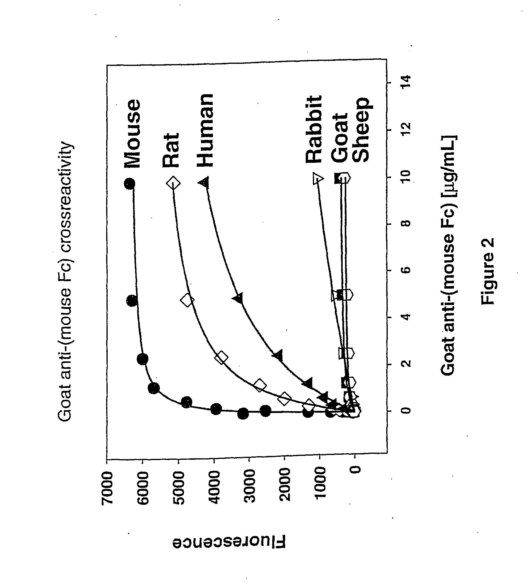
[0246] Polyclonal antibodies specific for the Fc region of an antibody were raised in goats against the purified FC region of an antibody from a different species (Example 1). Methods of immunizing animals are well known in the art, and suitable immunization protocols and immunogen concentrations can be readily determined by those skilled in the art (Current Protocols in Immunology 2.4.1-9 (1995); ILAR Journal 37, 93 (1995)). Briefly, individual goats were immunized with purified mouse Fc or purified rabbit Fc fragments. The initial immunization in 50% Freund's complete adjuvant (1000 μg conjugate (half subcutaneous, half intramuscularly)) was followed by 500 μg conjugate per goat in Freund's incomplete adjuvant two and four weeks later and at monthly intervals thereafter. Antibodies were purified from serum using protein A-Sepharose chromatography. Antibodies against mouse Fc isotypes can be prepared by starting with isotype-selected mouse Fc antige...

example 3

Preparation of Fab Fragments

[0247] Fragmentation of the goat anti-(mouse Fc) antibody to the monovalent Fab fragment was carried out using the proteolytic enzyme, papain, as described in Example 1. Following dialysis against PBS, the Fab fragment was purified on a protein A-Sepharose CL-4B column. The unbound fraction containing the Fab fragment and the papain was collected. This solution was then loaded onto a Sephacryl S-200 Superfine size-exclusion column and fractions corresponding to a molecular weight of ˜50 kDa were collected and analyzed by SDS-PAGE. The Fab fragments of goat anti-(rabbit Fc) can be prepared similarly.


PUM

PropertyMeasurementUnit
Molar densityaaaaaaaaaa
Coloraaaaaaaaaa
Ratioaaaaaaaaaa

Abstract

The present invention provides labeling reagents and methods for labeling primary antibodies and for detecting a target in a sample using an immuno-labeled complex that comprises a target-binding antibody and one or more labeling reagents. The labeling reagents comprise monovalent antibody fragments or non-antibody monomeric proteins whereby the labeling reagents have affinity for a specific region of the target-binding antibody and are covalently attached to a label. Typically, the labeling reagent is an anti-Fc Fab or Fab′ fragment that was generated by immunizing a goat or rabbit with the Fc fragment of an antibody. The present invention provides for discrete subsets of labeling reagent and immuno-labeled complexes that facilitate the simultaneous detection of multiple targets in a sample wherein the immuno-labeled complexes are distinguished by i) a ratio of label to labeling reagent, or ii) a physical property of said label, or iii) a ratio of labeling reagent to said target-binding antibody, or iv) by said target-binding antibody. This is particularly useful for fluorophore labels that can be attached to labeling reagents and subsequently immuno-labeled complexes in ratios for the detection of multiple targets.

Description

CROSS-REFERENCE TO RELATED APPLICATIONS [0001] This application claims priority to U.S. Ser. No. 60 / 329,068, filed Oct. 12, 2001; U.S. Ser. No. 60 / 369,418 filed Apr. 1, 2002, U.S. Ser. No. 10 / 118,204 filed Apr. 5, 2002, and PCT / US02 / 31416 filed Oct. 2, 2002, which disclosures are herein incorporated by reference.FIELD OF THE INVENTION [0002] The present invention relates to immuno-labeled complexes and methods for use in the detection and measurement of one or more targets in a biological sample. The invention has applications in the fields of molecular biology, cell biology, immunohistochemistry, diagnostics, and therapeutics. BACKGROUND OF THE INVENTION [0003] Immunolabeling is a method for qualitative or quantitative determination of the presence of a target in a sample, wherein antibodies are utilized for their specific binding capacity. The antibodies form a complex with the target (antigen), wherein a detectable label is present on the antibody or on a secondary antibody. The ...

Claims


Application Information

Patent Timeline
IPC IPC(8): G01N33/00C12Q1/68G01N33/53G01N33/58G01N33/68
CPCB82Y5/00B82Y10/00G01N33/6857G01N33/53G01N33/58B82Y30/00
Inventor BEECHEM, JOSEPHHAGEN, DAVIDJOHNSON, IAIN
Owner MOLECULAR PROBES
Learn More