Collagen scaffolds, medical implants with same and methods of use

a technology of collagen scaffold and medical implants, applied in the field of collagen scaffold coverings, can solve the problems of adverse reactions leading to blood clot formation in some patients, devices may provoke inflammation and/or fibrosis, other unwanted bioreactions, etc., and achieve the effect of improving biocompatibility and reducing the risk of adverse reactions

Inactive Publication Date: 2008-01-24
UNIV OF SOUTH FLORIDA
View PDF32 Cites 71 Cited by
  • Summary
  • Abstract
  • Description
  • Claims
  • Application Information

AI Technical Summary

Benefits of technology

[0010] Embodiments of the invention provide a biocompatible collagen covering for medical devices to provide improved biocompatibility and/or reduced risk

Problems solved by technology

Chronically implantable devices may provoke inflammation and/or fibrosis from tissue trauma or tissue response to the foreign body.
Implanted devices may also cause other unwanted bioreactions.
For example, recently, researchers have stated that drug-coated stents might cause adverse reactions leading to blood clot formation in some patients.
The pain (Lee et al., 2005), inconvenience, and discomfort of self-monitoring of blood gluc

Method used


Image

  • Collagen scaffolds, medical implants with same and methods of use
  • Collagen scaffolds, medical implants with same and methods of use
  • Collagen scaffolds, medical implants with same and methods of use

Examples


example 1

Preparation of Porous Cross-Linked Collagen Scaffolds

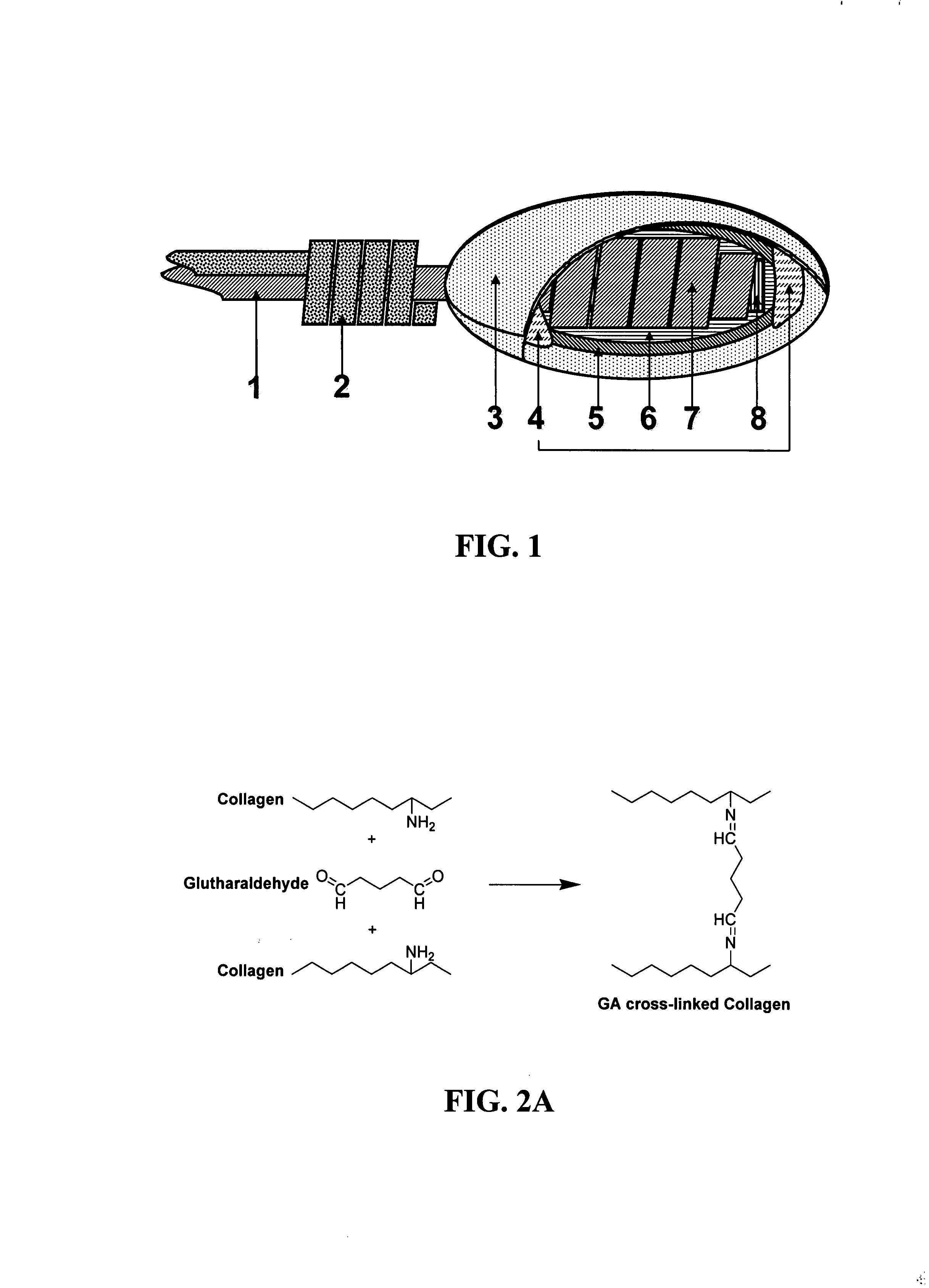
[0082] The chemistry of the NDGA cross-linking reaction differs from the reaction using the GA treatment (FIG. 2). GA is the most common cross-linking agent used for fixation of collagen scaffolds for tissue bioengineering. Both aldehyde functional groups of the GA molecule react with amine groups between two neighboring polypeptide chains, particularly lysine side chains. Unfortunately, GA cross-linking is encumbered with potential cytotoxicity problems caused by the presence of unreacted residual groups and / or the release of monomers and small polymers during enzymatic degradation (Huang-Lee et al., 1990; van Luyn et al., 1992).

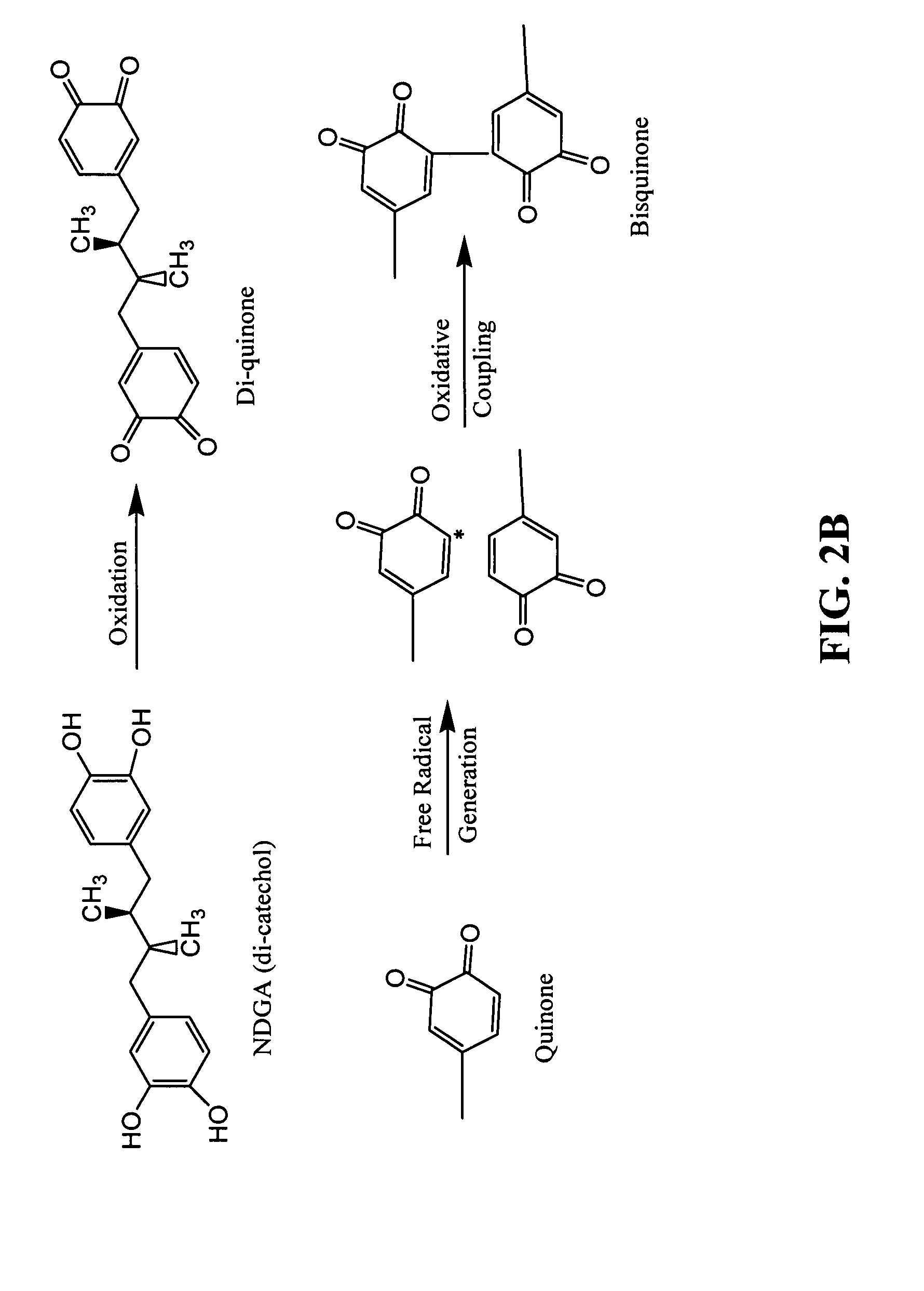
[0083] NDGA is an alternative cross-linking agent which possesses reactive catechols. Collagen cross-linking with NDGA mimics the quinine tanning mechanism in the skate egg capsule. Catechol-quinone tanning systems are prevalent in a wide variety of animals, which the process serves to strengthen vulner...

example 2

In Vitro and In Vivo Evaluation of Porous Collagen Scaffolds

[0085] The biological stability of the cross-linked collagen scaffolds was investigated by in vitro and in vivo biodegradation tests. Degradation in both uncross-linked (control) and cross-linked scaffolds was characterized by determining weight loss of the scaffold after enzymatic digestion. The uncross-linked scaffolds and scaffolds cross-linked with GA for 2 hours were completely degraded in the collagenase solution within several hours while NDGA or GA cross-linked (for 12 h) scaffolds were not degraded within 24 hours. A significant increase in resistance to enzymatic digestion could be shown after cross-linking. FIG. 5 shows long-term collagenase in vitro degradation test (weight remaining %) of the NDGA and GA cross-linked scaffolds. After one week exposure to collagenase, both types of scaffolds showed high resistance to enzymatic digestion (>80% weight remaining). After three and four weeks, all scaffolds retained...

example 3

Porous Collagen Scaffolds Around Implantable Glucose Sensors

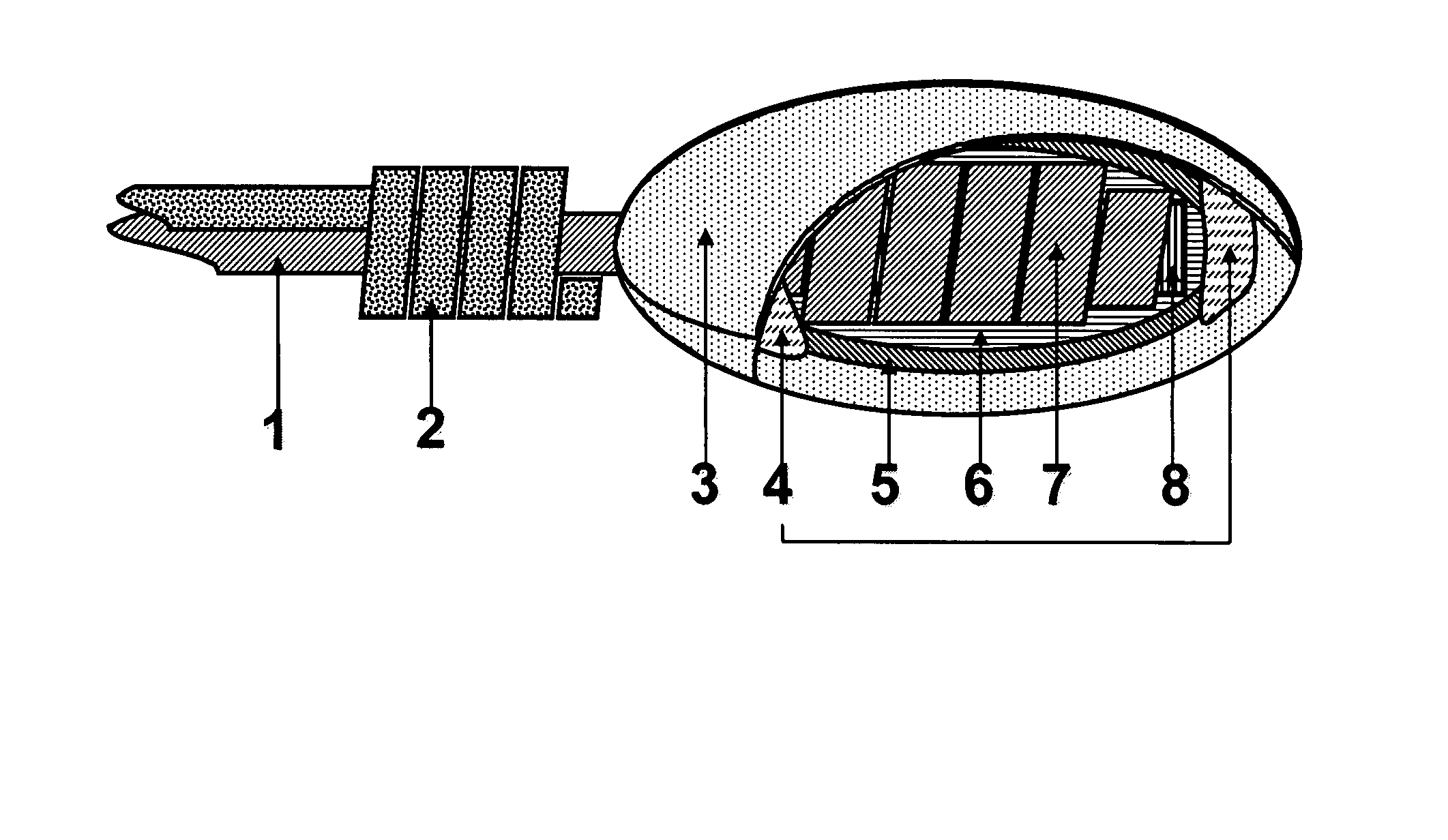
[0087] Coil-type glucose sensors loaded with cross-linked enzyme (GOD: Glucose Oxidase) were first fabricated by using Platinum-Iridium (Pt / Ir) wires. Then, bovine tendon type I collagen scaffolds were applied around the sensors (FIG. 1). Yu et al., 2006 previously reported that this “coil-type” sensor allows more GOD loading, provides a larger electrochemical surface area, and therefore increases the response current as compared to a “needle-type” sensor. The coil-type sensor of the invention is flexible and miniaturized (0.5 mm dia.) for subcutaneous implantation. It is composed of a two-electrode system with a glucose indicating platinum electrode and a Ag / AgCl reference-counter electrode. A sensor of the present invention utilizes a three-layer membrane configuration of cross-linked collagen scaffold, epoxy-polyurethane (Epoxy-PU) and GOD. The collagen scaffold (the outer layer in this case) can uptake 99% of its dry w...


PUM

PropertyMeasurementUnit
Pore sizeaaaaaaaaaa
Diameteraaaaaaaaaa
Diameteraaaaaaaaaa

Abstract

The subject invention concerns non-degradable three dimensional porous collagen scaffolds and coatings. These scaffolds can be prepared around sensors for implantation into a body. A specific embodiment of the invention concerns implantable glucose sensors. Sensors comprising a collagen scaffold of the invention have improved biocompatibility by minimizing tissue reactions while stimulating angiogenesis. The subject invention also concerns methods for preparing collagen scaffolds of the invention. The subject invention also concerns sensors that have a collagen scaffold of the invention around the exterior of the sensor.

Description

CROSS-REFERENCE TO RELATED APPLICATION [0001] The present application claims the benefit of U.S. Provisional Application Ser. No. 60 / 805,495, filed Jun. 22, 2006, which is hereby incorporated by reference herein in its entirety, including any figures, tables, nucleic acid sequences, amino acid sequences, and drawings.GOVERNMENT SUPPORT [0002] This invention was made with Government support under National Institutes of Health Grant No. 1R01 EB001640-01. The Government has certain rights in the invention.FIELD OF THE INVENTION [0003] This invention relates to implantable biosensors and medical devices. More specifically, this invention relates to collagen scaffold coverings for implantable sensors and / or other medical devices promoting biocompatibility. BACKGROUND OF THE INVENTION [0004] Chronically implantable devices may provoke inflammation and / or fibrosis from tissue trauma or tissue response to the foreign body. See, e.g., Reichert et al., Handbook of Biomaterial Evaluation, Ch. ...

Claims


Application Information

Patent Timeline
IPC IPC(8): A61K9/00A61B5/145
CPCA61B5/1486A61L27/24A61L27/34A61L31/044A61L31/10A61L27/54A61L27/48C08L89/06A61L31/129A61L2400/02A61L2400/18A61L2420/02A61B5/14542A61B5/14865A61L27/18A61L27/56
Inventor JU, YOUNG MINMOUSSY, FRANCISKOOB, THOMAS J.
Owner UNIV OF SOUTH FLORIDA
Why Eureka
Try Eureka