[0013] One of the objects of the present invention is to provide an ultraminiature (e.g., 350 μm
diameter)
endoscopic imaging apparatus / device / arrangement /
system with integrated
laser therapy capabilities for microsurgical applications inside the body, e.g., for safe and
effective treatment of twin-twin transfusion syndrome (TTTS). It is possible to overcome the limitations and deficiencies of the current TTTS therapy devices by providing a much smaller microsurgical
endoscope that provides more informative images with a better
image quality. This exemplary enhancement includes spectrally encoded
endoscopy (SEE) concepts that use
wavelength division
multiplexing to obtain high-resolution images through a single
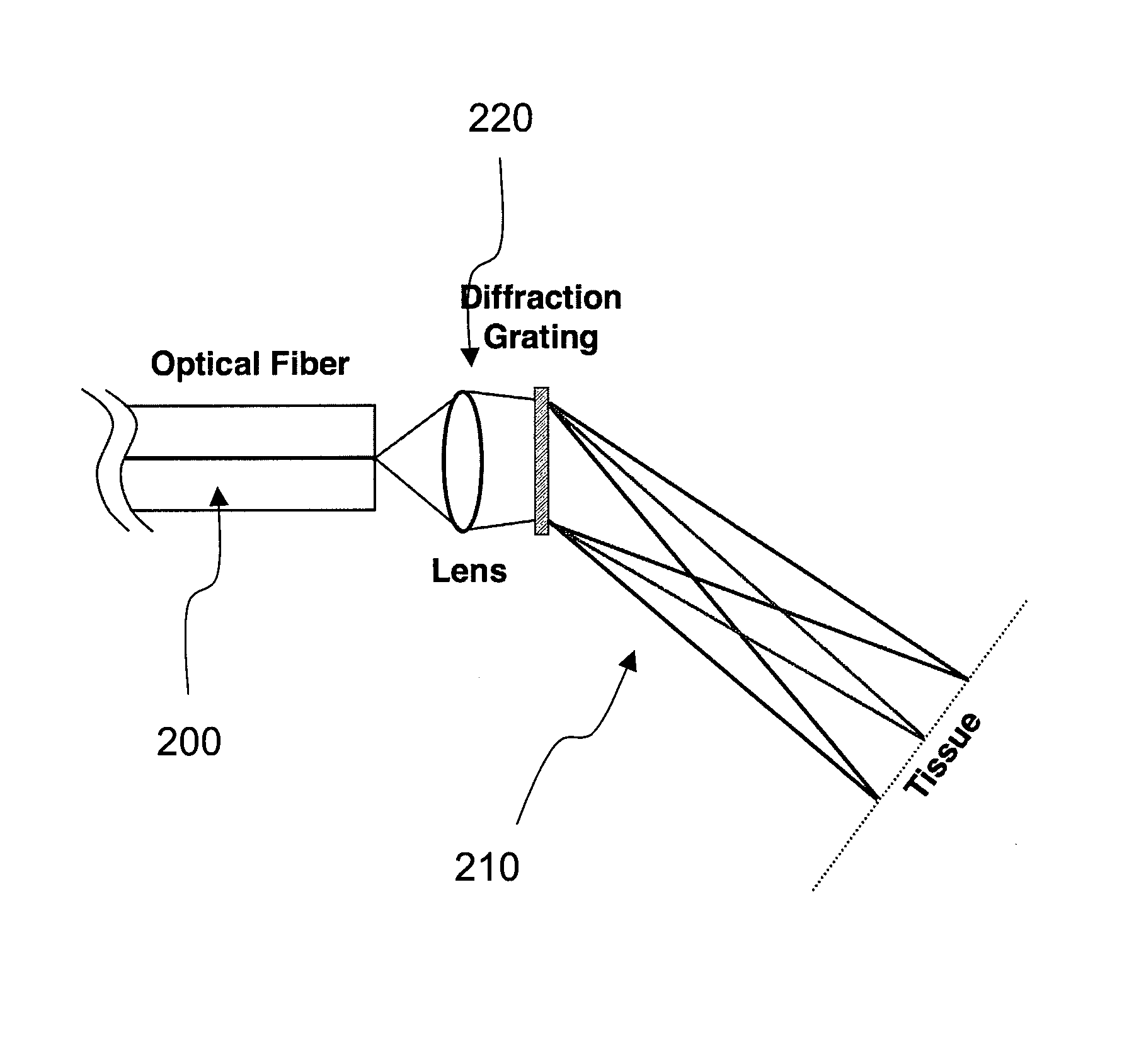
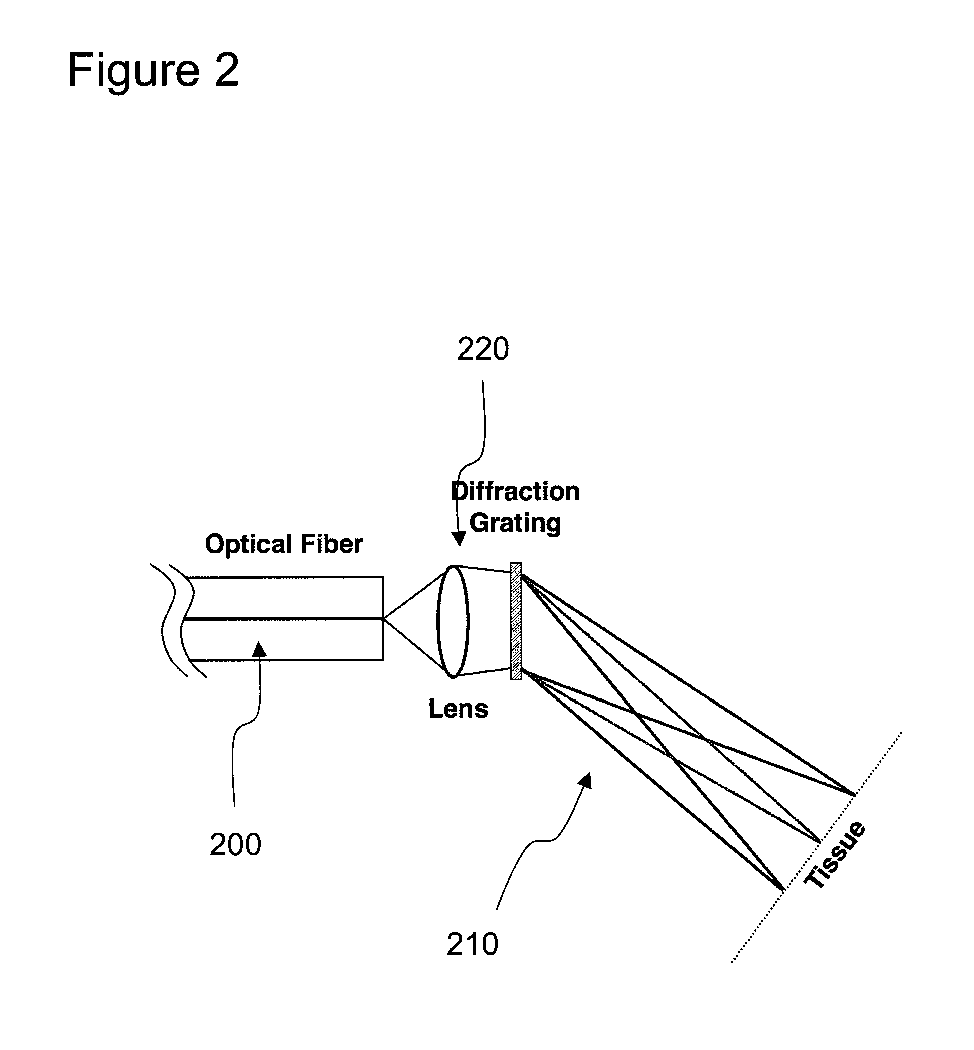
optical fiber. The exemplary SEE
system and probe can be provided for color differentiation of arterial and venous placental vessels and can add
Doppler imaging to quantify
blood flow. Additionally, it is possible to incorporate therapeutic
laser delivery through the same probe without increasing its size. exemplary total diameter of the exemplary apparatus / device /
system / arrangement can be, e.g., 350 μm, small enough to be introduced into the amniotic cavity through an amniocentesis needle (e.g., 22-gauge), which can provide, e.g., a 10-fold reduction in
complication rate.
[0014] Simultaneous imaging with more than one
wavelength band can provide color information from the sample. One such exemplary embodiment of apparatus and method to achieve such exemplary result may include a
coupling of visible (VIS) and near-
infrared (NIR) light into the SEE
fiber. If wavelengths are chosen correctly, for example 1064 and 532 nm, then the wavelength regions can overlap one another by diffracting at different orders. In another exemplary embodiment, the wavelengths may be selected such that the absorption properties of the sample can facilitate the differentiation and quantification of compounds within the sample, such as differentiation of arterial from
venous blood by measuring differences in oxy- and deoxy-
hemoglobin absorption. In yet another exemplary embodiment, the SEE probe may be situated in an interferometer, and a spectral
interferometric phase may be detected and analyzed to provide information on
blood flow and other motions of the sample. In another exemplary embodiment, the SEE probe may be configured by use of a
specialty fiber or at least one fiber adjacent to the imaging fiber to deliver therapeutic light to the sample to effect therapy. In yet another exemplary embodiment, the delivery of therapy light may be conducted in parallel with an imaging procedure.
[0016] According to still another exemplary embodiment, the SEE-guided therapy probe can be configured to effect coagulation of blood vessels such as those of the
placenta. In yet another exemplary embodiment, the SEE-guided therapy probe can be provided such that its diameter does not cause undue damage to the amniotic membranes or the
uterus, so to facilitate a safe therapy and place the
fetus and mother at
minimal risk. According to a further exemplary embodiment, the size of the SEE probe can be sufficiently small to fit within a narrow gauge needle, for example with a gauge of 18-25 Ga. In a still further exemplary embodiment, the needle can be an amniocentesis needle.
[0019] The exemplary embodiments of the present invention can overcome the deficiencies of the prior TTTS
laser coagulation devices by being significantly smaller and by facilitating further options for identifying
communicating vessels. This can be done using, e.g., spectrally encoded
endoscopy (SEE) procedures and systems. SEE procedures and systems can use wavelength-division
multiplexing to obtain high-resolution two- and three-dimensional endoscopic images through a single
optical fiber. High-resolution, 3D video-rate imaging
in vivo using a 350 μm diameter, monochromatic version of the SEE probe can be achieved with such procedures and systems. According to still other exemplary embodiments of the present invention, it is possible to obtain
color imaging, and the exemplary device may provide a measurement
blood flow. In yet another embodiment of the present invention, a therapeutic
laser light may be coupled through the same fiber so that imaging and intervention can be accomplished concurrently without increasing the
endoscope diameter.
[0020] For example, the size of the exemplary device can be, e.g., about 350 μm, so as to allow this device to be inserted into the amniotic cavity through a 22-gauge amniocentesis needle, which should lower the complication rates of placental
laser therapy by an
order of magnitude. Such exemplary embodiment of the device may facilitate a use thereof in other procedures where small size and highly capable image
guided intervention can decrease complication rates and improve
patient care.
[0021] According to a further exemplary embodiment of the present invention, the SEE procedures and apparatus can be utilized as follows. In particular, it is possible to provide a multifunctional SEE system and probe for discriminating arteries from veins. By incorporating high quality two- and three-dimensional imaging, e.g., color information and Doppler blood
flow mapping, this exemplary device / system / arrangement / apparatus can be used to identify A-V anastomoses. Further, it is possible to transmit high power Nd:YAG
laser light through the SEE probe to enable image-guided vessel photocoagulation without increasing the probe's size. Taken together, these exemplary features can facilitate the TTTS management to be enhanced by providing an endoscopic therapy system and apparatus with enhanced capabilities that can be inserted through an amniocentesis needle. This exemplary enhancement can make laser coagulation therapy for TTTS
safer and more effective. In addition to a treatment of TTTS, this exemplary device / apparatus can be used for an endoscopically-guided therapy in other areas of the body that have previously been difficult to access.


