Methods For Detecting Calcium Ion Influx

Inactive Publication Date: 2008-07-03
BABRAHAM INST
View PDF1 Cites 0 Cited by
  • Summary
  • Abstract
  • Description
  • Claims
  • Application Information

AI Technical Summary

Benefits of technology

[0133]An advantage of the methods of the invention is that the signal following calcium influx is

Problems solved by technology

A problem with many current methods is that the signal generated is of short duration and this restricts use of these assays in high throughput systems.
A further problem is that assay methods using fluorescent Ca2+ dyes in cell lines expressing a Ca2+ channel of interest are not generally suitable for targeting to a specific sub-cellular compartment of interest, e.g. under the plasma membrane.
Ca2+ dyes must be lo

Method used


Image

  • Methods For Detecting Calcium Ion Influx
  • Methods For Detecting Calcium Ion Influx
  • Methods For Detecting Calcium Ion Influx

Examples


example 1

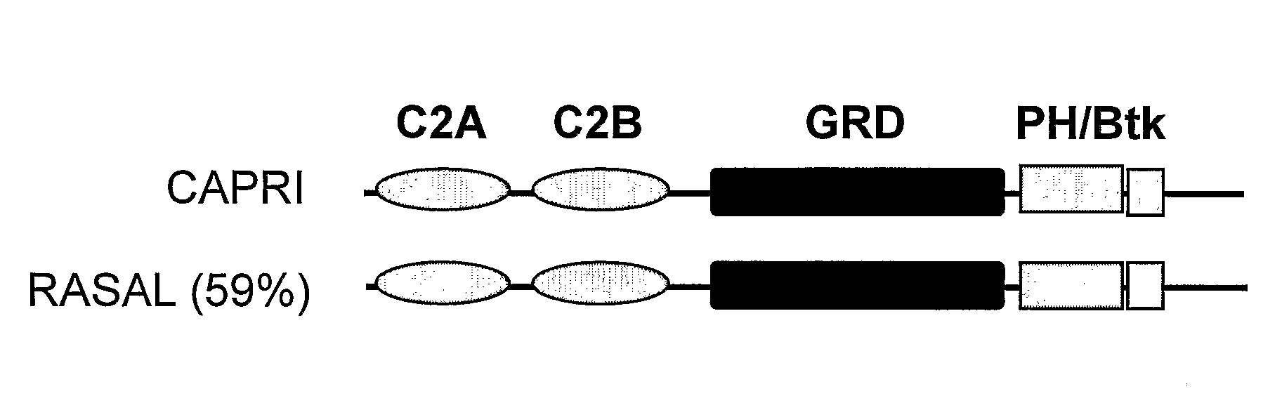
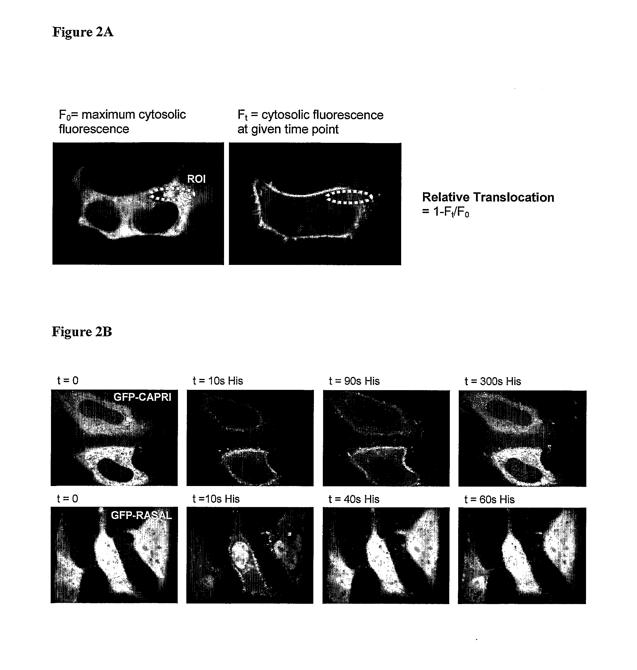
Ca2+ Influx Assay Using a Genetically-Encoded Fluorescent Reporter—Translocation of CAPRI and RASAL in Response to Agonist (100 μM Histamine) Stimulation of HeLa Cells

[0161]The day before transfection HeLa cells were seeded onto 22 mm glass coverslips in 6-well tissue culture dishes at such a density to reach 60-80% confluence within 24 hours. The following day (approximately 24 hours later) HeLa cells were transfected with green fluorescent protein GFP-CAPRI or GFP-RASAL using GeneJuice Transfection Reagent (Merck) according to the manufacturers instructions.

[0162]After 24 hrs incubation in transfection mix (complete media plus transfection reagent) at 37° C., 5% CO2) coverslips were transferred to holders containing 2 ml of KH buffer (10 mM HEPES, 118 mM NaCl, 4.7 mM KCl, 10 mM glucose, 1.2 mM KH2PO4, 4.2 mM NaHCO3, 1.2 mM CaCl2, pH 7.4) and monitoring performed by live imaging in a 37° C. heated chamber on an inverted Nikon TE-2000 microscope using a 40× oil objective lens. Indiv...

example 2

Screening to Detect a Receptor Antagonist Compound

[0170]The day before transfection COS-7, HEK293 and HeLa cells are seeded onto 22 mm glass coverslips in 6-well tissue culture dishes at such a density to reach 60-80% confluence within 24 hours. After approximately 24 hours, COS-7, HEK293 and HeLa cells are transiently transfected with GFP-CAPRI plasmid DNA using Lipofectamine (GIBCO BRL) or GeneJuice transfection reagent (Merck) according to manufacturers instructions and incubated for 24 hours as above. The cells are incubated with test compound in EM buffer (121 mM NaCl, 5.4 mM KCl, 1.6 mM MgCl2, 6 mM NaHCO3, 9 mM glucose, 1.3 mM CaCl2, 25 mM HEPES, pH 7.4) or KH buffer (10 mM HEPES, 118 mM NaCl, 4.7 mM KCl, 10 mM glucose, 1.2 mM KH2PO4, 4.2 mM NaHCO3, 1.2 mM CaCl2, pH 7.4) for up to one hour.

[0171]To stimulate calcium influx, the agonist histamine (HeLa; 1-100 μM)) or ATP (HeLa, HEK293 and COS; 50 μM) is applied by bulk addition (rapid mixing of 5 ml of agonist in appropriate im...

example 3

Screening for SOCE Channel Inhibitors

[0174]The day before transfection COS-7, HEK293 and HeLa cells are seeded onto 22 mm glass coverslips in 6-well tissue culture dishes at such a density to reach 60-80% confluence within 24 hours. After approximately 24 hours, COS-7, HEK293 and HeLa cells are transiently transfected with GFP-CAPRI plasmid DNA using Lipofectamine (GIBCO BRL) or GeneJuice transfection reagent (Merck) according to manufacturers instructions and incubated for 24 hours as above.

[0175]Prior to introduction of the test compound, cells are stimulated with 1-5 μM thapsigargin in 2 ml Ca2+-free media for 5 minutes. The cells are then incubated in the presence of the test compound for up to one hour.

[0176]The cells are imaged in Ca2+-free EM buffer (121 mM NaCl, 5.4 mM KCl, 1.6 mM MgCl2, 6 mM NaHCO3, 9 mM glucose, 0.5 mM EGTA, 25 mM HEPES, pH 7.4) or Ca2+-free KH buffer (10 mM HEPES, 118 mM NaCl, 4.7 mM KCl, 10 mM glucose, 1.2 mM KH2PO4, 4.2 mM NaHCO3, 0.5 mM EGTA, pH 7.4). ...


PUM

PropertyMeasurementUnit
Fluorescenceaaaaaaaaaa

Abstract

A method for detecting influx of calcium ions into a eukaryotic cell comprising providing a eukaryotic cell having a detectable reporter capable of translocation from the cytosol to associate with the plasma membrane in response to an influx of calcium ions, and, monitoring association of the detectable report with the plasma membrane and/or a decrease in the detectable report in the cytosol. The detectable report is preferably CAPRI or a derivative thereof and is preferably labelled with a fluorescent marker.

Description

TECHNICAL FIELD[0001]The invention relates to live, whole cell assays for detecting calcium ion (Ca2+) influx using a detectable reporter, particularly a fluorescent reporter. The methods are useful for detection of compounds that modulate calcium influx and can be performed in high throughput screening format.BACKGROUND TO THE INVENTION[0002]Calcium influx into the cell from the extracellular medium is vital for processes such as muscle contraction, secretion and gene activation. Calcium influx is mediated via calcium influx channels which can be divided into two groups on the basis of their activation mechanism: voltage-gated calcium channels and non-voltage-gated calcium-permeable calcium channels. Calcium influx can be stimulated or inhibited by factors that act on the calcium ion channel directly, or indirectly where modulation of a receptor results in a signal that acts on a calcium ion channel.[0003]Ion channels play an important role in numerous cell types and occur as large...

Claims


Application Information

Patent Timeline
IPC IPC(8): G01N33/53C07K19/00G01N33/50G01N33/68G01N33/84
CPCB82Y5/00B82Y10/00G01N2500/10G01N33/6872B82Y30/00
Inventor LOCKYER, PETERLOCKYER, JULIE
Owner BABRAHAM INST
Why Eureka
Try Eureka