Method of screening placental proteins responsible for pathophysiology of preeclampsia, and marker for early diagnosis and prediction of preeclampsia

a technology of placental proteins and pathophysiology, which is applied in the field of screening placental proteins responsible for the pathophysiology of preeclampsia, and a marker for early diagnosis and prediction of preeclampsia, can solve the problems of preeclampsia in pregnancy that can be a very serious health problem, fetal growth restriction, premature delivery,

Inactive Publication Date: 2009-01-22
HAN OU JIN
View PDF0 Cites 15 Cited by
  • Summary
  • Abstract
  • Description
  • Claims
  • Application Information

AI Technical Summary

Problems solved by technology

Preeclampsia in pregnancy can be a very serious health problem.
It can cause fetal growth restriction, fetal death and morbidity, premature deliveries, and death of the mother.
The exact cause of preeclampsia is not known, and treatments for efficiently curing or preventing preeclampsia are not also available yet.
Preeclampsia is known to cause several problems at the same time, such as high blood pressure (hypertension), pathological edema and leakage of protein into the urine (proteinuria).
Further, preeclampsia is one of the pregnancy complications that bring hypertension, proteinuria and traumatism to the mother.
It is known that preeclampsia occurs to only about 3-5% of pregnant women, but it can seriously affect both the mother and her unborn (or newborn) baby, and thus, acts as a major cause of increasing perinatal mortality and morbidity rates.
Early diagnosis and timely treatment of preeclampsia can remarkably reduce risks to the mother and her unborn baby, but such a monitoring method by using those symptoms as criteria is not effective for an early diagnosis of preeclampsia.
Further, no treatments are currently available to cure preeclampsia.
Preeclampsia can be mild, but potentially life-threatening depending on the severity of the disease.
Despite such clinical risks, however, it is difficult to find the cause or the pathogenesis of preeclampsia at an early stage, or to make an early diagnosis and prognosis.
Even if many researches have been conducted to monitor and predict the occurrence of preeclampsia, they are limited to using a specific protein or substance, which is not sufficient to explain the whole phenomenon about the occurrence of preeclampsia and the pathogenesis thereof.

Method used


Image

  • Method of screening placental proteins responsible for pathophysiology of preeclampsia, and marker for early diagnosis and prediction of preeclampsia
  • Method of screening placental proteins responsible for pathophysiology of preeclampsia, and marker for early diagnosis and prediction of preeclampsia
  • Method of screening placental proteins responsible for pathophysiology of preeclampsia, and marker for early diagnosis and prediction of preeclampsia

Examples


example 1

Separation of Placental Proteins

[0022]Normal placenta and preeclamptic placenta were prepared for 2D E-proteomics analysis. Placental tissues were ground to fine powder in presence of liquid nitrogen, and a buffer (tissue: 0.2 g / 10 ml) was added thereto. The samples were then divided into tubes, boiled for five minutes, put into the ice bath for five minutes, and centrifuged at 8,000 rpm and 4° C. for 10 minutes. Each of the upper phases was transferred to a new tube by 800 μl, treated with enzymes (DNase / RNase), and put into the ice bath for 10 minutes. Next, 200 μl of 10% TCA / acetone preparation-50% TCA / acetone was added to each tube. The tubes were placed into the ice bath for a period of 1 hour. The samples were then centrifuged at 12,000 rpm and 4° C. for 10 minutes, and the resulting pallets were washed with acetone. The remaining dry powders were kept at −20° C.

example 2

Separation of Proteins by 2D E-Proteomics

[0023] Primary Separation of Proteins by Isoelectric Point

[0024]Proteins are primarily separated based on isoelectric point. Dry immobilized pH gradient (IPG) strips of 13 cm were added with 250 μl isoelectric point marker containing 50 μg protein and rehydrated over 10 hours. The rehydrated IPG strips were subjected to isoelectric point separation in an IPG phore (GE Healthcare, USA). The isoelectric point separation was carried out for 1 hour at 500 V, for 1 hour at 1,000 V, and finally at 8,000 V until the final accumulated voltage becomes 60,000 V. At this time, the highest current was set to 50 μA per strip. The strips separated by isoelectric point were slowly stirred over a period of 15 minutes in presence of a primary phase equilibrium solution (50 mM Tris-HCl containing 6M urea, 30% glycerol, 2% SDS, bromophenol blue and 1% DTT, pH 8.8). These primary phase equilibrated strips were soaked in a secondary phase equilibrium solution (50...

example 3

Gel Scanning and Gel Image Analysis

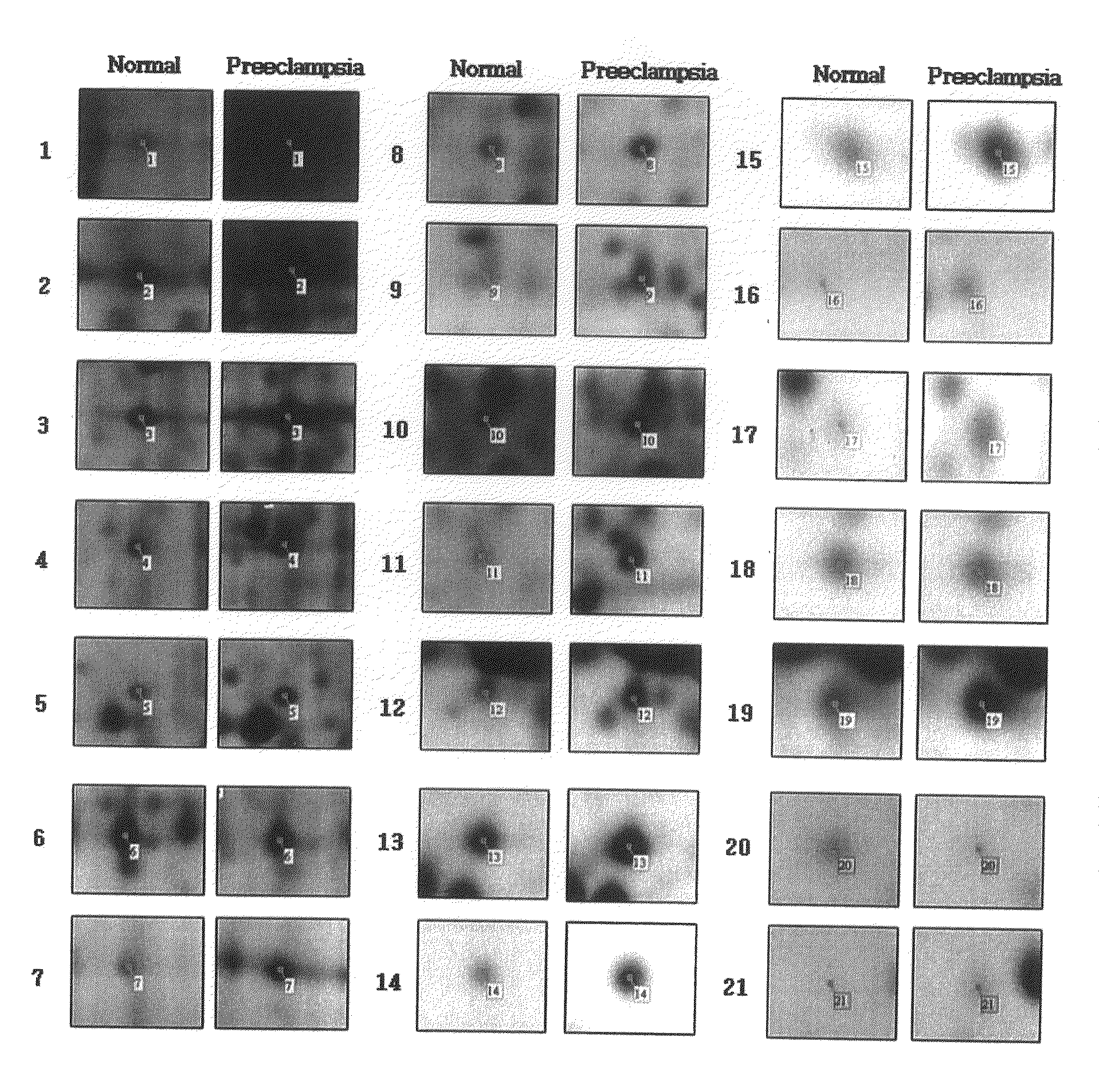
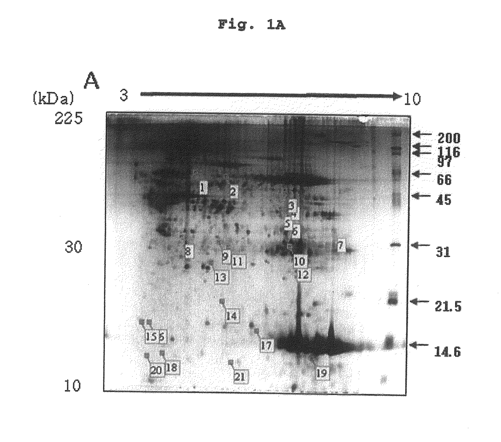
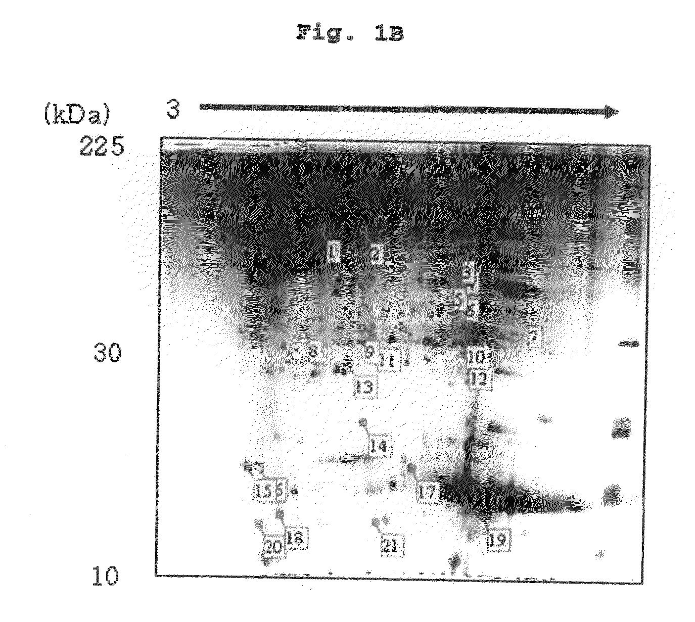
[0027]The stained gel was scanned through a flatbed scanner (UMAX PowerLook 1100, USA). During scanning, the option of a transmissive type with 300 dpi resolution was chosen. The scanned gel images were analyzed through an image analysis program (Image Master 2D Platinum, GE Healthcare, USA). Based on the image analysis result, proteins showing differences up to 140% or more in two groups of placental samples were selected and finally identified. In result, the inventors checked proteins showing big differences between normal placenta and preeclamptic placenta by electrophoresis shown in FIG. 1 (In FIG. 2, left-hand side: normal cell; right-hand side: gastric cancer cell), and identified 21 proteins with differences in expression between normal placenta and preeclamptic placenta.


PUM

PropertyMeasurementUnit
pHaaaaaaaaaa
sizeaaaaaaaaaa
pHaaaaaaaaaa

Abstract

The present invention relates to a method of screening placental proteins responsible for pathophysiology of preeclampsia, and a marker for early diagnosis and prediction of preeclampsia. In accordance with one aspect of the present invention, there is provided a method of screening placental proteins responsible for pathophysiology of preeclampsia by 2D E-proteomics analysis, comprising: isolating placental proteins from a placental tissue; separating the isolated proteins two-dimensionally through 2D electrophoresis; and comparing and analyzing the separated proteins based on scanned gel images and differences in the images between normal placental proteins and preeclamptic placental proteins, wherein the comparison and analysis of the placental proteins based on the scanned gel images and differences in the images are accomplished by selecting proteins with differences of 140% or more between two placentas.

Description

FIELD OF THE INVENTION[0001]The present invention relates to a method of screening placental proteins responsible for pathophysiology of preeclampsia, and a marker for early diagnosis and prediction of preeclampsia.BACKGROUND OF THE INVENTION[0002]Preeclampsia in pregnancy can be a very serious health problem. It can cause fetal growth restriction, fetal death and morbidity, premature deliveries, and death of the mother. The exact cause of preeclampsia is not known, and treatments for efficiently curing or preventing preeclampsia are not also available yet. Preeclampsia is known to cause several problems at the same time, such as high blood pressure (hypertension), pathological edema and leakage of protein into the urine (proteinuria). Further, preeclampsia is one of the pregnancy complications that bring hypertension, proteinuria and traumatism to the mother. It is known that preeclampsia occurs to only about 3-5% of pregnant women, but it can seriously affect both the mother and h...

Claims


Application Information

Patent Timeline
Patent Type & Authority Applications(United States)
IPC IPC(8): C12Q1/48G01N33/00C12Q1/32C12N9/02C12N9/14C12N9/10C12Q1/34C12Q1/26
CPCG01N33/689G01N33/6893G01N2800/368G01N2550/00G01N33/53C12Q1/37G01N33/543G01N33/561
Inventor PARK, WON SUNKIM, NA RIHAN, JIN
Owner HAN OU JIN
Why Eureka
Try Eureka