
Method for Detecting Transferase Enzymatic Activity

Inactive Publication Date: 2010-01-28
PROMEGA
View PDF6 Cites 18 Cited by
  • Summary
  • Abstract
  • Description
  • Claims
  • Application Information

AI Technical Summary

Benefits of technology

[0017]In one embodiment of the invention, a method for detecting kinase activity of a sample is provided. In a preferred embodiment, the sample is contacted with a kinase substrate, and at least one of a phosphate group donor (specifically ATP) and a phosphate group acceptor substrate or the enzyme itself (autophosphorylation) to form a reaction mixture. Kinase activity or the effect of a compound on kinase activity in a sample would result in a decrease or increase in the ATP levels in the sample. Thereafter, a luminescent reporter with contacted with the reaction mixture. ATP interacts with a luminescent reporter compound and produces a luminescence signal that is directly proportional to the amount of ATP present. The luminescence output of the reporter compound is then detected, typically reported as Relative Light Units (“RLU”). An advantage of the present invention is that the kinase activity detection methods can be performed in a single well in a multi-well plate, making them suitable for use as high throughput screening methods. The method of the present invention may be optimized by altering the amounts of ATP and kinase substrate. In addition, increasing the reaction temperature may improve kinase activity.
[0018]The method of the present invention can be utilized to detect kinase activity over a wide range of ATP concentrations, generally from about 1 to about 100 μM of ATP. The method of present invention may be used to detect kinase activity at low concentration levels of ATP, generally below 5 μM of ATP, more preferably in the range of about 1 to about 3 μM of ATP.
[0019]In another embodiment of the invention, the method of detecting kinase activity comprises contacting a sample with a kinase substrate, at least one of a phosphate group donor (specifically ATP) and a phosphate group acceptor for a first predetermined time period to allow for sufficient opportunity for the kinase to interact with the kinase substrate. The resulting kinase reaction mixture is then contacted with a composition (“reagent composition”) for a second predetermined time period. The reagent composition comprises a bioluminescence generating enzyme, a luminogenic molecule and a transferase quenching agent. Thereafter, the luminescence produced in the resulting reaction mixture is then detected. The luminescence is produced by the conversion of the luminogenic molecule into a luminescing compound by bioluminescence generating enzyme such as luciferase. This method can be used to measure a distinct end-point of a kinase reaction. The reagent composition allows, in a single step, for the simultaneous quenching or termination of transferase activity and generation of a luminescent signal that is directly proportional to the amount of ATP present.
[0020]The method is homogeneous and can be used for a wide variety of transferases such as protein kinases and lipid kinases and substrates such as amino acids, peptides, proteins (including fusion proteins and other kinases), sugars and lipids. The regent is robust and resulting luminescence is much less suspectible to interference by library compounds than other luciferase-based ATP detection reagents. In addition, the reagent composition facilitates measurement of transferase activity in a single sample over a long period of time as well as measurement of transferase activity in many samples in a high throughput format over a long period of time, thus eliminating the need for luminometers with reagent injectors and allowing for batch-mode processing of multiple samples.
[0021]In general, the methods comprise adding a composition (“reagent composition”) comprising a bioluminesce generating enzyme such as a luciferase (such as exemplified by, but not limited to, SEQ ID NOs: 1-4), a luminogenic substrate such as luciferin or luciferin derivative, and one or more transferase quenching agents to a sample and detecting luminescence, wherein the activity of the reagent composition has enhanced stability [i.e., the reagent composition is capable of maintaining at least about 30%, more preferably at least about 60% activity (as measured by luminescence when the reagent composition is combined with the sample) for at least one hour, even more preferably at least 70%, 80%, 90%, 95%, 99% or greater activity for at least one hour, still more preferably for at least two hours and even more preferably for at least four hours relative to the reagent composition's activity when it is created, i.e., just after (0 to 10 minutes)], the luciferase enzyme is combined with a transferase quenching agent, and wherein the transferase quenching agent is present in the reagent composition at a concentration sufficient to reduce transferase activity endogenous to the sample by at least about 25%, more preferably at least about 30%, more preferably at least about 40%, even more preferably 50%, 60%, 70%, 80%, 90%, 95%, or 99% or greater relative to the sample's transferase activity in the absence of the transferase quenching agent. The reagent composition may be admixed before use by adding a solution comprising one or more transferase quenching agents to a lyophilized luciferase.
[0022]Loss of stability is defined as irreversible loss of activity. The reagent composition loses stability over time and the amount of activity lost varies depending on the particular luciferase, transferase quenching agent and, when present, enzyme stabilizing agent used. Preferably the stability of the reagent composition is demonstrable in the temperature range of about 20° C. to about 37° C. Although the methods of the invention may be used with a sample containing any amount of ATP, it is preferable to use a sample containing a non-saturated amount of ATP (i.e., a range where luminescence is linearly proportional to the concentration of ATP).

Problems solved by technology

A lack of controlled activity of kinases in cells is believed to lead to the formation of tumours.
Current types of assays used to measure kinase activity and to detect potential kinase inhibitors are cumbersome and costly.
These requirements make a FRET based assay cumbersome and costly.
The time consuming and costly optimization of antibody binding with specific fluorescent labeled molecules such as peptides is required where antibodies are used.
Additionally, the FP assay there is the potential for phosphorylated protein and other reaction components, e.g., lipids and detergents, to interfere with the polarization.
However, a SPA requires radio-labeled ligands, which have disposal costs and possible health risks.

Method used


Image

  • Method for Detecting Transferase Enzymatic Activity
  • Method for Detecting Transferase Enzymatic Activity
  • Method for Detecting Transferase Enzymatic Activity

Examples


example 1

Determining the Activity of Lipid Dependent Serine / Threonine Kinases

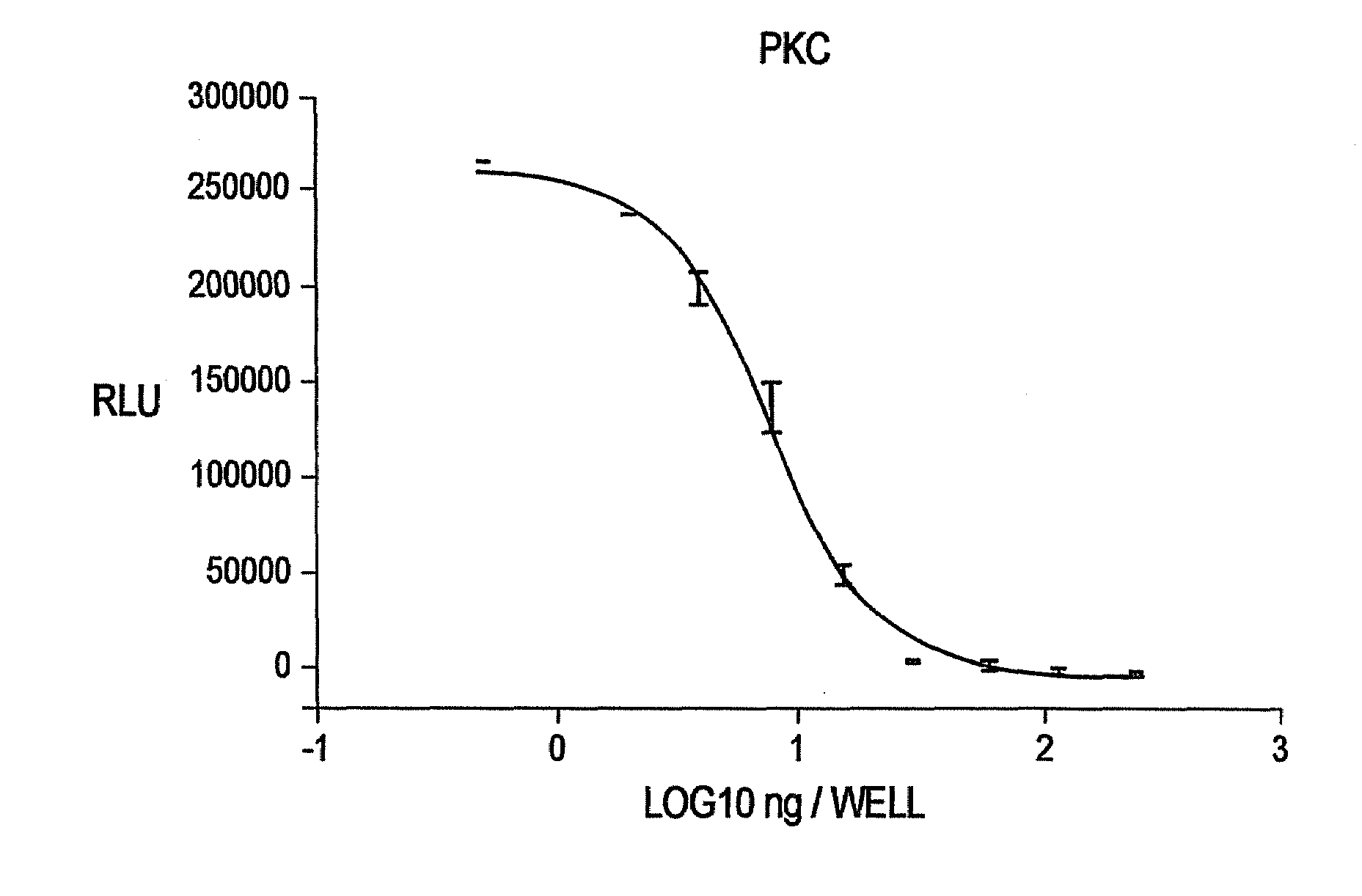
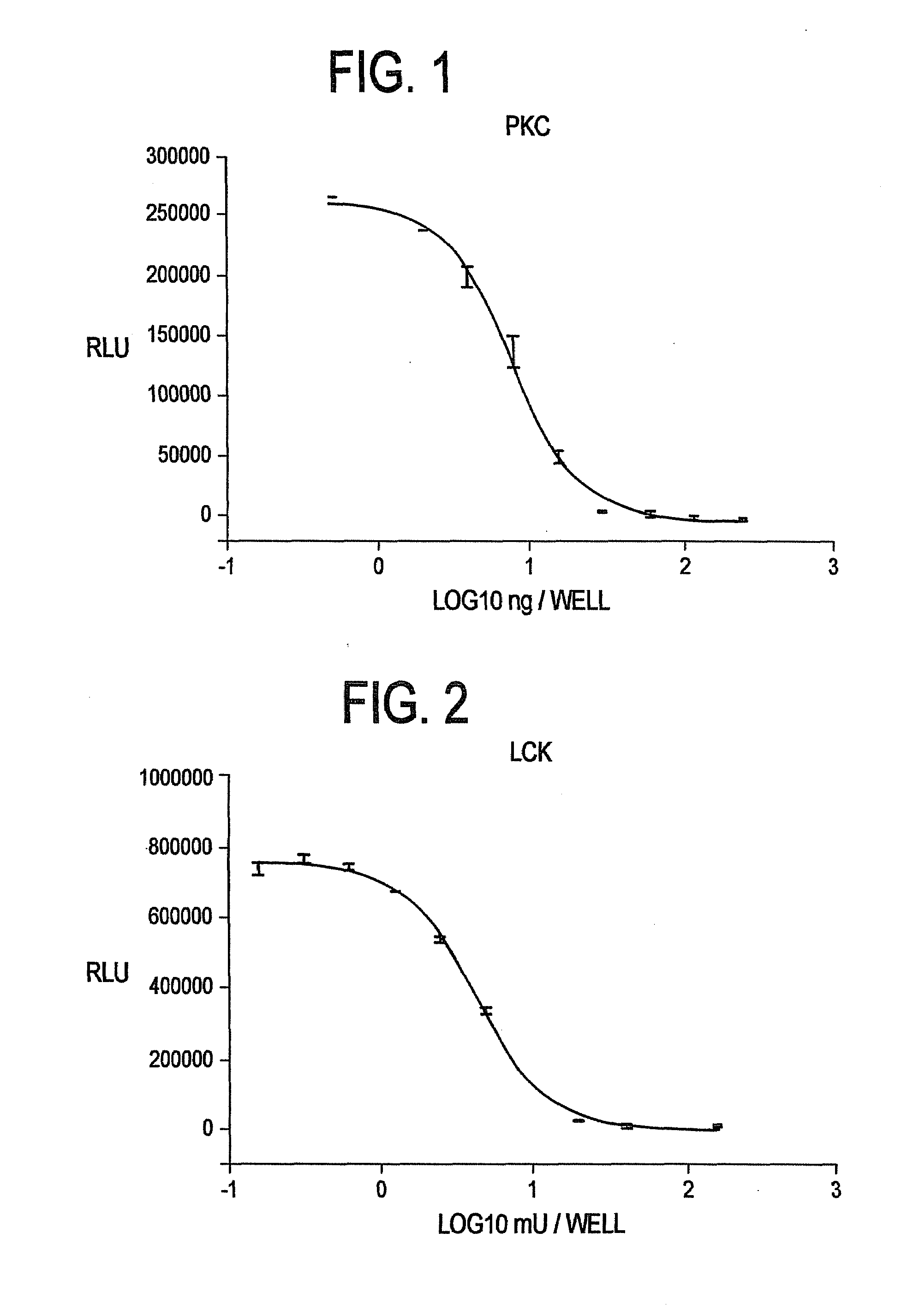
[0140]Commercially available Protein Kinase C (“PKC”) was titrated in a 96 well plate (n=2). Kinase reactions were performed in 20 mM Tris-HCl (pH 7.5), 10 mM MgCl2, 0.1 mg / ml bovine serum albumin (“BSA”), 250 μM EGTA, 400 μM CaCl2, 0.32 mg / ml phosphatidylserine, 0.032 mg / ml diacylglycerol, 10 μM biotinylated peptide (AAKIQASFRGHMARKK), and 1 μM ATP with the indicated amount of PKC (Promega, Catalog #V5621). Final kinase reaction volume was 50 μl. Following a 90 minute kinase reaction, 50 μl of a luminescent buffer reagent (pH 6.0+0.15) containing 20 mM citrate; 55 mM MES; 10.5 mM magnesium sulfate; 0.6 mM CDTA; 225 mM potassium buffer (pH 6.0); 1 mM NaF; 0.0125 uM sodium pyrophosphate; 0.5% DTAB; 1.0% Thesit; 0.1% Mazu DF 204; 2.5 mM luciferin; and 0.2% 2-(N-morpholino) ethanesulfonic acid containing 10 μg / ml [TRUE?] of a thermostable firefly luciferase reporter 146-1H2 (see SEQ ID NO.: 4 and SEQ ID NO.: 8) was add...

example 2

Determining the Activity of Tyrosine Kinases

[0141]Commercially available Lck was titrated in a 96 well plate (n=2). Lck is a gene family encoding nonreceptor tyrosine kinases of the Src family. Kinase reactions were performed in 8 mM imidazole hydrochloride (pH 7.3), 8 mM β-glycerophosphate, 200 μM EGTA, 20 mM MgCl2, 1 mM MnCl2, 0.1 mg / ml BSA, 250 μM biotinylated peptide substrate (AEEEIYGELEA), and 3 μM ATP with the indicated amount of Lck (Upstate, Catalog #14-442). Final kinase reaction volume was 50 μl. Following a 60 minute kinase reaction, 50 μl of a luminescent buffer reagent as described in Example 1 was added to each well and allowed to incubate for 10 minutes prior to reading luminescence. FIG. 2 clearly shows that a consistent titration curve is obtained using the method of the present invention.

example 3

Determining the Activity of Cyclic-AMP Dependent Serine / Threonine Protein Kinase

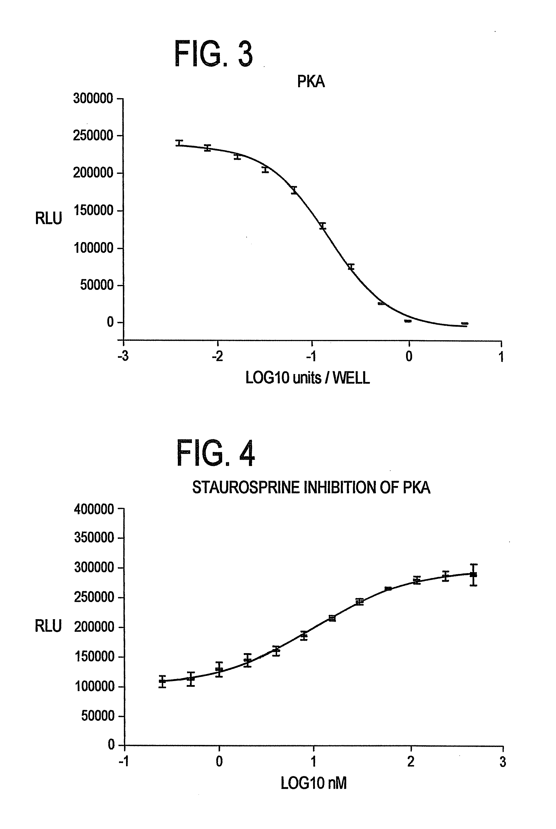
[0142]Commercially available Protein Kinase A (“PKA”) was titrated in a 96 well plate (n=8). Kinase reactions were performed in 40 mM Tris-HCl (pH 7.5), 20 mM MgCl2, 0.1 mg / ml BSA, 5 μM kemptide (LRRASLG), and 1 μM ATP with the indicated amount of PKA (Promega, Catalog #V5161). Final kinase reaction volume was 50 μl. Following a 20 minute kinase reaction, 50 μl of a luminescent buffer reagent as described in Example 1 was added to each well and allowed to incubate for 10 minutes prior to reading luminescence. FIG. 3 clearly shows that using the method of the present invention, a reliable titration curve is obtained.


PUM


Abstract

Methods and kits for detecting transferase activity in a sample by measuring ATP using a composition comprising an ATP-dependent bioluminescence-generating enzyme, a luminogenic molecule and one or more transferase quenching agents.

Description

CROSS REFERENCE TO RELATED APPLICATIONS[0001]This non-provisional application claims the benefit of priority to U.S. provisional application No. 60 / 408,662, filed Sep. 6, 2002 entitled METHOD FOR DETECTING TRANSFERASE ENZYMATIC ACTIVITY.FIELD OF THE INVENTION[0002]The present invention relates generally to the fields of enzymology and molecular biology. In particular, this invention relates to methods, compositions and kits for improving the detection and quantitation of transferase activity.BACKGROUND OF THE INVENTION[0003]Advances in the biological, biomedical and pharmaceutical sciences have accelerated the pace of research and diagnostics unparalleled to the past. With whole genome sequences becoming quickly and successively available, the assembly of large libraries of small molecules, and the ability to move pharmaceutical development, clinical diagnostic tests and basic research from a reductionist to a whole system approach demands assays that facilitate high throughput anal...

Claims


Application Information

Patent Timeline
IPC IPC(8): C12Q1/48C12P13/16C12Q1/66G01NG01N33/53
CPCC12Q1/66C12Q1/485
Inventor SOMBERG, RICHARDGOUELI, SAID A.
Owner PROMEGA
Learn More