Monitoring and manipulating cellular transmembrane potentials using nanostructures

a technology of cellular transmembrane potential and nanostructure, which is applied in the direction of fluid pressure measurement, liquid/fluent solid measurement, peptide measurement, etc., can solve the problems of complex role of cell electrical properties in generating a defined disease phenotype, serious complications to health, and change of cell electrical properties

Inactive Publication Date: 2010-05-13
LIFE TECH CORP
View PDF1 Cites 11 Cited by
  • Summary
  • Abstract
  • Description
  • Claims
  • Application Information

AI Technical Summary

Problems solved by technology

Transport of ions across the membrane through ion channels will lead to disturbance of the existing equilibrium of ion concentrations on both sides of membrane and, thus, to changes of electrical properties of the cell.
In humans, inherited or induced changes in ion channel function could result in serious complications to health.
Voltage-gated calcium ion channels are involved in numerous cellular functions, and their role in generating a defined disease phenotype is complex.
Most notably, mutations of the HERG potassium ion channels expressed in cardiac tissues or pharmacological blockage of HERG channels cause heart disease (long Q-T syndrome), which leads to increased risk of ventricular tachycardia and sudden death.
Several drugs affect these channels and can lead to life threatening cardiac arrhythmias.
Although this technique allows detailed biophysical characterization of ion channel activation, inactivation, gating, ion selectivity, and drug interactions, throughput is quite low and ease-of-use of patch-clamp instrumentation is generally unsatisfactory for effective mass screening.
The search for molecules that modulate ion channel function has been hindered by the lack of direct electrical measurements in HTS formats.
Real-time measurements of transmembrane potential kinetics that accurately reflect ion channel activity are fundamental to cell physiology, but are difficult to measure in existing HTS format methods.
Competition-binding assays, although successfully used for other target classes, often fail to identify ligands that modulate specific ion channel states.
Unfortunately binding assays only detect binding of compounds to ion channels an...

Method used


Image

  • Monitoring and manipulating cellular transmembrane potentials using nanostructures
  • Monitoring and manipulating cellular transmembrane potentials using nanostructures
  • Monitoring and manipulating cellular transmembrane potentials using nanostructures

Examples


example 1

Preparation of Cells on Coverslips

[0131]Experiments were performed on A431 (a human cell line from an epidermoid carcinoma) cells or CHO (Chinese hamster ovary) cells stably expressing M1 muscarinic Gq-protein coupled receptor using nanoparticles commercially available from Quantum Dot Corp. (a wholly owned subsidiary of Invitrogen Corp.; Carlsbad, Calif.) and Evident Technologies (Troy, N.Y.). The intracellular (pipette) solution (pH 7.3) was composed of 140 mM CsCl, 10 mM EGTA, 10 mM HEPES. The extracellular solution (pH 7.4) was composed of 140 mM NaCl, 5 mM KCl, 1.8 mM CaCl2, 1 mM MgCl2, 10 mM EGTA, 10 mM glucose, 10 mM HEPES.

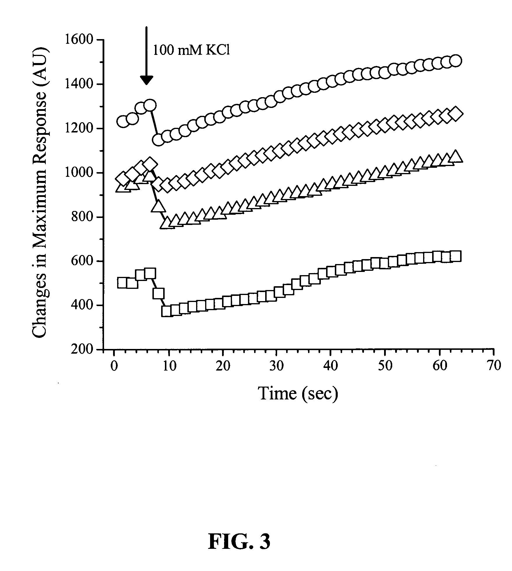
[0132]In experiments with extracellular delivery of nanoparticles, several types of commercially available nanocrystals were used. In one series of experiments, streptavidin-functionalized QD605 (Quantum Dot Corp.) in the buffer solution B from the QDot® kit were added to extracellular solution in concentrations from 25 to 500 μg / ml. In another experimental...

example 2

Use of Patch Pipette

[0136]Glass micropipettes for patch-clamp experiments were pulled from borosilicate glass capillaries (1.2 mm no-capillary glass, Sutter Instruments; Novato, Calif.) using a Sutter 2000™ pipette puller (model Sutter 2000; Sutter Instruments; Novato, Calif.) using the prerecorded 4-step patch pipette pulling protocol. The open diameter of the pipette tip was 1.5-2.2 μm with a resistance of 2-3 MΩ. The micropipettes were filled with intracellular solution.

[0137]Experiments were performed at room temperature in whole-cell patch-clamp configuration using a Axopatch200B patch-clamp amplifier (Molecular Devices; Sunnyvale, Calif.). After successful giga-seal formation, brief pulses of suction were used to rupture the cellular membrane to achieve whole-cell patch-clamp configuration.

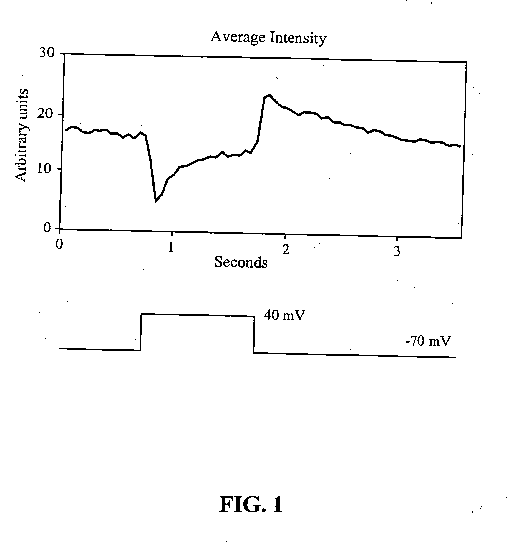
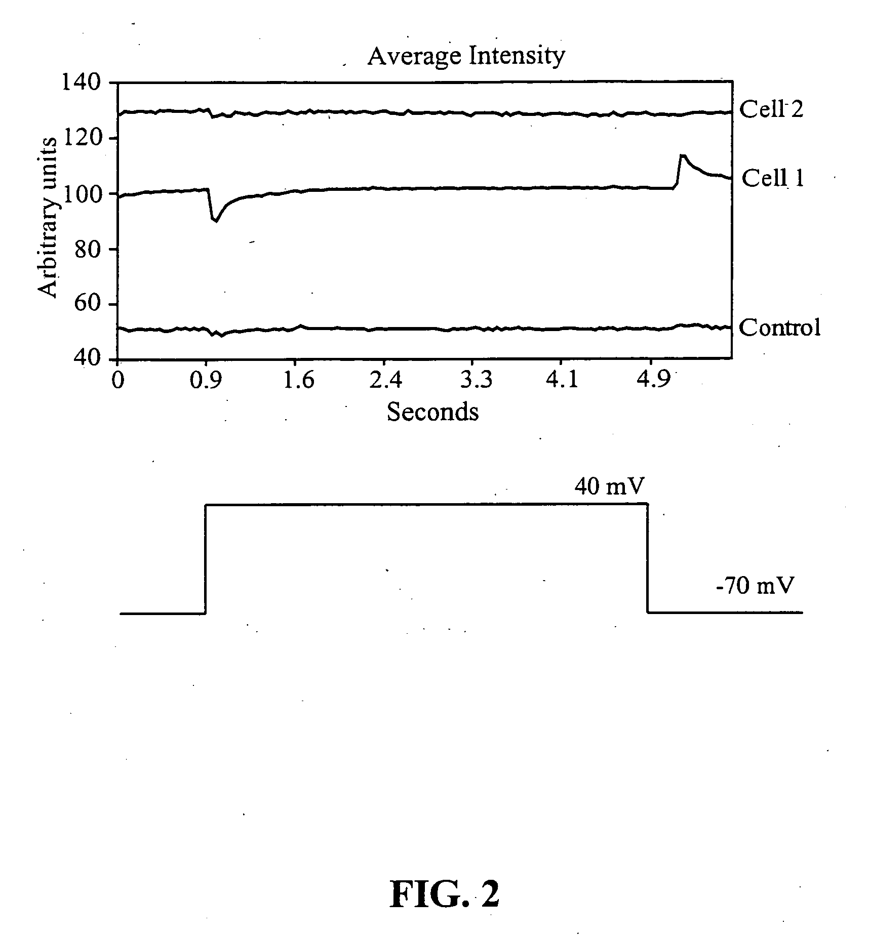
[0138]The following test protocol was used for cell stimulation. The membrane potential was set at −70 mV. A depolarizing pulse necessary to take the cell to +40 mV was applied to the interi...

example 3

External loading of streptavidin-coated nanoparticles

[0139]The emission intensity of externally applied streptavidin-functionalized nanoparticles occurring in response to voltage stimulation of the cell (QD605-streptavidin, Quantum Dot Corp.) was visualized using a cooled CCD Optronics Tec 470 camera (Optronic Engineering, Goleta, Calif.) linked to a computer. Voltage changes elicited across the cellular membrane via patch pipette attached to a cell did not result in changes in the emission intensity of these particular nanoparticles. Nine cells were tested in this series, and none exhibited changes in emission intensity to the voltage stimulation protocol described in the previous example. The streptavidin coating of the nanoparticles used in this example may have prevented the nanocrystals from being strategically placed inside the cellar membrane, the site of the highest membrane gradient. The streptavidin coating of the nanoparticles used in this example may have prevented the n...


PUM

PropertyMeasurementUnit
Electric potential / voltageaaaaaaaaaa

Abstract

The use of nanostructures to monitor or modulate changes in cellular membrane potentials is disclosed. Nanoparticles having phospholipid coatings were found to display improved responses relative to nanoparticles having other coatings that do not promote localization or attraction to membranes.

Description

CROSS REFERENCE TO RELATED APPLICATIONS[0001]The present application claims priority to U.S. Provisional Patent Application Ser. No. 60 / 659,975 filed Mar. 8, 2005, the contents of which are incorporated herein by reference.FIELD OF THE INVENTION[0002]The invention relates to compositions and methods useful for monitoring and manipulating cellular transmembrane voltages. In particular, nanoparticles and their use in monitoring and manipulating transmembrane voltages is disclosed.DESCRIPTION OF RELATED ART[0003]All cells have phospholipid membranes that serve as bimolecular barriers and to separate cell contents from the extracellular environment. The purpose of the plasma membrane is to maintain the necessary difference in composition between two compartments by restricting or permitting the passage of materials through the membrane as a function of intracellular signaling.[0004]Each cell has a resting membrane potential originating from the so-called “separation of charge” across th...

Claims


Application Information

Patent Timeline
IPC IPC(8): G01N27/26
CPCB82Y5/00B82Y15/00G01N33/588G01N33/6872
Inventor IGNATIUS, MICHAEL J.MOLOKANOVA, ELENASAVTCHENKO, ALEX
Owner LIFE TECH CORP
Why Eureka
Try Eureka