
Method and System for Nuclear Imaging Using Multi-Zone Detector Architecture

a detector architecture and multi-zone technology, applied in the field of radiographic imaging, can solve the problems of having the lowest spatial resolution, affecting the detection accuracy of nuclear imaging, and limiting the approach to detecting at most three valid events, so as to reduce the effective size of the photodetectors, reduce the effect of removing the glass walls of the pmt, and increase the packing fraction

Inactive Publication Date: 2011-03-31
WOLDEMICHAEL TILAHUN WOLDESELASSIE
View PDF14 Cites 11 Cited by
  • Summary
  • Abstract
  • Description
  • Claims
  • Application Information

AI Technical Summary

Benefits of technology

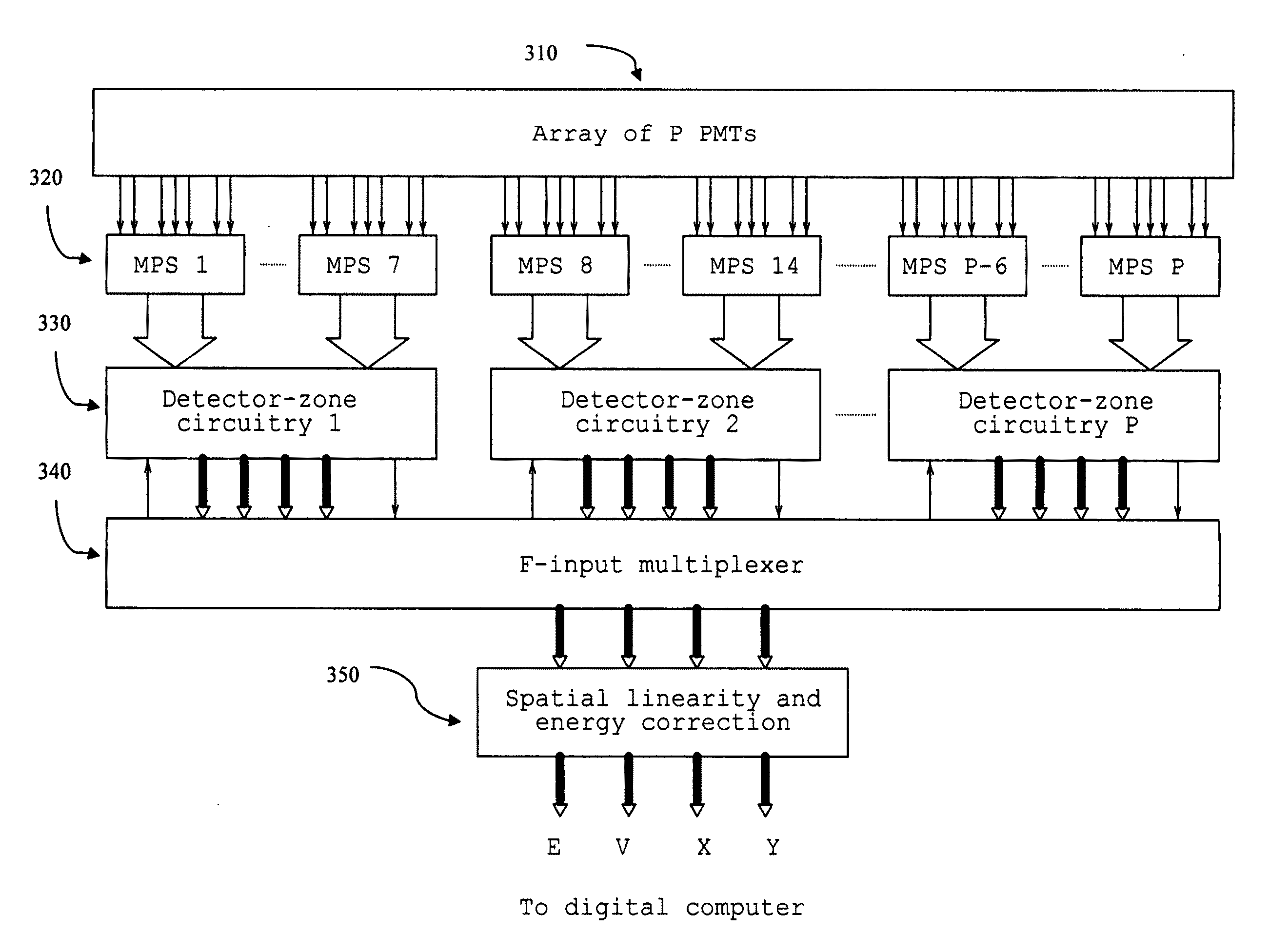
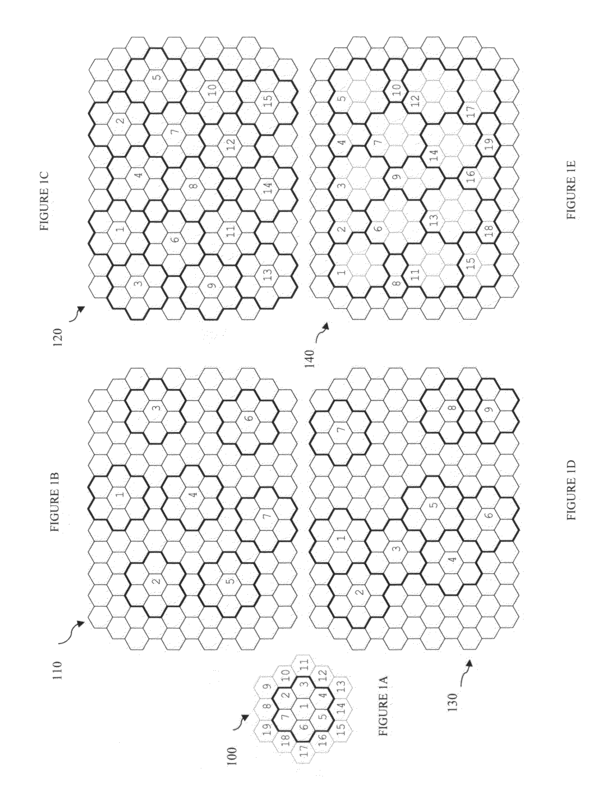
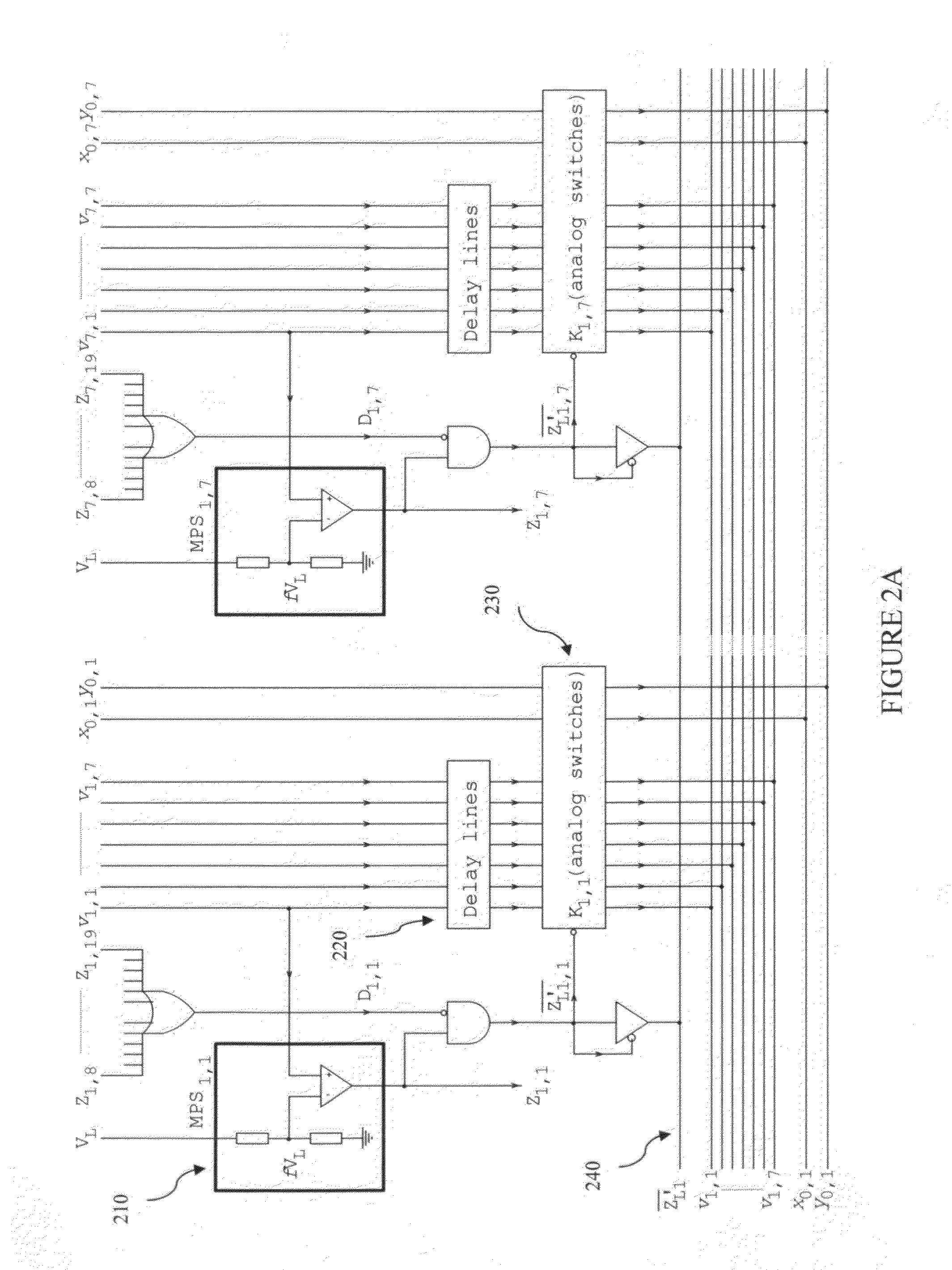
[0034]In one embodiment of MZDA a system includes at least one energy detector for producing bursts of optical photons in response to events including photons of the incident radiation. MZDA includes F number of sharing groups of seven photodetectors arranged in a honeycomb array for viewing zones of the at least one energy detector and converting the bursts of photons into signal outputs, where each of the central groups is associated with a zone of the at least one energy detector. A misplaced pile-up suppression circuitry is associated with each of the central groups for rejecting signal outputs that are due to scattered photons of the incident radiation. A detector-zone circuitry is associated with each of the F number of sharing groups for generating energy and position signals in detection of F number of the events during a period of one detector deadtime. Another embodiment further includes pileup prevention circuitry, which the circuitry enables the paralyzable detector to operate as nonparalyzable, and prevents the deadtime from increasing with count rate and leading to detector paralysis. Yet another embodiment further includes analog to digital converters (ADCs) for the energy and position signals. A multiplexer with F inputs transfers the digital energy and position signals of the F number of the events to computer memory. Still another embodiment further includes spatial linearity correction hardware, which operates on the digitized signals to mitigate distortions arising from nonlinearities. Another embodiment further includes energy correction hardware for mitigating errors arising from inequalities in gains of the photodetectors for image display and data analysis. The above techniques of MZDA to improve the sensitivity, spatial resolution, and signal-to-noise ratio (SNR) of nuclear images have been applied to exemplary designs for both planar and tomographic (PET / SPECT) imaging.
[0035]In another embodiment of MZDA a method includes steps for using fiber optics to couple the bursts of photons from a continuous scintillator to photodetectors for reducing an effective size of the photodetectors, thereby effectively removing the glass walls from PMTs and increase packing fraction to 100%, enabling to substantially increase the number of photodetectors, thereby further reducing effective detector deadtime, which increases both the detector sensitivity / count-rate response, and spatial resolution in the image. This increases the spatial sampling of the scintillation detector, hence enabling more accurate determination of the energy and position signals and reducing image blurring due to noise. Another embodiment further includes steps for coupling independent adjacent radiation detectors to photodetectors. This enables adjacent radiation detectors, normally provided with separate signal processing circuitry, to be arranged in detector zones and to share the circuitry to reduce cost while maintaining the spatial resolution capabilities of the independent detectors.

Problems solved by technology

Although nuclear imaging systems are unique in their ability to provide functional information, in contrast to the other imaging modalities, they have the disadvantage of having the lowest spatial resolution, spatial resolution being the size of the smallest object that can be resolved in the image.
It is therefore inevitable that the determination of the photon energy and the spatial coordinates becomes susceptible to noise from PMTs at large distances from the interaction point.
This approach, however, is also limited to detecting at most only three valid events at a time.
The large number of valid interactions that continues to be rejected, therefore, still remains a significant disadvantage.
Mispositioning of scintillation events in this way leads to image blurring referred to as depth-of-interaction (DOI) error.
Thus, thick detectors for PET have the attendant problems of lower spatial resolution and significant DOI blurring of the image.
Detector segmentation, however, has no effect on DOI error since the high energy gamma rays are able to penetrate the segments regardless.
Imaging applications can involve patient movement or fast redistribution of the radiopharmaceutical within the body, which can lead to blurred images unless an image can be built up in the shortest possible time.
The count rate for a paralyzable detector therefore increases only up to a peak value, which is proportional to the reciprocal of its deadtime τ0, and then decreases as count rate increases, eventually leading to detector paralysis.
This occurs because, when one scintillation event follows immediately after the previous one, they merge together and the energy signals pile up in the SA, leading to the rejection of both by the PHA.
The inefficient use of the detector and PMT array has long been recognized and has been receiving considerable attention.
However, as noted earlier, it has not been practical to increase detector sensitivity by more than a factor of two or three times, as only two or three events per deadtime period can be recorded.
This means that rapid redistribution of radioactivity within the body, which may take place in many investigations, cannot be observed in real time, but only after the fact, since the data needs to be subsequently processed using image reconstruction software.
Intrinsic spatial resolution and detector sensitivity, however, have conflicting thickness requirements, since the greater the detector's thickness, the greater the divergence of the scintillation light that emerges on the PMT side.
BGO detectors for 511-keV gamma rays typically have 20-30 mm thicknesses to have adequate detection efficiency, and a continuous BGO detector of 30 mm thickness would have an intrinsic resolution of 31 mm, which would be unacceptable.
Further improvement of spatial resolution would require greater reduction of the segment size, which would make it progressively more difficult to identify individual segments within the detector.
However, theoretical analysis of the dependence of resolution on crystal geometry indicates that even this would be impractical.
Although detector block segmentation is effective in restricting divergence of the scintillation light once an interaction in a segment has taken place, it has no effect on the ability of obliquely incident gamma rays to penetrate the detector segments.
Image distortion due to gamma rays penetrating the detector segments, or DOI error, requires detector technology that involves either multiple layers of scintillator or the use of avalanche photodiodes (APDs) as photodetectors along with the PMTs, which increases both detector cost and complexity without improving resolution.
Although positron tomographs with correction for DOI errors are becoming available, improvement in image quality has only been marginal.
Image quality in nuclear imaging, nevertheless, remains significantly inferior to those of CT and MRI.
Currently available detector architecture is unlikely to address the problem in any significant manner.
Moreover, despite the large numbers of publications and patents awarded for the improvement of sensitivity in nuclear imaging systems, little attention has been devoted to improving the spatial resolution of nuclear images.
It is also to be noted that identification of valid coincidences in current PET systems is often carried out using software operating on large quantities of raw data as opposed to real-time validation using hardware, which leads to an inefficient use of storage space and computing time.
Discrete detectors in these arrays, however, require independent readout using Si photodiodes, which makes them expensive due to their large numbers.
HPGe detectors require cryogenic cooling, however, while ZnCdTe detectors are currently too difficult to fabricate in large sizes.
As a result, despite its low quantum efficiency, unit-to-unit variations in quantum efficiency, and poor packing fraction from the dead zone due to the glass walls, the PMT still remains the photodetector of choice in nuclear imaging systems.

Method used


Image

  • Method and System for Nuclear Imaging Using Multi-Zone Detector Architecture
  • Method and System for Nuclear Imaging Using Multi-Zone Detector Architecture
  • Method and System for Nuclear Imaging Using Multi-Zone Detector Architecture

Examples


Embodiment Construction

[0037]The present invention is best understood by reference to the detailed figures and description set forth herein.

[0038]Embodiments of the invention are discussed below with reference to the Figures. However, those skilled in the art will readily appreciate that the detailed description given herein with respect to these figures is for explanatory purposes as the invention extends beyond these limited embodiments. For example, it should be appreciated that those skilled in the art will, in light of the teachings of the present invention, recognize a multiplicity of alternate and suitable approaches, depending upon the needs of the particular application, to implement the functionality of any given detail described herein, beyond the particular implementation choices in the following embodiments described and shown. That is, there are numerous modifications and variations of the invention that are too numerous to be listed but that all fit within the scope of the invention. Also, ...


PUM


Abstract

A method and system for nuclear imaging normally involve detection of energy by producing at most two or three bursts of photons at a time in response to events including incident gamma radiation. F number of sharing central groups of seven photodetectors, depending on the photodetector array size, is arranged in a honeycomb array for viewing zones of up to F bursts of optical photons at a time for each continuous detector and converting the bursts of optical photons into signal outputs, where each of the central groups is associated with a zone. This enables the detector sensitivity to be increased by as much as two orders of magnitude, and to exchange some of this excess sensitivity to achieve spatial resolution comparable to those in CT and MRI, which would be unprecedented. Signal outputs that are due to scattered incident radiation are rejected for each of the central groups to reduce image blurring, thereby further improving image quality. For planar imaging, the energy and position signals of up to the F number of valid events are generated once every deadtime period and transferred to computer memory for image display and data analysis. The number of valid events detected is up to 6F for SPECT and up to 3F for PET imaging.

Description

CROSS-REFERENCE TO RELATED APPLICATIONS[0001]The present Utility patent application claims priority benefit of the U.S. provisional application for patent Ser. No. 61,246,918 filed on Sep. 29, 2009 under 35 U.S.C. 119(e). The contents of this related provisional application are incorporated herein by reference for all purposes.FEDERALLY SPONSORED RESEARCH OR DEVELOPMENT[0002]Not applicable.REFERENCE TO SEQUENCE LISTING, A TABLE, OR A COMPUTER LISTING APPENDIX[0003]Not applicable.COPYRIGHT NOTICE[0004]A portion of the disclosure of this patent document contains material that is subject to copyright protection. The copyright owner has no objection to the facsimile reproduction by anyone of the patent document or patent disclosure as it appears in the Patent and Trademark Office, patent file or records, but otherwise reserves all copyright rights whatsoever.FIELD OF THE INVENTION[0005]The present invention relates generally to the field of radiation imaging with emphasis on medical app...

Claims


Application Information

Patent Timeline
IPC IPC(8): G01T1/164G01T1/208
CPCA61B6/037G01T1/1647
Inventor WOLDEMICHAEL, TILAHUN WOLDESELASSIE
Owner WOLDEMICHAEL TILAHUN WOLDESELASSIE
Learn More