
Active Microfluidic Membranes

a microfluidic membrane and active technology, applied in the field of biofabricated active microfluidic membranes, can solve the problems of limited enzymatic conversion efficiency of such devices, difficult to efficiently immobilize catalytically active enzymes in microfluidic networks, and limited conversion efficiency in conventional techniques, so as to achieve controllable thickness and permeability

Inactive Publication Date: 2011-04-07
UNIV OF MARYLAND
View PDF13 Cites 26 Cited by
  • Summary
  • Abstract
  • Description
  • Claims
  • Application Information

AI Technical Summary

Benefits of technology

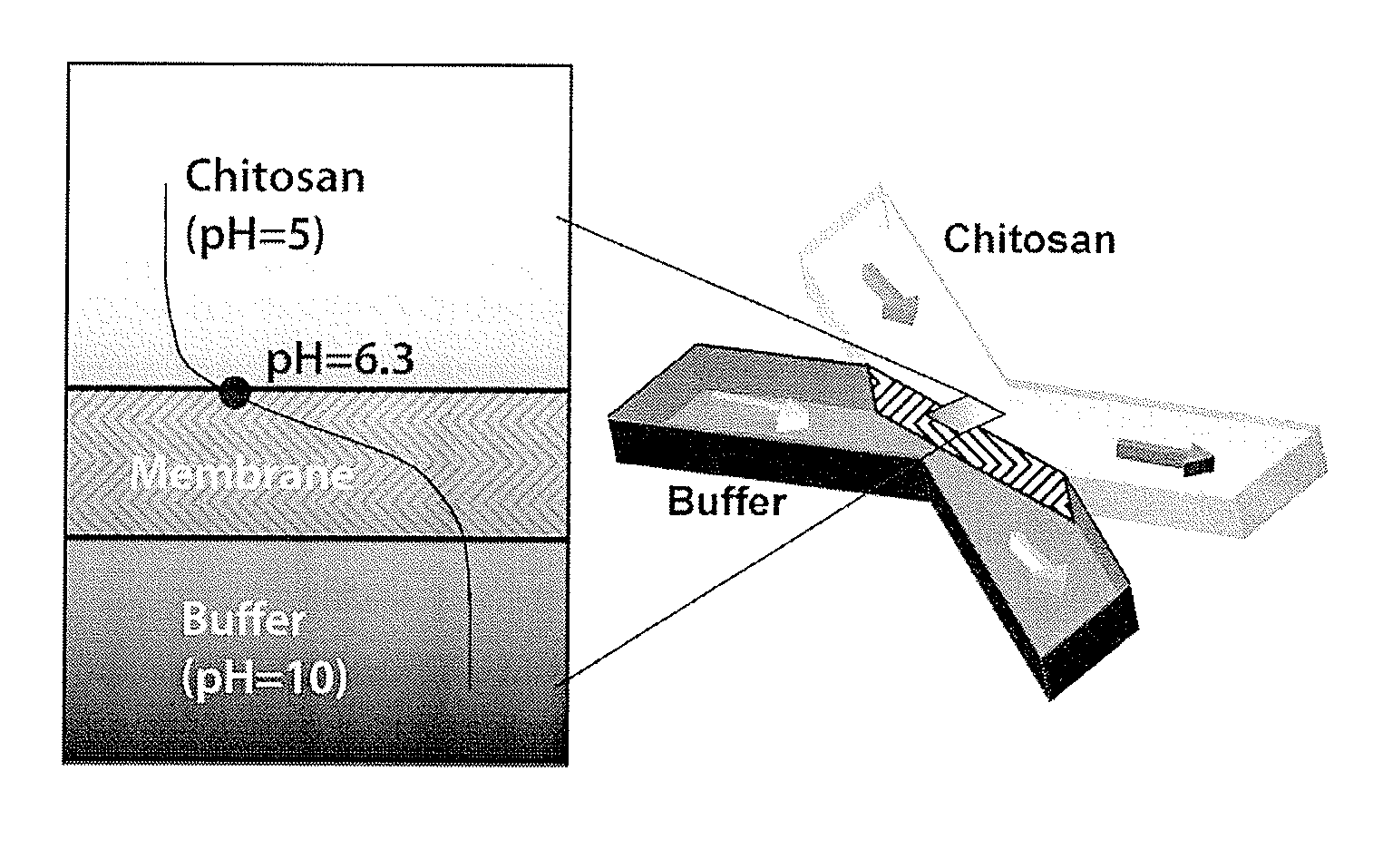
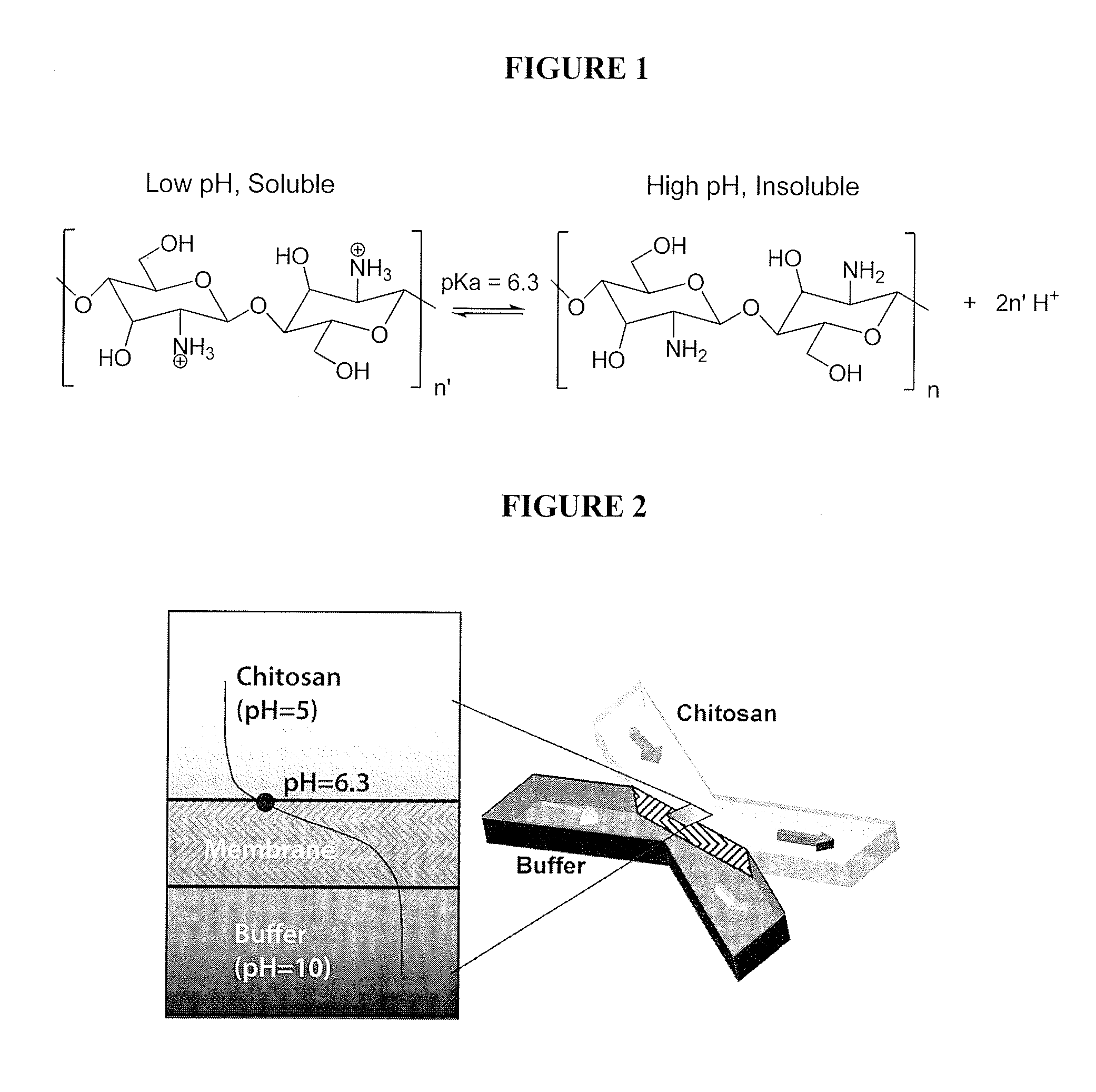
[0015]According to one embodiment, the invention provides a method of in situ biofabrication of a freestanding chitosan membrane in a sealed microfluidic device. In a preferred sub-embodiment, the chitosan membrane is provided by tuning the pH gradient at the interface of two laminar flow streams in microfluidics. The biofabricated chitosan membrane may be formed from the biopolymer chitosan via a natural process, which does not need an initiator as in a polymer chain reaction. The formation process of the chitosan membrane is controllable to allow for real-time fabrication with controllable thickness and permeability. Further, the formation process is versatile, allowing for complex intersecting membranes, such as T-shaped interfaces and sequential membrane interfaces as well as for simple membranes.
[0016]Further, the present invention provides for the removal of the AMM in a sealed microfluidic device, preferably using acid-base dissolution. The dissolution process is controllable for real-time membrane thinning and / or removal. The dissolution process thus allows for reusability of the device.
[0018]The present invention provides the ability to fabricate microfluidic reactors having high enzymatic activity by permitting the direct interaction of the substrate species in the perfusion flow with the enzyme on the permeable AMM to obtain product species. The membrane will preferably be semi-permeable to enzymatic substrate / product, allowing the fluidic streams to either flow through or flow by the membrane. This provides a microfluidic reactor having dramatically enhanced enzymatic conversion efficiency.

Problems solved by technology

However, the capability to efficiently immobilize catalytically active enzymes in a microfluidic network remains challenging.
Thus, the conversion efficiency in conventional techniques is limited given much of the introduced substrate passes the enzyme site un-reacted.
However, the enzymatic conversion efficiency of such devices is subject to the limitations associated with having the enzyme immobilized onto the microchannel surface.
In particular, enzyme located at the side of a flow channel is a geometry that prevents transport of substrate species to the enzyme, thus reducing efficiency of substrate-enzyme interaction.
Approaches for membrane integration include direct incorporation of commercial membranes or forming membranes as part of the bioMEMS chip fabrication process, both of which pose difficulty in packaging the microfluidic chips, or require additional complexity and cost in fabrication (de Jong, J et al.
Moreover, the composition and properties of animal-derived collagen by thermo-gelation has been difficult to control (Tan W. et al., supra, Biomedical Microdevices, 5(3):235-244); Shibata, K. et al., supra, Japanese Journal of Applied Physics, 47(6, Pt.2):5208-5211; Sundararaghavan, H. G. et al., supra, Biotechnology and Bioengineering, 102(2):632-643).
In the case of in situ membrane microfabrication, the lingering initiators and monomer residues from either photopolymerization or polymer chain reactions may be toxic to subsequent biological applications, and subsequent modification of the formed membrane is required for biomolecule assembly (Hisamoto, H. et al., supra, Analytical Chemistry, 75(2):350-354).

Method used


Image

  • Active Microfluidic Membranes
  • Active Microfluidic Membranes
  • Active Microfluidic Membranes

Examples


example 1

Biofabrication of an Active Microfluidic Membrane (AMM) in a Microfluidic Device

Materials, Preparations and Tests

[0119]Chitosan (medium molecular weight, average molecular weight 300,000 g / mol), phosphate buffered saline tablets (10 mM phosphate buffer, 2.7 mM KCl and 137 mM NaCl, pH 7.4), fluorescein (for fluorescence, free acid, λex=490 nm / λem=514 nm in 0.1 M Tris pH 8.0) and universal pH indicator (pH 4-10) were purchased from Sigma-Aldrich Corporation, St. Louis, Mo. Sodium hydroxide and hydrochloric acid were purchased from Fisher Scientific, Pittsburgh, Pa. Polydimethylsiloxane (“PDMS”) kits (Sylgard 184 and curing agent) were purchased from Dow Corning, Greensboro, N.C. Microbore PTFE tubing (0.022″ ID / 0.042″ OD) was purchased from Cole-Parmer, Vernon Hills, Ill. Genie syringe pumps were purchased from Kent Scientific Corporation, Torrington, Conn. Micro glass slides and single-use syringes were purchased from VWR International, LLC, West Chester, Pa. 5-(and 6-)-Carboxyfluore...

example 2

Biofabrication of a Dual Membrane Active Microfluidic Membrane (AMM) in a Microfluidic Device

[0145]3D microenvironments are crucial for in vitro study of cell biology, especially for mammalian cells with limited tolerance to hydrodynamic forces of 2D cell culture systems. In 2D cell culture, cells are displaced as a monolayer on a flat substrate. In 3D cell culture, cells are supported in all directions either by neighboring cells or an extracellular matrix (ECM). Moving from 2D to 3D cell culture systems in microfluidics improves the biological relevance of analyses (Ong, S M et al. (2008) “A gel-free 3D microfluidic cell culture system,” Biomaterials 29(22):3237-3244). Various natural and synthetic hydrogels have been incorporated into microfluidic cell culture systems to support cells in 3D. However, in many cases ultraviolet photo-polymerization and thermo-initiative gelation are cytotoxic to cells (Sundararaghavan, H G et al. (2009) “Neurite growth in 3D collagen gels with grad...

example 3

Viability and Signaling Response of Cells Incorporated into a Dual Membrane Active Microfluidic Membrane (AMM) of a Microfluidic Device

[0151]To demonstrate that cells incorporated into an AMM retained viability, red E. coli cells were evaluated in vitro for their ability to fluoresce in response to Autoinducer-2 (AI-2). E. coli BL21 (DsRed) cells were assembled within an alginate scaffold membrane of an alginate-chitosan dual AMM (formed as described above). Luria Both (LB) supplemented with 60 μM AI-2 (signal molecule to stimulate RFP production) and 10 mM CaCl2 (to maintain alginate gel stability) was introduced into the microchannel at a flow rate of 5 μL / min (in contrast to the 0.2 μL / min flow rate employed for in vivo experiments). FIG. 32A shows that cell density was very high inside the alginate gel after culturing for 5 hours, indicating that the cells remained viable and proliferated dramatically within the alginate scaffold membrane of the dual AMM. As shown in FIG. 32B, t...


PUM

PropertyMeasurementUnit
Sizeaaaaaaaaaa
Solubility (mass)aaaaaaaaaa
Permeabilityaaaaaaaaaa

Abstract

The present invention relates to a biofabricated Active Microfluidic Membrane (AMM) in a microfluidic network of a microfluidic device and a method for the in situ biofabrication of such a microfluidic network. More specifically, the invention relates to devices exhibiting (and methods of) positioning (i.e., erecting, modifying or removing a membrane matrix in situ in a microchannel of a microfluidic network of a microfluidic device. In one embodiment, the membrane comprises a single type of matrix constituent, such as chitosan, alginate, etc. Alternatively, the membrane may be composed of two or more matrix constituents, which may be integrated into one another or layered adjacent to one another.

Description

CROSS-REFERENCE TO RELATED APPLICATIONS [0001]This application claims priority to U.S. Patent Application Ser. No. 61 / 247,341 (filed Sep. 30, 2009, pending), which application is herein incorporated by reference in its entirety.STATEMENT REGARDING FEDERALLY SPONSORED RESEARCH OR DEVELOPMENT [0002]The U.S. Government has a paid-up license in this invention and the right in limited circumstances to require the patent owner to license others on reasonable terms as provided for by the terms of NSF SCO35224414 awarded by the National Science Foundation.BACKGROUND OF THE INVENTION[0003]1. Field of the Invention[0004]The present invention relates to a biofabricated Active Microfluidic Membrane (AMM) in a microfluidic network of a microfluidic device and a method for the in situ biofabrication of such a microfluidic network. More specifically, the invention relates to devices exhibiting (and methods of) positioning (i.e., erecting, modifying or removing a membrane matrix in situ in a microc...

Claims


Application Information

Patent Timeline
IPC IPC(8): C12M1/00C08B37/04C08B37/08C12N11/10C07K1/04C12P1/00B01L3/00
CPCB01L3/502753B01L2200/0636B01L2200/0647B01L2300/0681C12N11/10B01L2300/0867C08B37/003C08L5/04C08L5/08B01L2300/0864C12M21/18C12M23/16
Inventor LUO, XIAOLONGRUBLOFF, GARY W.BERLIN, DEAN L.BENTLEY, WILLIAM E.BUCKHOUT-WHITE, SUSANCHENG, YIBETZ, JORDAN
Owner UNIV OF MARYLAND
Learn More