Elisa kit for detecting lincomycin

Active Publication Date: 2011-05-12
BEIJING KWINBON BIOTECH
3 Cites 2 Cited by

AI-Extracted Technical Summary

Problems solved by technology

Those techniques are not suitable for on-site detection and screen of large numbers of samples due to...

Benefits of technology

[0004]One objective of the present invention is to provide an ELISA kit for detecting lincomycin. The kit of the present invention has advantages of simple operation and less time consuming. The kit o...

Abstract

The present invention provides an ELISA kit for detecting lincomycin comprising a coating antigen and an enzyme labeled reagent, wherein the coating antigen is selected from the group consisting of a lincomycin hapten-carrier protein conjugate, a lincomycin antibody and a lincomycin anti-antibody; when the coating antigen is the lincomycin hapten-carrier protein conjugate, the enzyme labeled reagent is an enzyme-labeled lincomycin anti-antibody; when the coating antigen is the lincomycin antibody, the enzyme labeled reagent is an enzyme-labeled lincomycin hapten-carrier protein conjugate; and when the coating antigen is the lincomycin anti-antibody, the enzyme labeled reagent is an enzyme-labeled lincomycin hapten-carrier protein conjugate; and the lincomycin hapten is obtained through the condensation reaction between lincomycin and succinic anhydride. The ELISA kit according to the present invention can be used for detecting the content of lincomycin remained in a sample such as an animal tissue (muscle, liver), honey, etc.

Application Domain

Technology Topic

LincomycinChemistry +9

Image

  • Elisa kit for detecting lincomycin
  • Elisa kit for detecting lincomycin
  • Elisa kit for detecting lincomycin

Examples

  • Experimental program(4)

Example

EXAMPLE 1
Preparation of the Components of the Kit
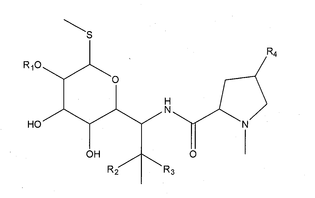
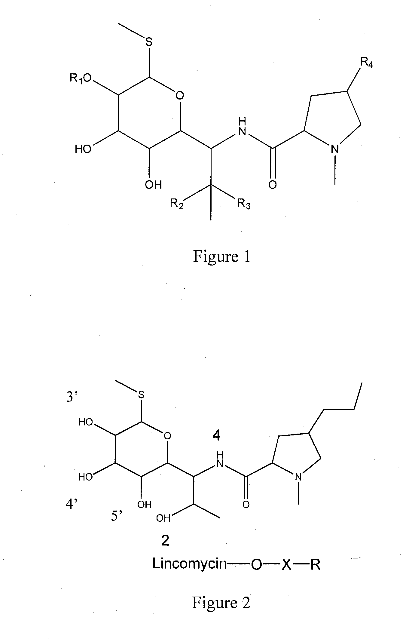
[0066]1. Synthesis of antigen
[0067]a. Synthesis of Hapten
[0068]Lincomycin hapten having carboxyl group is prepared according to succinic anhydride method.
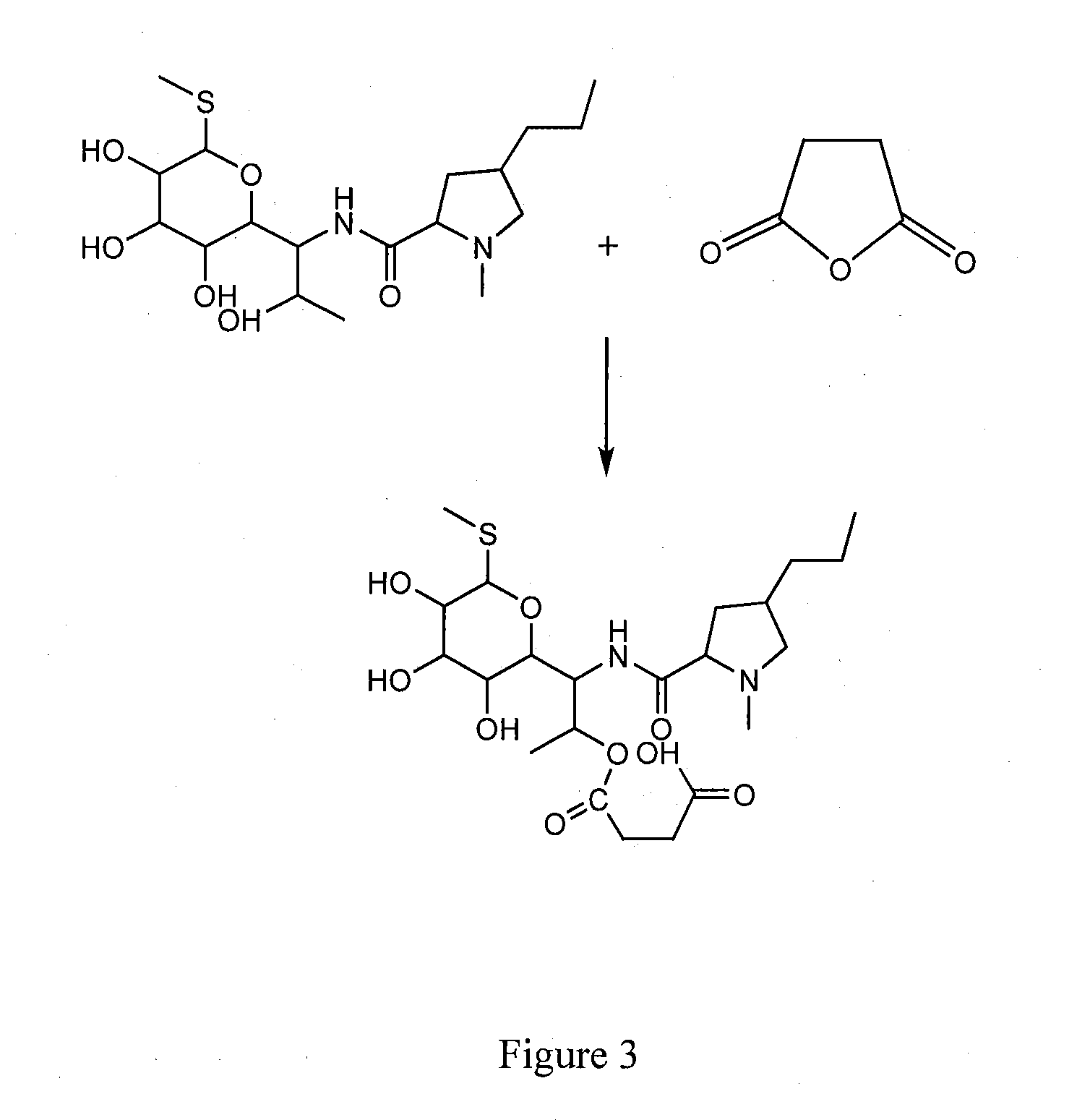
[0069]Hapten LIN-S1 is obtained by the steps of: weighting 2.3 g lincomycin hydrochloride (purchased from Dr Corn, Germany, Cat no. C14635000) and 0.5 g succinic anhydride, placing into a 50 ml round flask, adding anhydrous pyridine till the lincomycin hydrochloride and succinic anhydride are completely dissolved, and reacting at 70° C. for 24 h under stirring; removing the solvent via distillation under reduced pressure after the reaction is finished, washing the residues with acetone for several times and then with 2-3 times volume of ethyl acetate/n-hexane (2:1, v/v), adequately vibrating, and crystallizing to obtain the lincomycin hemisuccinate hapten.
[0070]The hapten LIN-S2 is obtained by the steps of:
[0071]Weighting 1.59 g lincomycin, placing into a 25 ml round flask, adding 6 ml N,N-dimethyl formamide (DMF) to dissolve the lincomycin, successively adding 1.41 g imidazole and 1.56 g t-butyl dimethylchlorosilane (t-BuMe2Si—Cl) under stirring, further stirring at 30° C. for 2 h, extracting the product with ethyl acetate, concentrating the extract with a rotary evaporator, and then drying in vacuum for 24 h to obtain 4-O-t-BuMe2Si-LIN.
[0072]Placing 0.56 g 4-O-t-BuMe2Si-LIN and 0.1 g succinic anhydride into a 50 ml round flask, adding anhydrous pyridine till 4-O-t-BuMe2Si-LIN and succinic anhydride are completely dissolved, and reacting at 70° C. under stirring for 24 h, removing the solvent via distillation under reduced pressure after the reaction is finished, washing the residues with acetone for several times, washing the residues with ethyl acetate which is 2-3 times volume of the residues, vibrating adequately, concentrating the ethyl acetate extract with a rotary evaporator, and drying in vacuum for 24 h to obtain 4-O-t-BuMe2Si-2-O-lincomycin hemisuccinate;
[0073]Placing 0.3 g 4-O-t-BuMe2Si-2-O-lincomycin hemisuccinate into a 50 ml round flask, adding 17 mL methanol for dissolving 4-O-t-BuMe2Si-2-O-lincomycin hemisuccinate, adding dropwise 0.26 g p-toluenesulfonic acid hydrate in 10 ml methanol solution, continuously stirring for 25 min, removing methanol via rotary evaporation, after partitioning of the product between the aqueous layer and the ethyl acetate layer, concentrating the ethyl acetate layer with a rotary evaporator, and then drying in vacuum for 24 h to obtain hapten LIN-S2.
[0074]b. Synthesis of Immunogen
[0075]By the way of illustration, as described below, an immunogen is obtained from lincomycin hapten and bovine serum albumin through mixed anhydride method.
[0076]The procedure specifically comprises the following steps:
[0077]Dissolving 5.8 mg lincomycin hapten into 0.1 ml N,N′-dimethyl formamide (DMF), cooling to 10° C., adding 2 μl isobutyl chloroformate, reacting at 10° C. for 30 min under stirring; dissolving 60 mg bovine serum albumin in 2 ml 50 mmol/L Na2CO3, reacting at 10° C. for 4 h, staying at 4° C. overnight, loading onto a Sephadex G-25 column, equilibrium and eluting with a buffer solution (0.2 mol/L phosphate buffer, pH 7.4), pooling the fractions containing bovine serum albumin, placing the pooled fractions into a dialysis tubing, and dialyzing in 0.2 mol/L phosphate buffer (pH 7.4) for 7 days while changing the dialysis buffer 3-4 times per day. The immunogen thus obtained is freeze-dried for storage. Through detection, the binding molar ratio of lincomycin hapten to the carrier protein is 13:1.
[0078]c. Preparation of the Coating Antigen of Lincomycin-Conjugated Antigen
[0079]By the way of illustration, as described below, a coating antigen is obtained by conjugating lincomycin hapten with ovalbumin.
[0080]The procedure specifically comprises the following steps:
[0081]Dissolving 5.8 mg lincomycin hapten in 0.1 ml DMF, cooling to 10° C., adding 2 μl isobutyl chloroformate, reacting at 10° C. for 30 min under stirring; dissolving 30 mg ovalbumin in 2 ml 50 mmol/L Na2CO3, reacting at 10° C. for 4 h, staying at 4° C. overnight, loading onto a Sephadex G-25 column, balancing and eluting with a buffer solution (0.2 mol/L phosphate buffer, pH 7.4), pooling the fractions containing ovalbumin, placing the pooled the fractions into a dialysis tubing, and dialyzing in 0.2 mol/L phosphate buffer (pH 7.4) for 7 days while changing the dialysis buffer 3-4 times per day. The immunogen thus obtained is freeze-dried for storage. Through detection, the binding molar ratio of lincomycin hapten to the carrier protein is 15:1.
[0082]2. Preparation of monoclonal antibody
[0083]a. Immunization of Animals
[0084]Healthy Balb/c mice 6-8 weeks old are selected and immunized at a dosage of 100 μg per one mice. For the first immunization, the immunogen is well mixed with complete Freund's adjuvant (purchased from Sigma Inc., —Cat no. F5881) at equal amount. For the follow-up immunization, the immunogen is mixed with incomplete Freund's adjuvant (purchased from Sigma Inc., Cat no. F5506).
[0085]b. Cell Fusion and Cloning
[0086]When the mouse serum titer is determined as high, the spleen cells are collected and fused with SP2/0 myeloma cells at an amount ratio of 7:1. The cell supernatant is detected by indirect competitive ELISA for screening the positive wells. The cells in the positive wells are cloned via limiting dilution method until a hybridoma cell secreting monoclonal antibody is obtained.
[0087]c. Identification of the Preferred Cell Strains of Monoclonal Antibody
[0088]The 50% inhibitory concentration and specificity of the monoclonal antibodies prepared from the two hapten schemes above are detected. The results are shown in the table below.
TABLE 1 Detecting results of the properties of the antibodies obtained from the two hapten schemes Technical schemes Technical indexes LIN-S1 LIN-S2 IC50 0.4 μg/kg 489 μg/kg Cross reaction Lincomycin 100% 100% Clindamycin <1% 13.1% Erythromycin <1% <1% Tylosin <1% <1% Streptomycin <1% 9.2% Kanamycin <1% 14.7%
[0089]As shown in Table 1, the antibody prepared from hapten LIN-S2 exhibits cross reaction with several antibiotics, has low specificity with IC50 of 489 μg/kg, and can not satisfy the requirement for detection of residual lincomycin.
[0090]Therefore, according to the present invention, lincomycin monoclonal antibody prepared from hapten LIN-S1 is selected as the preferred antibody. This antibody is secreted by hybridoma cell strain C-1-3 (CGMCC No. 2398), and has high specificity to lincomycin with IC50 of 0.4 μg/kg.
[0091]d. Freezing and Thawing of the Cells
[0092]The monoclonal hybridoma cell strain against lincomycin may be prepared into suspensions with cryopreservation solution at 1×10⁶ cells/ml, and stored in liquid nitrogen for a long term. When the cells are to be recovered, the cryopreservation tube is taken out from liquid nitrogen, and immediately placed into a water bath at 37° C. for thawing. The cryopreservation solution is removed via centrifuge, and the cells are removed into a flask for culture.
[0093]e. Production and Purification of the Monoclonal Antibody
[0094]Sterile paraffin oil is injected intraperitoneally into several Balb/c mice 6-8 weeks old at 0.5 mg/one mice. The monoclonal hybridoma cell strain against lincomycin is intraperitoneally injected at 0.5×10⁷ cells/one mice after 7 days. The ascites are collected after another 7 days, purified according to caprylic acid-saturated ammonium sulfate method, and stored at −20° C.
[0095]2. Preparation of polyclonal antibody
[0096]New Zealand rabbit is selected as the animal for immunization, and immunized with the conjugate of lincomycin hapten and bovine serum albumin as the immunogen at a dosage of 1.5 mg/kg. For the first immunization, the immunogen is mixed with complete Freund's adjuvant (for the source, see ibid) at equal amount to obtain an emulsion, and subcutaneously injected into neck and back at multiple sites. After an interval of 3-4 weeks, the immunogen is mixed with incomplete Freund's adjuvant (for the source, see ibid) at equal amount to obtain emulsion and then used for booster immunization. The animals receive 5 immunizations in total. For the last immunization, no adjuvant is added. Ten days after the last immunization, the antibody titer in the serum is detected. The blood is collected from heart, and the polyclonal antibody is obtained by purification according to caprylic acid-saturated ammonium sulfate method.
[0097]3. The goat-anti-mouse antibody is prepared by the steps of: using goat as the animal to be immunized, and immunizing pathogen-free goat with the immunogen of mouse antibody. The goat-anti-rabbit antibody is prepared by the steps of: using goat as the animal to be immunized, and immunizing pathogen-free goat with the immunogen of rabbit antibody.
[0098]4. Preparation of the ELISA plate
[0099]a. Preparation of Solutions
[0100]Coating buffer: 0.2 mol/L boric acid buffer, pH 8.6. The coating buffer is prepared by weighting 10.49 g Na2B4O7.10H2O and 5.57 g H3BO3, and dissolving in 1 L deionized water. Blocking solution: 0.05 mol/L phosphate buffer (pH 7.2) containing 10 wt % calf serum and 0.05 wt % sodium azide. The blocking solution is prepared by weighting 12.90 g Na2HPO4.12H2O, 2.18 g NaH2PO4.2H2O and 5 g sodium azide, dissolving in deionized water, adding 100 mL calf serum, and fixing the volume to 1 L.
[0101]Concentrated washing solution: 0.2 mol/L phosphate buffer (pH 7.4) containing 1.0 wt % Tween-20 and 0.5 wt % thiomersal. The concentrated washing solution is prepared by weighting 14.51 g Na2HPO4.12H2O, and 1.48 g NaH2PO4.2H2O, dissolving in deionized water, adding 10 mL Tween-20 and 5 g thiomersal, and fixing the volume to 1 L with deionized water. Diluted washing solution: an appropriate amount of the concentrated washing solution is mixed with deionized water at a volume ratio of 1:19 for further use.
[0102]Concentrated redissolving solution: 0.2 mol/L phosphate buffer (pH, 7.4) containing 5% bovine serum albumin. The concentrated redissolving solution is prepared by weighting 14.51 g Na2HPO4.12H2O, and 1.48 g NaH2PO4.2H2O, dissolving in deionized water, adding 50 g bovine serum albumin, and fixing the volume to 1 L with deionized water.
[0103]b. Coating of the ELISA Plate
[0104]Diluting the lincomycin-conjugated antigen, lincomycin antibody or lincomycin anti-antibody with the coating buffer to 0.05-2 μg/ml, adding into the microtiter wells at 100 μl/well, incubating at 37° C. for 2 hours or at 4° C. overnight, removing the coating buffer, washing with the 20-time-diluted concentrated washing solution twice for 30 seconds each time, removing the liquid in the wells, and absorbing the residual liquid with absorbent paper, adding 200 μl blocking solution into each well, incubating at 37° C. for 2 hours, removing the liquid in the wells, drying and then sealing the wells with Al film, and storing at 4° C. in dry places for further use.
[0105]5. Enzyme-labeled goat-anti-mouse anti-antibody
[0106]The horseradish peroxidase-labeled goat-anti-mouse anti-antibody of the present invention is obtained by conjugating the labeling enzyme with the anti-antibody according to the glutaraldehyde method or sodium periodate method.

Example

EXAMPLE 2
Construction of ELISA Kit for Detecting Lincomycin
[0107]The ELISA kit for detecting lincomycin is constructed to comprise the following components: [0108] (1) an ELISA plate coated with lincomycin-conjugated antigen; [0109] (2) horseradish peroxidase-labeled goat-anti-mouse anti-antibody; [0110] (3) working solution of lincomycin monoclonal antibody; [0111] (4) 6 vials of lincomycin standard solution at concentrations of 0 μg/L, 0.2 μg/L, 0.6 μg/L, 1.8 μg/L, 5.4 μg/L and 16.2 μg/L, respectively; [0112] (5) color developing solution consisting of developing solution A and developing solution B, wherein the developing solution A is carbamide peroxide solution, and the developing solution B is tetramethylbenzidine solution; [0113] (6) stopping solution: 2 mol/L hydrochloric acid; [0114] (7) concentrated washing solution: 0.2 mol/L phosphate buffer (pH 7.4) containing 1.0 wt % Tween-20 and 0.5 wt % thiomersal; and [0115] (8) concentrated redissolving solution: 0.2 mol/L phosphate buffer (pH 7.4) containing 5% bovine serum albumin.

Example

EXAMPLE 3
Detection of Lincomycin in Real Samples
[0116]1. Pre-treatment of the samples
[0117]a) Preparation of the Solutions
[0118]0.015 mol/L hydrochloric acid solution (special use for animal tissue samples) is prepared by adding 1.25 ml concentrated hydrochloric acid solution into a container containing deionized water, and fixing the volume to 1 L.
[0119]Methanol-hydrochloric acid solution (special for animal tissue samples) is prepared by uniformly mixing 10 ml methanol and 60 ml 0.015 mol/L hydrochloric acid solution.
[0120]Redissolving solution is prepared by diluting the concentrated redissolving solution with deionized water at a volume ratio of 1:1 and mixing for further use.
[0121]b) Animal Tissues (Chicken Meat, Chicken Liver, Pork Meat, Pork Liver, etc.)
[0122]The animal tissues are pre-treated by adding 2.0 g homogenized sample into a 50 ml polystyrene centrifugal tube, adding 10 mL methanol-hydrochloric acid solution, vibrating for 5 min, centrifuging at >3000 g for 5 min at room temperature, collecting 200 μl supernatant, adding 600 μl redissolving solution, mixing uniformly, and taking 50 μl for analysis.
[0123]c) Honey Samples
[0124]The honey samples are pre-treated by adding 2.0 g honey into a 50 ml polystyrene centrifugal tube, adding 5 mL deionized water, vibrating with an eddy vibrator till the honey are completely dissolved, and then vibrating with the eddy vibrator for 5 min, centrifuging at >3000 g for 5 min at ambient temperature, collecting 1 ml supernatant, adding 1 ml redissolving solution, vibrating with the eddy vibrator for 30 s, and taking 50 μl for analysis.
[0125]2. Detection using the kit
[0126]The detection process comprises the steps of: adding 50 μl lincomycin standard solution or sample solution into the microtiter wells of the ELISA plate coated with lincomycin-conjugated antigen, adding 50 μl working solution of lincomycin monoclonal antibody, sealing the plate with a cover film, reacting in a incubator at 25□ for 30 min, removing the liquid from the wells, adding 250 μl washing solution into each well and removing the liquid after 30 s, repeating the washing step for 5 times, and removing the residual liquid with absorbent paper; adding 100 μl working solution of horseradish peroxidase-labeled goat-anti-mouse anti-antibody, reacting in the thermostatic container at 25□ for 30 min, removing the liquid in the wells, repeating the washing step, adding developing solution A of carbamide peroxide and developing solution B of tetramethylbenzidine (TMB), moderately vibrating, developing color in the thermostatic container at 25□ in dark for 15 min, adding the 50 μl stopping solution of 2 mol/L hydrochloric acid into each well, moderately vibrating, and detecting the absorbance (OD value) for each well at 450 nm using Microplate Reader.
[0127]3. Analysis of the detecting result
[0128]The percent absorbance is calculated by dividing the average absorbance (B) of the standard solution at each concentration by the absorbance (B0) of the first standard solution (blank standard) and multiplying by 100%. The standard curve is plotted with the semi-log value of the concentration of the lincomycin standard solution (μg/L) as X axis, and the percent absorbance as Y axis. The percent absorbance of the sample solution is calculated according to the same method. The concentration corresponding to each sample is obtained from the standard curve. T he amount of residual linxomycin can be calculated from the standard curve.

PUM

PropertyMeasurementUnit
Acidity7.3pH
Acidity7.4pH
Dissociation constant0.1 ~ 0.3mol / L
tensileMPa
Particle sizePa
strength10

Description & Claims & Application Information

We can also present the details of the Description, Claims and Application information to help users get a comprehensive understanding of the technical details of the patent, such as background art, summary of invention, brief description of drawings, description of embodiments, and other original content. On the other hand, users can also determine the specific scope of protection of the technology through the list of claims; as well as understand the changes in the life cycle of the technology with the presentation of the patent timeline. Login to view more.

Similar technology patents

Dry polymer hydration apparatus and methods of use

InactiveUS20060107998A1Reduce air introductionSimple operationFlow mixersMixing methodsInjectorImpeller
Owner:SCHLUMBERGER TECH CORP

Car Navigation Apparatus

Owner:MITSUBISHI ELECTRIC CORP

Mobile communication terminal

Owner:MITSUBISHI ELECTRIC CORP

Classification and recommendation of technical efficacy words

  • Simple operation
  • Less time-consuming

Headgear equipped with laser hair care apparatus

InactiveUS20100242155A1High degree of attentionLess time-consumingLight therapyHatsBiomedical engineeringHands free
Owner:CARULLO JR JOHN F

Articles and methods for applying color on surfaces

InactiveUS20060165979A1Permanent effectLess time-consumingCovering/liningsDecorative surface effectsEngineeringAdhesive
Owner:KINSEY VON ADONIRAM +5

Power control in mobile radio communications systems

InactiveUS20050143112A1Reduce complexityLess time-consumingPicture reproducers using cathode ray tubesPicture reproducers with optical-mechanical scanningEngineeringTransmitted power
Owner:OPTIS WIRELESS TECH LLC
Why Eureka
Try Eureka