Method for culturing and subculturing primate embryonic stem cell, as well as method for inducing differentiation thereof

a technology of embryonic stem cells and subculturing methods, which is applied in the field of culturing and subculturing primate embryonic stem cells, can solve the problems of inability to meet the needs of clinical application, risky drug administration might be overlooked, and the method of continuously culturing and subculturing embryonic stem cells derived from primate including humans cannot be established, so as to achieve safe and efficient preparation, safe culture, and low cost

Inactive Publication Date: 2011-06-23
NAT CENT FOR GLOBAL HEALTH & MEDICINE +2
View PDF0 Cites 10 Cited by
  • Summary
  • Abstract
  • Description
  • Claims
  • Application Information

AI Technical Summary

Benefits of technology

[0037]According to the method for culturing and subculturing primate embryonic stem cells of the present invention, primate embryonic stem cells can be safely cultured with keeping the cells in the undifferentiated state, using a simple device and a method without inducing a cellular disorder such as chromosome aberration and the like. In addition, according to the present method, primate embryonic stem cells can be cultured to be maintained in an undifferentiation state at low cost, and it is possible to generally meet to a demand in regeneration medicine and research field.
[0038]According to the present invention, a “specific precursor cell” which is identified as a common precursor cell regarding a vascular endothelial cell, a blood cell and the like can be safely and very efficiently prepared.
[0039]In addition, according to the present invention, it is possible to prepare in large quantity, vascular endothelial cells, blood cells, hematopoietic stem cells and myeloid lineage cells, each of which have high re-productivity and allow stable subculture and freeze-thaw.
[0040]Further, according to the present invention, various cells can be easily supplied as a material for safe and secure blood products for transfusion (including hematopoietic stem cell transplantation, glanulocyte transfusion, myeloid lineage cell administration), as a material for a treatment of a vascular damage and improvement of a topical blood stream or as a material suitable for clinical use in medical care for the purpose of promoting regeneration of other various tissues.
[0041]Further, according to the present invention, it firstly enables to supply in a large quantity, a cell population having a nature which correctly mimics a living body tissue of a primate, especially a human. Since these cells can be also adequately used for a test for efficacy or toxicity of a drug, they can greatly contribute to development of not only clinical practice but also medical industry.

Problems solved by technology

However, a method for continuously culturing and subculturing embryonic stem cells derived from a primate including a human, which keeps the cells undifferentiated, has not been established yet.
Because a toxicity test of a drug or the like on a fetus (pregnant mother's body) is not currently conducted from an ethical point of view and only accumulation of accidental cases gives drug information, a risky drug administration might be overlooked due to lack of information on drug safety.
However, since this method uses a large amount of four kinds of expensive human-derived extracellular matrices in addition to a large amount of cocktail of synthetic cytokines (Non-Patent Document 2 (Ludwig et al., Nature Biotechnology, Vol. 24, pp.
185-187, (2006))), it is not suitable for clinical application from an economical point of view.
Although these human embryonic stem cells can be subcultured and maintained without a feeder cells, a special equipment and a special skill are required for culture / subculture / freeze preservation of the cells and thus handling of a large amount of cells is substantially difficult (Non-Patent Document 3 (Buzzard, Natural Biotechnology, Vol. 22, pp.
In addition, because use of a large amount of a synthetic cytokine is required for these feeder-free cultures in which a conditioned medium of cells derived from a heterogeneous animal is not used, great economical burden has to be involved in the research itself.
Therefore, regarding a human embryonic stem cell, the technique of stable feeder-free culture has not been established yet.
Therapeutic angiogenesis using a vascular endothelial precursor cell obtained from a living body tissue (peripheral blood, bone marrow blood) has been tried, but regeneration of the vascular endothelial cell from a transplanted cell has not been directly confirmed so far.
In addition, a mature vascular endothelial cell obtained from a living body has already lost the proliferating ability and it is difficult to prepare a large amount of samples, regarding a primate.
Thus, a basic research on the vascular endothelial cell is very delayed.
Further, as described later, since primary cultured vascular endothelial cells obtained from a human living body (particularly, commercially available freeze-thawed primary cultured vascular endothelial cells) have often lost during in vitro expansion of the culture, an inherent nature possessed by a cell in living body, a drawback is the point that a nature of the vascular endothelial cell in a living body is not correctly reflected.
However, in the case of using conventional method, one problem is that an efficiency of differentiation of a primate embryonic stem cell into a vascular endothelial precursor cell is as extremely low as 2% or less (Non-Patent Document 4 (Sone et al., Circulation, Vol. 107, pp.
In addition, regarding not only a primate embryonic stem cell but also a mouse embryonic stem cell, a method for preparing vascular endothelial precursor cells which can be subcultured using feeder-free culture has not been established.
However, since it is almost impossible to in vitro expand hematopoietic stem cells (bone marrow blood, umbilical blood etc.) obtained from a living body, an amount of the hematopoietic stem cells to be used for medical transplantation is limited (for example, a sample from one donor can be administered to only one patient), and use for basic medical research including a cell culture experiment is substantially impossible.
In addition, regarding a mature hemocyte of a primate, because it is difficult to prepare a large amount of samples as compared with an experimental animal such as a mouse, fundamental research is much delayed.
However, in the case of using feeder free culture, an efficacy of inducing differentiation of an embryonic stem cell of a primate including a human into a hemocyte is not sufficiently high, and an amount of prepared hemocytes is limited.
Moreover, in order to maintain a sufficient amount of hemocytes, it is necessary to conduct an experiment by repeating it tens times or hundreds times, but induction of differentiation using a conventional method is transiently effective, and it is impossible to actually apply these methods to regeneration / transplantation medicine.
In addition, a stroma cell is indispensible for hematopoiesis in a living body, but use of stroma cells for transplantation / reproduction medicine is very limited and it is also substantially impossible to culture stroma cells in vitro because a hematopoietic stroma cell obtained from a living body (fetal liver, adult bone marrow etc.) rapidly loses the function due to subculturing or freeze-thaw.
However, preparation of hematopoietic stroma cells from an embryonic stem cell of any species including a mouse has not been successful yet.
Even now, there is lack of information regarding the hematopoietic mechanism, particularly, initial hematopoiesis during development.

Method used


Image

  • Method for culturing and subculturing primate embryonic stem cell, as well as method for inducing differentiation thereof
  • Method for culturing and subculturing primate embryonic stem cell, as well as method for inducing differentiation thereof
  • Method for culturing and subculturing primate embryonic stem cell, as well as method for inducing differentiation thereof

Examples


example 1

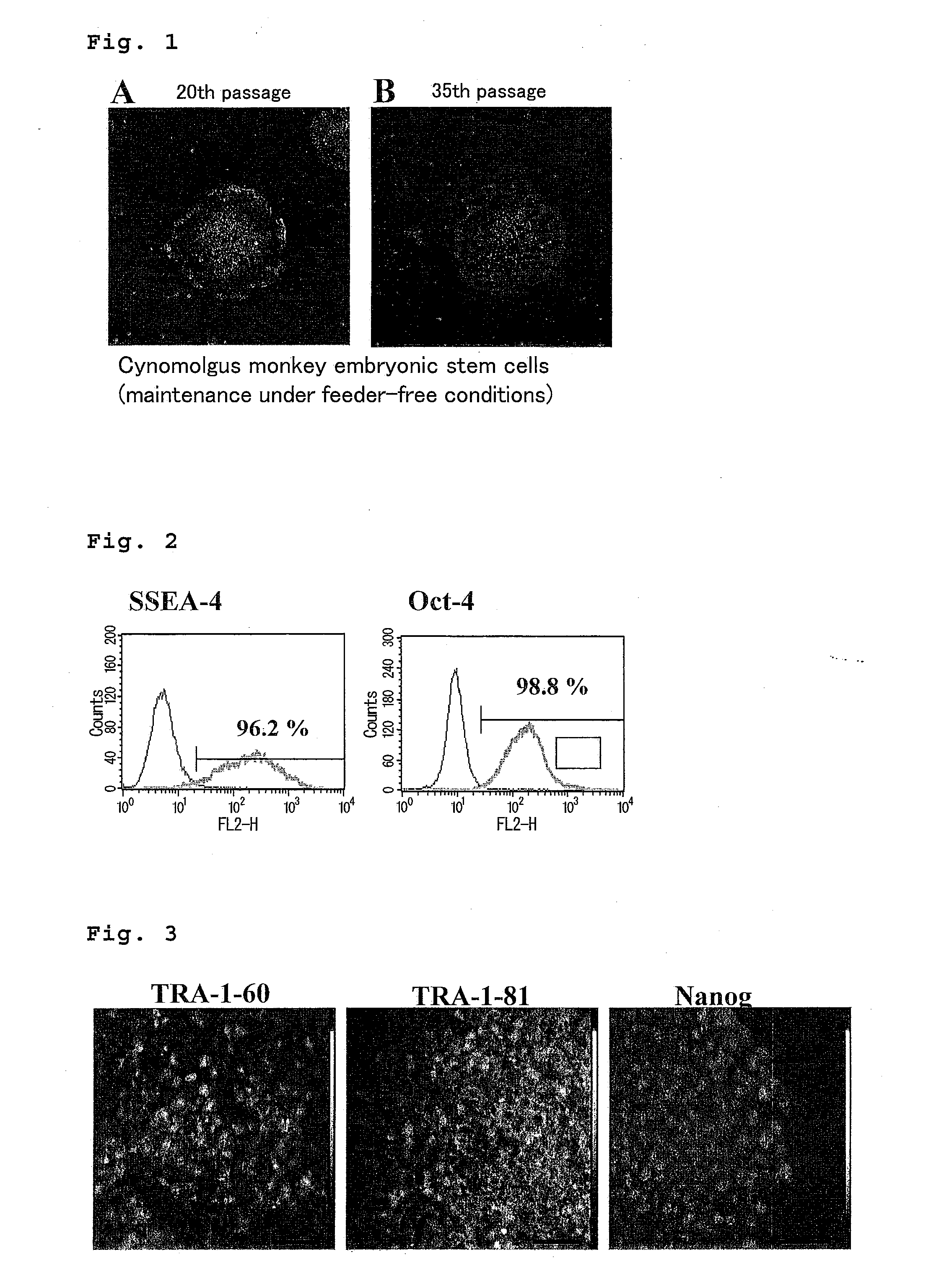
Undifferentiation-Maintenance Culture of Cynomolgus Monkey Embryonic Stem Cells without Using Feeder Cells and Cytokines

(1) Preparation of Undifferentiation-Maintenance Culture Solution

[0208]Cynomolgus monkey embryonic stem cells were cultured at 37° C. and 5 volt CO2 in a CO2 incubator on a 10 cm culture dish or a 78 cm2 culture dish which was coated at room temperature for around 15 minutes to 30 minutes with Matrigel© matrix [manufactured by BS (BD Biosciences)] diluted by 30-fold with an undifferentiation-maintenance culture solution 1-1 (composition: DMEM / Ham'S F-12 [manufactured by Kohjinbio Co., Ltd.], 20 vol % KNOCKOUT® SR [manufactured by Invitrogen Corp.], 1 mM L-glutamine [manufactured by Invitrogen Corp.], 2 mM non-essential amino acid solution [manufactured by Invitrogen Corp.], 1 mM sodium pyruvate [manufactured by Invitrogen Corp.], final concentration 100 U / ml of penicillin [manufactured by Invitrogen Corp.], final concentration 100 μg / ml of streptomycin [manufacture...

example 2

Undifferentiation-Maintenance Culture of Human Embryonic Stem Cells without Using Feeder Cells and Cytokines (Method Using Culture Container Coated with Matrigel® Matrix)

(1) Preparation of Undifferentiation-Maintenance Culture Solution

[0219]Cynomolgus monkey embryonic stem cells were cultured at 37° C. and 5 volt CO2 in a CO2 incubator on a 10 cm culture dish or a 78 cm2 culture dish coated at room temperature for around 15 minutes to 30 minutes, with Matrigel® matrix [manufactured by BS (BD Biosciences)] diluted by 30-fold with an undifferentiation maintenance culturing solution 1-1 (composition: DMEM / Ham'S F-12 [manufactured by Kohjinbio Co., Ltd.], 20 vol % KNOCKOUT® SR [manufactured by Invitrogen Corp.], 1 mM L-glutamine [manufactured by Invitrogen Corp.], 2 mM non-essential amino acid solution [manufactured by Invitrogen Corp.], 0.1 μM 2-mercaptoethanol [manufactured by Sigma Chemical Co.], final concentration 100 U / ml of penicillin [manufactured by Invitrogen Corp.], final con...

example 3

Undifferentiation-Maintenance Culture of Human Embryonic Stem Cells without Using Feeder Cells and Cytokines (Method Using a Culture Container Coated with One Kind of Human-Derived Protein Component)

(1) Preparation of Undifferentiation-Maintenance Culture Solution

[0227]The medium as described in Example 2 was used.

(2) Technique of Undifferentiation-Maintenance Culture

[0228]The human embryonic stem cells are plated at the density of about 2 to 3 within the microscope's field of view using a phase-contrast microscope equipped with 4× objective lens and 10× ocular lens in the condition that the colony is relatively uniformed in size to have the diameter of 500 μm, and thereafter, medium exchange is performed every day. Since the size of the colony becomes about 1,000 μm after 3 to 4 days, the cells are detached using a cytodetachment solution 1 (composition: 0.25% trypsin solution [manufactured by Invitrogen Corp.], 1 mg / ml collagenase IV [manufactured by Invitrogen Corp.], 1% KNOCKOUT...


PUM

PropertyMeasurementUnit
diameteraaaaaaaaaa
diameteraaaaaaaaaa
diameteraaaaaaaaaa

Abstract

The present invention provides a method for subculturing primate embryonic stem cells, and a method for inducing differentiation of the same cell into a vascular endothelial cell and a blood cell. The present invention provides a method comprising culturing primate embryonic stem cells in a medium containing a protein component without using feeder cells and cytokines in a container coated with an extracellular matrix, detaching colonies of the resulting embryonic stem cells in the presence of a cytodetachment agent, and plating the colonies in the similar medium, and a method comprising culturing primate embryonic stem cells in a serum-containing or not containing medium in the presence of cytokine, adhesion-culturing the resulting embryoid body or embroyid body-analogous cellular aggregate in the presence of a cytokine to obtain specific precursor cells, and separating non-adherent cells and adherent cells from the specific precursor cells to obtain blood cells and vascular endothelial precursor cells.

Description

TECHNICAL FIELD[0001]The present invention relates to a method for culturing and subculturing primate embryonic stem cells wherein said method keeps the primate embryonic stem cells in an undifferentiated state, a method for inducing differentiation of the primate embryonic stem cell into various cells such as a blood cell, a vascular endothelial precursor cell and the like, a method for expanded reproduction of the resulting cell, as well as a hemocyte, a vascular endothelial precursor cell and the like.BACKGROUND TECHNIQUE[0002]An embryonic stem cell having a pluripotency, which can be differentiated into a variety of cells, was established in 1980's regarding a mouse. A method for culturing and subculturing it is already known. However, a method for continuously culturing and subculturing embryonic stem cells derived from a primate including a human, which keeps the cells undifferentiated, has not been established yet.[0003]A human embryonic stem cell is extremely important for i...

Claims


Application Information

Patent Timeline
Patent Type & Authority Applications(United States)
IPC IPC(8): C12N5/0735C12N5/078C12N5/071C12N5/0789
CPCC12N5/0606C12N5/0634C12N2500/84C12N2501/105C12N2501/115C12N2501/125C12N2533/90C12N2501/165C12N2501/22C12N2501/23C12N2501/26C12N2506/02C12N2509/00C12N2501/155
Inventor YUO, AKIRATOBE, KUMIKOSAEKI, KOICHINAKAHARA, MASAKONAKAMURA, NAOKOYOGISASHI, YOSHIKOMATSUYAMA, SATOKOYONEDA, ASAKO
Owner NAT CENT FOR GLOBAL HEALTH & MEDICINE
Why Eureka
Try Eureka