

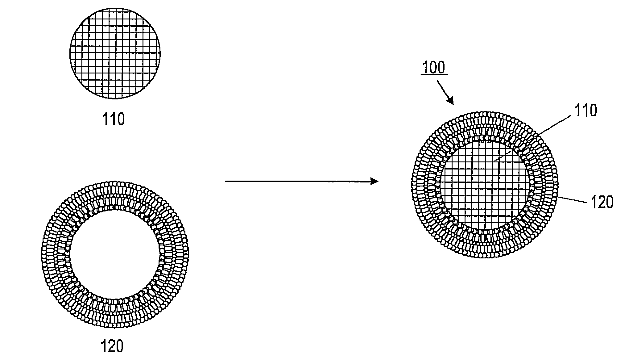
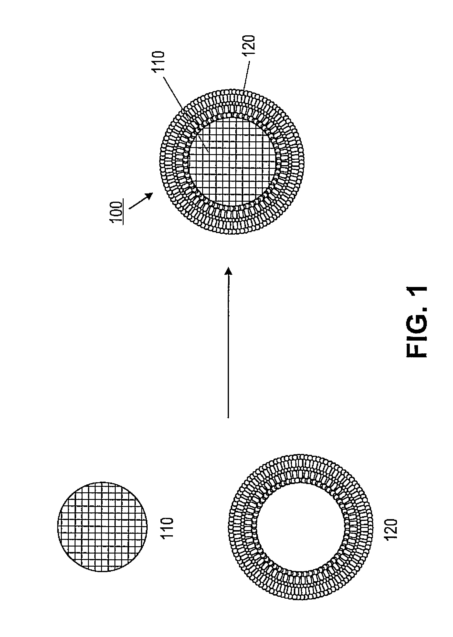
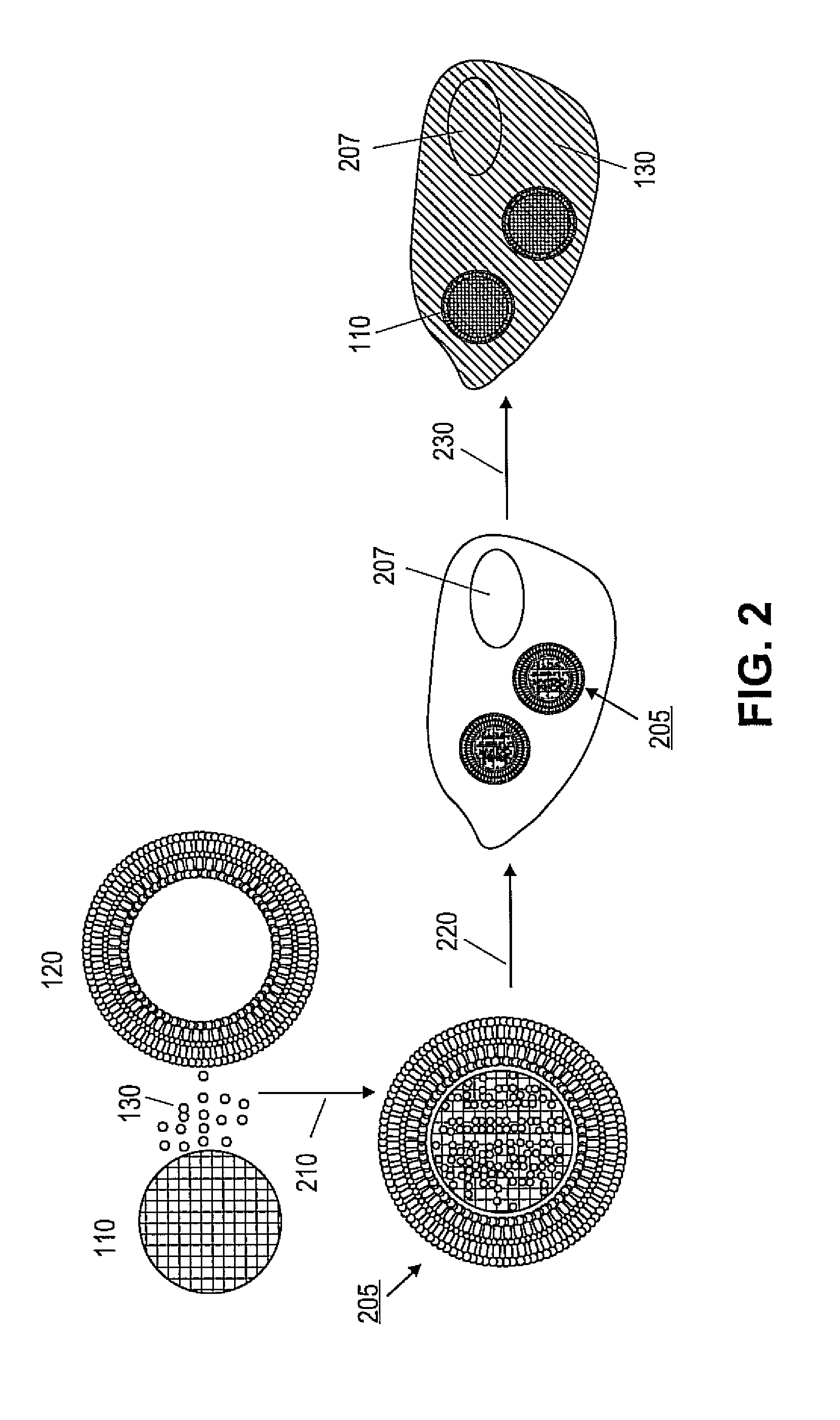
Porous nanoparticle supported lipid bilayer nanostructures
a nanostructure and nanoparticle technology, applied in the field of nanostructures, can solve the problems of limited cargo concentration and category, low stability of liposomes, and inability to load cargo under, and achieve the effects of reducing the rate significantly increasing the fraction of calcein dye releas
- Summary
- Abstract
- Description
- Claims
- Application Information
AI Technical Summary
Benefits of technology
Problems solved by technology
Method used
Image

Examples
example 1
Materials
[0052]Exemplary phospholipids were obtained from Avanti Polar Lipids Inc. (Alabaster, Ala.). Exemplary cholesterol was obtained from Sigma (St. Louis, Mo.). Texas Red-labeled DHPE lipid and fluorescein isothiocyanate (FITC) were obtained from Invitrogen (Carlsbad, Calif.). All silanes and calcein were obtained from Aldrich Sigma (St. Louis, Mo.). Chinese Hamster Ovary (CHO) cells and cell culture related chemicals and media were obtained from American Type Culture Collection (ATCC).
[0053]All UV-vis absorption data were collected on a Perkin-Elmer spectrophotometer; all fluorescence data were obtained on a Horiba Jobin Yvon Fluoromax-4 fluorometer; and all light scattering data were collected on a Zetasizer Nano dynamic light scattering instrument (Malvern).
[0054]Lipids and other chemicals used in the examples included the following:
DOTAP: 1,2-Dioleoyl-3-Trimethylammonium-Propane (Chloride Salt)
[0055]
DOPC: 1,2-Dioleoyl-sn-Glycero-3-Phosphocholine
[0056]
DOPS: 1,2-Dioleoyl-sn-G...
example 2
Preparation of Mesoporous Silica Nanoparticles
[0061]Mesoporous silica nanoparticles were prepared by the aerosol-assisted self-assembly method, wherein silica / surfactant aerosols were generated using a commercial atomizer (Model 9302A, TSI, Inc., St Paul, Minn.) operated with nitrogen as a carrier / atomization gas. The reaction was started with a homogeneous solution of soluble silica precursor tetraethyl orthosilicate (TEOS), HCl, and surfactant prepared in an ethanol / water solution with an initial surfactant concentration much less than the critical micelle concentration. The pressure drop at the pinhole was about 20 psi. The temperature for the heating zones was kept at about 400° C. Particles were collected on a durapore membrane filter maintained at about 80° C. cetyltrimethylamonium bromide (CTAB) was selected as the structure directing template.
[0062]In a typical synthesis of mesoporous silica nanoparticles, about 55.9 mL H2O, about 43 mL ethanol, about 1.10 mL 1N HCl, about 4...
example 3
Preparation of Liposomes
[0065]Phospholipids were dissolved in chloroform at concentrations of about 10 to about 25 mg / mL. Aliquots were dispensed into scintillation vials so that each vial contained 2.5 mg lipids. For mixed lipids, the total amount of lipids was also controlled to be about 2.5 mg per vial. Some lipids were mixed with a small fraction (2-5%) of Texas Red-labeled DHPE. The chloroform in the vials was evaporated under a nitrogen flow in a fume hood and lipid films were formed. The vials were then stored in a vacuum oven at room temperature overnight to remove any residual chloroform. The samples were frozen at about −20° C. before use.
[0066]To prepare lipid bilayers or liposomes, the vials were brought to room temperature and rehydrated by adding 1 mL of 0.5×PBS with occasional shaking for at least 1 hr, forming a cloudy lipid suspension. The suspension was extruded with a mini-extruder purchased from Ananti Polar Lipids. A membrane with pore diameter of 100 nm was use...
PUM
| Property | Measurement | Unit |
|---|---|---|
| Pore size | aaaaa | aaaaa |
| Pore size | aaaaa | aaaaa |
| Particle diameter | aaaaa | aaaaa |
Abstract
Description
Claims
Application Information