
Lipophilic drug carrier

Inactive Publication Date: 2012-06-14
EPITARGET
View PDF1 Cites 7 Cited by
  • Summary
  • Abstract
  • Description
  • Claims
  • Application Information

AI Technical Summary

Benefits of technology

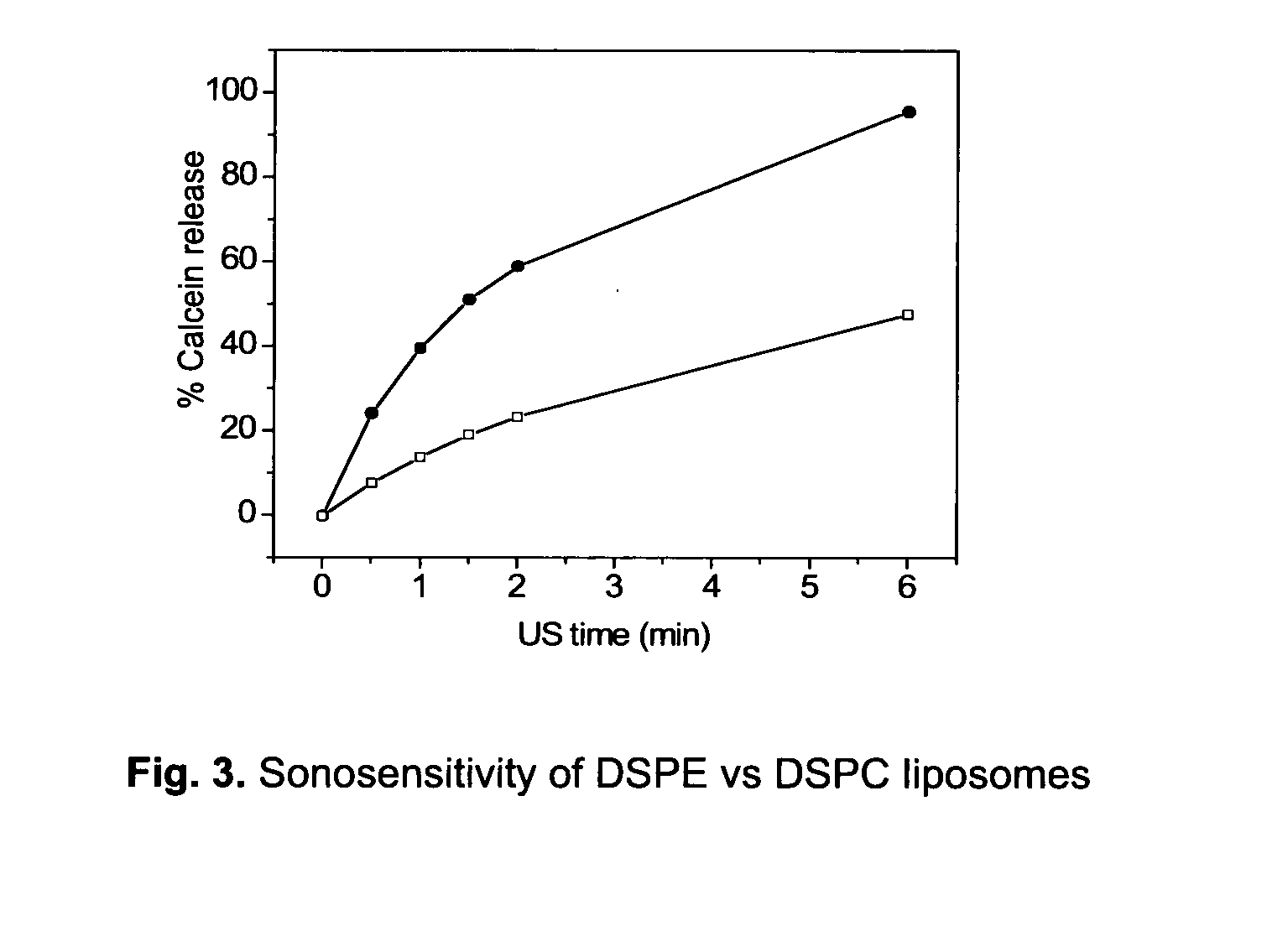
[0029]The current inventors have found that incorporation of lipophilic drugs in liposomal formulations comprising an inverted structure forming (ISF) lipid enhances delivery, particularly in combination with acoustic energy.
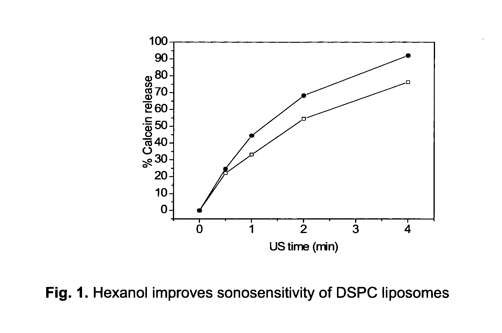
[0038]The current particulate material may comprise a suitable ISF PE and / or ISF PC phospholipid as the sole phospholipid or in combination with other lipids or phospholipids. In preferred embodiments of the current invention the particulate material comprises 25 mol % or more ISF PE and / or PC, more preferably 47 mol % or more, even more preferably 52 mol % or more, even more preferably 54.5 mol % or more, even more preferably 58 mol % or more, even more preferably 62 mol % or more, even more preferably 67 mol % or more, and yet even more preferably 77 mol % or more ISF PE and / or PC lipid. In general, a higher concentration of ISF lipid yields higher sonosensitivity.
[0043]Components for improving blood circulation time and / or further modulate sonosensitivity may be included in the material, like e.g. polyvinyl alcohols, polyethylene glycols (PEG), dextrans, or polymers. PEG or a derivate thereof, at any suitable concentration, is preferred. However, PEG concentrations are preferably up to 15 mol %, more preferably within the range 3 to 10 mol %, even more preferably in the range 3 to 8 mol %, and even more preferably within the range 5.5 to 8 mol %. In embodiments of the current invention the PEG concentration is 3, 5.5, 8, or 10 mol %. The PEG moiety may be of any molecular weight or type, however, it is preferred that the molecular weight is within the range 350 to 5000 Da, more preferably within 1000-3000 Da. In a preferred embodiment the molecular weight is 2000 Da. The PEG moiety may be associated with any molecule allowing it to form part of the particulate material. Preferably, the PEG moiety is conjugated to a sphingolipid (e.g. ceramide), a glycerol based lipid (e.g. phospholipid), or a sterol (e.g. cholesterol), more preferably to a ceramide and / or PE, and even more preferably to PE, like DMPE, DPPE, or DSPE. The acyl chain length should be the same as that of the main phospholipid of the membrane. The lipid-grafted PEG is preferably DPPE-PEG 2000 and / or DPPE-PEG 5000. In a particularly preferred embodiment lipid-grafted PEG is DSPE-PEG 2000.
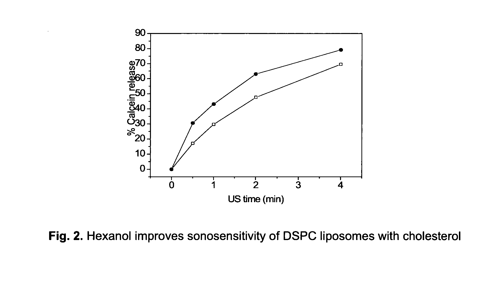
[0048]The particulate material may also comprise a sterol, wherein the sterol may be cholesterol, a secosterol, or a combination thereof. The secosterol is preferably vitamin D or a derivate thereof, more particularly calcidiol or a calcidiol derivate. The particulate material may comprise any suitable sterol concentration, preferably cholesterol, depending on the specific particle properties. In general, 50 mol % sterol is considered the upper concentration limit in liposome membranes. However, the particulate material preferably comprises up to 20 mol % cholesterol, more preferably up to 30 mol %, and even more preferably up to 40 mol % cholesterol, and most preferably within the range 20 to 40 mol %. In preferred embodiments of the current invention the particulate material comprises 20, 26, 30, 35, or 40 mol % cholesterol. Accordingly, the cholesterol concentration is preferably within any of the possible ranges constituted by the mentioned embodiment concentrations. Higher concentration ranges are, however, preferred. Sterols may have a therapeutic effect, as well as improve stability and reduce blood clearance rates.
[0064]The use or methods may further comprise the step of administering or activating said particulate material by means of acoustic energy or ultrasound. Hence, the active drug is released or administrated from the particulate material by means of acoustic energy. In this way the patient is protected against potential toxic effects of the drug en route to the target tissue and high local concentrations of the drug are obtainable in short time. Preferably, only the diseased volume is exposed to acoustic energy or ultrasound, but whole body exposures are also possible. The acoustic energy or ultrasound should preferably have a frequency below 3 MHz, more preferably below 1.5 MHz, more preferably below 1 MHz, more preferably below 0.5 MHz, more preferably below 0.25 MHz, and even more preferably below 0.1 MHz. In preferred embodiments of the current invention the frequency is 1.17 MHz, 40 kHz or 20 kHz. It should, however, be noted that focused ultrasound transducers may be driven at significantly higher frequencies than non-focused transducers and still induce efficient drug release from the current sonosensitive material. Without being limited to prevailing scientific theories, the current inventors believe that the level of ultrasound induced cavitation in the target tissue is the primary physical factor inducing drug release from the particulate material of the invention. A person skilled in the art of acoustics would know that ultrasound at any frequency may induce so-called inertial or transient cavitation.

Problems solved by technology

Lack of targeted drug delivery reduces the therapeutic-to-toxicity ratio thus limiting medical therapy.
This limitation is particularly evident within oncology where systemic administration of cytostatic drugs affects all dividing cells imposing dose limitations.
However, development of such drug delivery particles has faced two opposing challenges: efficient release of the encapsulated drug at the diseased site while maintaining slow non-specific degradation or passive diffusion in healthy tissue.
At present, this constitutes the main challenge in drug delivery (Drummond, Meyer et al.
Micelle formation and disruption is therefore an equilibrium process controlled by concentration, making these particles rather unstable and less suitable for drug delivery.
In addition, limited drug types can be encapsulated.
Gas-filled liposomes and microbubbles are highly US responsive but too large (˜1 μm) for efficient accumulation in e.g. tumour tissue.
However, reports on ultrasound sensitive liposomes are scarce.
However, liposomal doxorubicin (Caelyx® or Doxil®) is not engineered for ultrasound mediated drug release and shows a rather low drug release in vitro (see e.g. WO2008120998, incorporated herein in its entirety by reference).
Due to the gas bubble, such microbubbles are too large for passive in target tissues and are therefore less suited for e.g. cancer treatment.

Method used


Image

  • Lipophilic drug carrier
  • Lipophilic drug carrier
  • Lipophilic drug carrier

Examples


example 1

Preparation of Liposomes Containing Fluorescent Drug Marker Calcein

[0080]DSPC, DSPE, DOPE and DSPE-PEG 2000 were purchased from Genzyme Pharmaceuticals (Liestal, Switzerland). Cholesterol, calcein, HEPES, TRITON-X100 (10% solution), sodium azide and sucrose were obtained from Sigma Aldrich. Hexanol was supplied by BDH Chemicals Ltd. (Poole, England).

[0081]Calcein carrying liposomes (liposomal calcein) of different membrane composition were prepared using the thin film hydration method (Lasic 1993). The nominal lipid concentration was 16 mg / ml. Liposomes were loaded with calcein via passive loading, the method being well known within the art. The hydration liquid consisted of 10 mM

[0082]HEPES (pH 7.4) and 50 mM calcein. For the preparation of liposomal calcein containing hexanol, the hydration liquid was supplemented with a given amount of hexanol 2 days prior to usage in the lipid film hydration step.

[0083]After three freeze-thaw cycles, the liposomes were down-sized to 80-90 nm by ...

example 2

Characterisation of Calcein Containing Liposomes

[0085]Liposomes were characterised with respect to key physicochemical properties like particle size, pH and osmolality by use of well-established methodology.

[0086]The average particle size (intensity weighted) and size distribution were determined by photon correlation spectroscopy (PCS) at a scattering angle of 173° and 25 deg C (Nanosizer, Malvern Instruments, Malvern, UK). The width of the size distribution is defined by the polydispersity index. Prior to sample measurements the instruments was tested by running a latex standard (60 nm). For the PCS measurements, 10 μL of liposome dispersion was diluted with 2 mL sterile filtered isosmotic sucrose solution containing 10 mM HEPES (pH 7.4) and 0.02% (w / v) sodium azide. Duplicates were analysed.

[0087]Osmolality was determined on non-diluted liposome dispersions by freezing point depression analysis (Fiske 210 Osmometer, Advanced Instruments, MA, US). Prior to sample measurements, a r...

example 3

US Mediated Release Methodology and Quantification For Calcein Containing Liposomes

[0088]Liposome samples were exposed to 20 or 40 kHz ultrasound up to 6 min in a custom built sample chamber as disclosed in Huang and MacDonald (Huang and Macdonald 2004). The US power supply and converter system was one of two systems: (1) ‘Vibra-Cell’ ultrasonic processor, VC 750, 20 kHz unit with a 6.35 cm diameter transducer or (2) ‘Vibra-Cell’ ultrasonic processor, VC754, 40 kHz unit with a 19 mm cup horn probe, both purchased from Sonics and Materials, Inc. (USA). Pressure measurements were conducted with a Bruel and Kjaer hydrophone type 8103.

[0089]Both systems were run at the lowest possible amplitude, i.e. 20 to 21% of maximum amplitude. For the 20 kHz system this translates to a transducer input power of 0.9-1.2 W / cm2 and a peak-to-peak transducer pressure of about 460 kPa.

[0090]For the US measurements, liposome dispersions were diluted in a 1:500 volume ratio, with isosmotic sucrose solutio...


PUM

PropertyMeasurementUnit
Fractionaaaaaaaaaa
Molar densityaaaaaaaaaa

Abstract

Novel acoustically sensitive drug carrying particles comprising non-lamellar forming lipids are disclosed, as well as uses and methods thereof. The drug carrying particles accumulate in the diseased target tissue and efficiently release their pay-load upon exposure to acoustic energy.

Description

FIELD OF THE INVENTION[0001]The present invention is related to particles comprising non-lamellar forming amphiphilic lipids for controlled drug delivery and release at a defined volume in an animal. Specifically, the invention relates to acoustically sensitive drug carrying particles, e.g. liposomes, as well as compositions, methods and uses thereof.BACKGROUND OF THE INVENTION[0002]Lack of targeted drug delivery reduces the therapeutic-to-toxicity ratio thus limiting medical therapy. This limitation is particularly evident within oncology where systemic administration of cytostatic drugs affects all dividing cells imposing dose limitations. Hence, it exists a clear need for more efficient delivery of therapeutic drugs at the disease target with negligible toxicity to healthy tissue. This challenge has to a certain extent been accommodated by encapsulating drugs in a shell protecting healthy tissue en route to the diseased volume. Such protective shells may include a number of diffe...

Claims


Application Information

Patent Timeline
IPC IPC(8): A61K9/127A61K31/7068A61P31/00A61P37/02A61P29/00A61P35/00A61K31/704
CPCA61K9/0009A61K9/0019A61K31/704A61K9/1272A61K9/1271A61P29/00A61P31/00A61P35/00A61P37/02
Inventor NILSSEN, ESBEN A.FOSSHEIM, SIGRID L.EVJEN, TOVE JULIE
Owner EPITARGET
Learn More