Organic colored microparticles, diagnostic reagent kit containing the same, and in vitro diagnosis method

a technology of colored microparticles and diagnostic reagents, which is applied in the field of organic colored microparticles, diagnostic reagent kits containing the same, and in vitro diagnosis methods, can solve the problems of inhibition of adsorption in the presence of surfactants, limited color to a single, and absence of fixed binding sites, etc., to achieve rapid diagnosis, improve color properties, and reduce the level of erroneous diagnosis

Inactive Publication Date: 2012-09-06
ASAHI KASEI FIBERS CORPORATION
View PDF12 Cites 7 Cited by
  • Summary
  • Abstract
  • Description
  • Claims
  • Application Information

AI Technical Summary

Benefits of technology

[0030]The organic colored microparticles according to the present invention have extraordinarily superior coloring properties in comparison with the coloring properties of latex particles of the prior art, and since they are able to adsorb antigen and other ligands, they can be applied to immunochromatography. The organic colored microparticles according to the present invention are able to provide a highly sensitive immunochromatography kit as a result of actualizing coloring in the case of being captured with a selective and specific reaction, and as a result of enabling multicoloration, are useful for simultaneous measurement of multiple test substances. In addition, since the organic colored microparticles of the present invention enable the selection of an arbitrary method used to bind a ligand such as chemical bonding in addition to physical adsorption attributable to a dye, they can be applied to various test substances. Thus, the present invention enables rapid diagnoses with a low level of erroneous diagnosis, thereby greatly contributing to rapid diagnosis while also considerably expanding the application range of immunochromatography.EMBODIMENTS OF THE INVENTION
[0032]The following provides a detailed explanation of the invention of the present application.
[0033]The microparticles in the present invention refer to organic colored microparticles having an average particle size of 10 nm to 1000 nm and a color intensity of 1.0 to 5.0. The preferable range of average particle size is 100 nm to 900 nm, and more preferably 200 nm to 800 nm. If the average particle size exceeds 1000 nm, development becomes slow when using in an immunochromatography kit, rapid evaluation is prevented, the microparticles are easily captured on the developing film, and the background per se becomes colored, thereby resulting in a tendency for the expected coloring at the detected location to be ambiguous. At a detected location in particular, there are many cases in which the pore size of the developing film becomes smaller due to coating of the capture reagent. Consequently, the label tends to be captured easily, or in other words, the rate of false positives increases. As a result, the test kit cannot be considered to be reliable.<Color Intensity>
[0034]Color intensity in the present invention is defined as the value obtained by measuring visual absorbance of a dispersion of the organic colored microparticles at an optical path length of 10 mm using an integrating sphere in the range of 400 nm to 800 nm, subtracting the background component of the dispersion medium, obtaining an absorbance curve of the dispersion matrix per se, dividing the maximum value (ABS) thereof by the weight percentage of the dispersion matrix, and calculating per 0.01% by weight. Since the use of an integrating sphere makes it possible to reduce the effects of diffuse light on the particles, the resulting value can serve as an indicator of the degree of coloring of the microparticles, and a larger value thereof can be judged to indicate more distinct coloring. Although the color intensity of the microparticles of the present invention is 1.0 or more, the color intensity is preferably as high as possible. Color intensity can be increased either by using a dispersed dye or pigment that demonstrates a high degree of coloring, or selecting means for increasing the number times staining is carried out. However, since color intensity of 5.0 or more cannot be achieved with several rounds of staining using ordinary dyes, in consideration of economy, color intensity is preferably 1.0 to 5.0, more preferably 1.5 to 5.0 and even more preferably 2.0 to 5.0. In the case color intensity is lower than 1.0, visibility of a detected site becomes inferior when using in an immunochromatography kit due to weak coloring, thereby impairing reliability of test results.<Material of Organic Colored Microparticles>
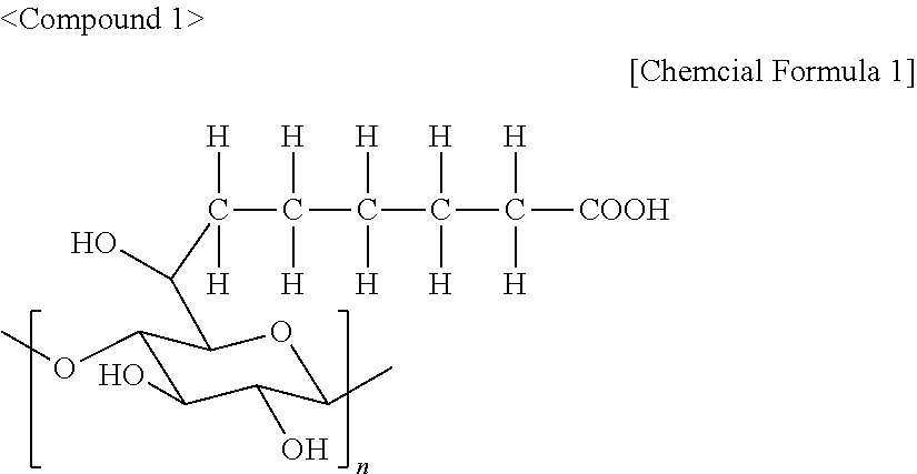
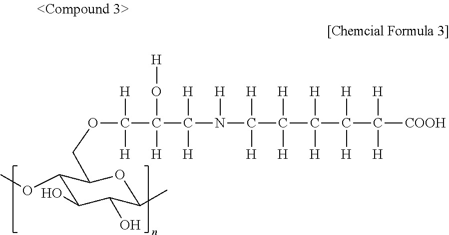
[0035]There are no particular limitations on the material of the organic colored microparticles in the present invention provided it has high color intensity and is stably dispersed. Although materials that can be deeply colored using dye or pigment can be applied, the realization of deep dyeing and strong dyeing is preferable when testing by immunochromatography and because this contributes to stabilization of kit quality during long-term storage. In order to achieve strong dyeing, for example, a covalently bonding reactive dye can be used, and a material derived from cellulose can be used that can be dyed with a reactive dye. Since microparticles composed of a material derived from cellulose have a large number of hydroxyl groups, not only are they able to retain numerous reactive dyes by covalent bonding, but they also are able to maintain a stable dispersion in water and the like after being deep dyed. For this reason, although the use of cellulose for the material of the organic colored microparticles is preferable, there are no particular limitations on the type thereof. For example, recycled cellulose, purified cellulose or natural cellulose can be used. Partially derivatized cellulose may also be used. Preferably 20% by weight to 90% by weight of the organic colored microparticles is derived from cellulose. More preferably, 20% by weight to 80% by weight of the organic colored microparticles is derived from cellulose. Even more preferably, 20% by weight to 70% by weight is derived from cellulose.<Production Method of Material of Colored Organic Microparticles>
[0036]There are no particular limitations on the method used to produce the material of the organic colored microparticles in the present invention. Although the fine particles of a desired average grain size may be obtained by sizing, in the present invention, cellulose microparticles are prepared by using a congealing liquid obtained by dissolving cellulose in a good solvent thereof and mixing with water, organic solvent or ammonia and the like. The use of this method enables the grain size of the cellulose microparticles to be adjusted according to the composition of the congealing liquid. Although not intended to limit the production method of the material of the organic colored microparticles of the present invention, the following provides a detailed explanation thereof using a specific example.

Problems solved by technology

In the case of metal colloids, since color is generated due to plasmon effects corresponding to the type of metal thereof, the resulting color is limited to a single color.
However, in the case of physical adsorption, there are cases in which problems such as the absence of a fixed binding site and inhibition of adsorption in the presence of a surfactant can occur.
In addition, there are also cases in which an adequate amount of ligand cannot be bound.
However, according the examples disclosed in Patent Document 2, the dyeing capacity of the particles is low at about 6% by weight, and the resulting color intensity is weak.
Consequently, in the case of using for immunochromatography, it is not possible to obtain distinct coloring effects, thereby resulting in a lack of reliability.
Although these microparticles are used in immunochromatography by imparting an antibody by physical adsorption or chemical bonding as described in Patent Document 4 indicated below, since the amount of antibody bound is insufficient and coloring of the microparticles per se is weak, distinct coloring results are unable to be obtained.

Method used


Image

  • Organic colored microparticles, diagnostic reagent kit containing the same, and in vitro diagnosis method
  • Organic colored microparticles, diagnostic reagent kit containing the same, and in vitro diagnosis method
  • Organic colored microparticles, diagnostic reagent kit containing the same, and in vitro diagnosis method

Examples


example 1

[0065]Linter cellulose was dissolved in a cuprammonium solution followed by diluting with water and ammonia to prepare a cuprammonium solution cellulose solution having a cellulose concentration of 0.37% by weight. The copper concentration of that solution was 0.13% by weight, and the ammonia concentration was 1.00% by weight. Next, a congealing liquid was prepared having a tetrahydrofuran concentration of 90% by weight and water concentration of 10% by weight. 500 g of the preliminarily prepared cuprammonium cellulose solution having a cellulose concentration of 0.37% by weight were then added while slowly stirring 5000 g of the congealing liquid using a magnetic stirrer. After continuing to stir for about 5 seconds, 1000 g of 10% by weight sulfuric acid were added to carry out neutralization and regeneration and obtain 26500 g of a slurry containing the target cellulose microparticles. The resulting slurry was then centrifuged for 10 minutes at a speed of 10000 rpm. The precipitat...

example 2

[0068]Although the unstained cellulose microparticles obtained in Example 1 were stained using the same procedure, the procedure was carried out for a total of 10 cycles to obtain stained microparticles. The results of measuring average particle size and color intensity before and after staining are shown in the following Table 1.

example 3

[0069]Cellulose microparticles and stained cellulose microparticles were obtained using the same method as Example 1 with the exception of using Levafix Rubine CA Gr. (Registered Trade Mark) manufactured by Dystar GmbH Corp. (to also be referred to as red dye B) as a reactive stain to stain the unstained cellulose microparticles obtained in Example 1. The results of measuring average grain size and color intensity before and after staining are shown in the following Table 1.


PUM

PropertyMeasurementUnit
particle sizeaaaaaaaaaa

Abstract

Provided are an immunochromatography kit that is highly sensitive and capable of multicoloration, and organic colored microparticles that are ideal as an element of the immunochromatography kit. Organic colored microparticles having an average grain size between 10 and 1,000 nm and a color intensity between 1.0 and 5.0 are prepared using cellulose as the starting material. When the organic colored microparticles are used as a label in an immunochromatography kit, the immunochromatography kit is of a high sensitivity than conventional technology. The immunochromatography kit is also capable of multicoloration and is useful for rapid diagnosis.

Description

TECHNICAL FIELD[0001]The present invention relates to organic colored microparticles derived from an organic polymer, a reagent kit using the microparticles, and an in vitro diagnosis method.BACKGROUND ART[0002]Microparticles composed of a polymer are used in various fields due to the ease of controlling their particle size, mechanical strength, particle size distribution, shape and degree of aggregation, examples of which include toner, anti-blocking materials of packing materials, insulating fillers, crystal nucleating agents, chromatographic fillers and abrasives. More recently, microparticles have also been applied to applications such as carriers for immunodiagnostic reagents, spacers of liquid crystal displays, standard particles for calibration of analytical equipment and standard particles for testing of porous films.[0003]The amount of microparticles composed of a polymer used in immunodiagnostic reagent carrier applications in particular is increasing, and the amount used ...

Claims


Application Information

Patent Timeline
Patent Type & Authority Applications(United States)
IPC IPC(8): G01N33/558B82B1/00B82Y15/00
CPCG01N33/548Y10T428/2982G01N33/585G01N33/558G01N33/543G01N33/52G01N33/587G01N2333/59G01N2458/00
Inventor YOSHIDA, SATORU
Owner ASAHI KASEI FIBERS CORPORATION
Why Eureka
Try Eureka