
Glucose sensor

a glucose sensor and sensor technology, applied in the field of glucose sensors, can solve the problems of inadequate changes in fluorescence characteristics of known glucose sensors upon glucose binding, problems such as sensitivity and response, and achieve excellent fluorescence intensity change upon glucose binding, accurate and reliable readout of glucose concentrations, and excellent responsiveness to glucose binding

Inactive Publication Date: 2012-09-13
KINGS COLLEGE LONDON
View PDF1 Cites 7 Cited by
  • Summary
  • Abstract
  • Description
  • Claims
  • Application Information

AI Technical Summary

Benefits of technology

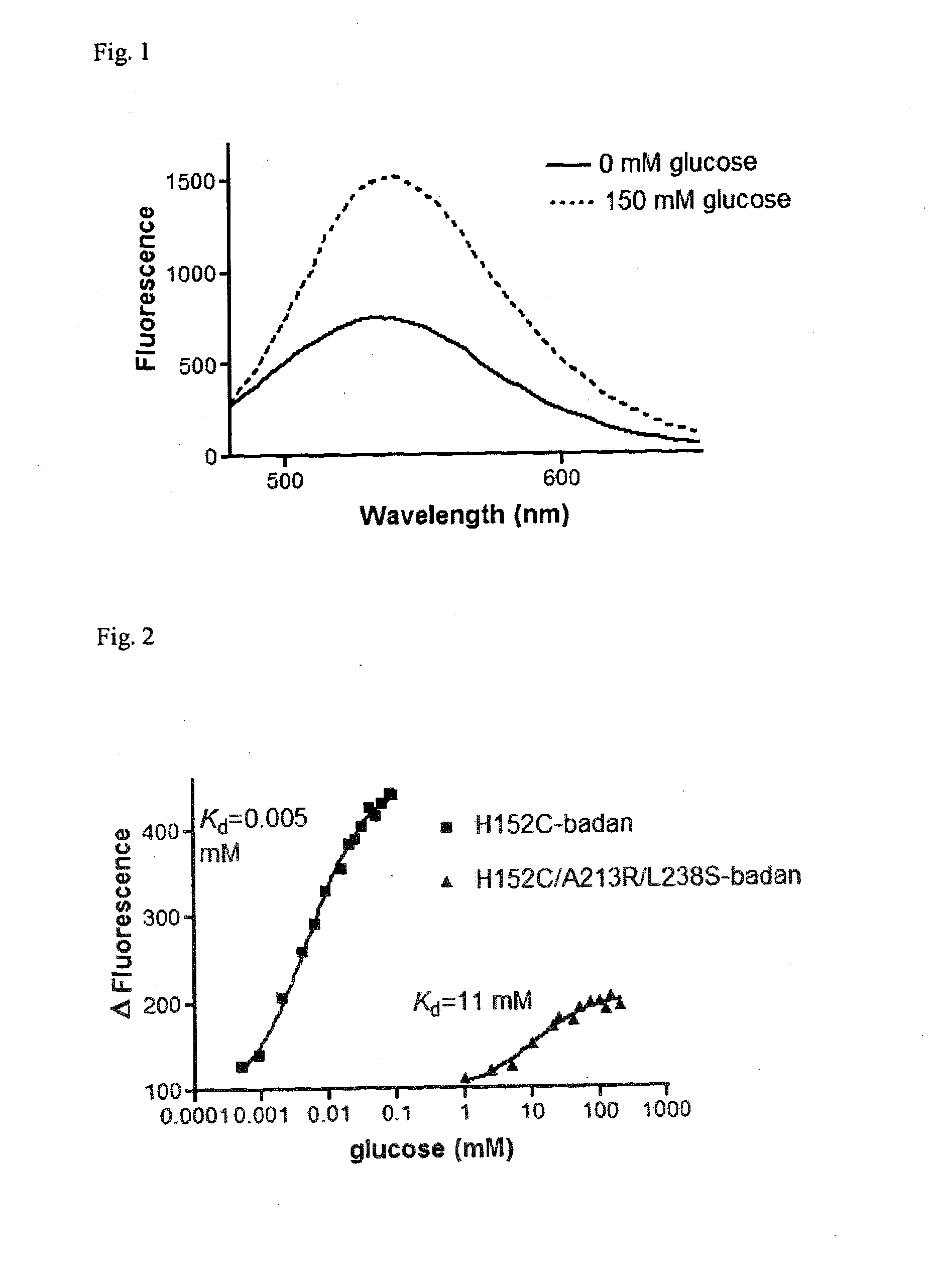
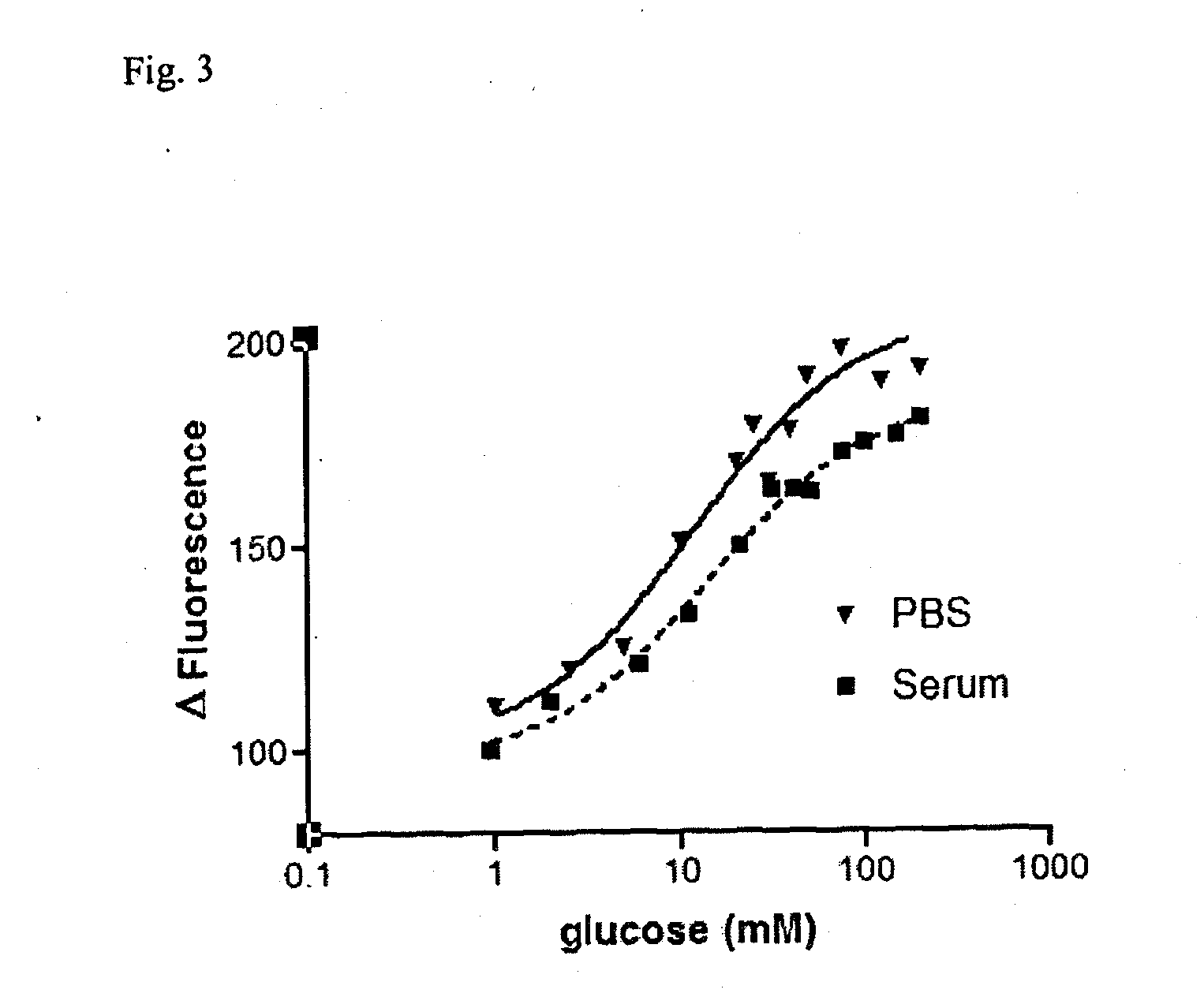
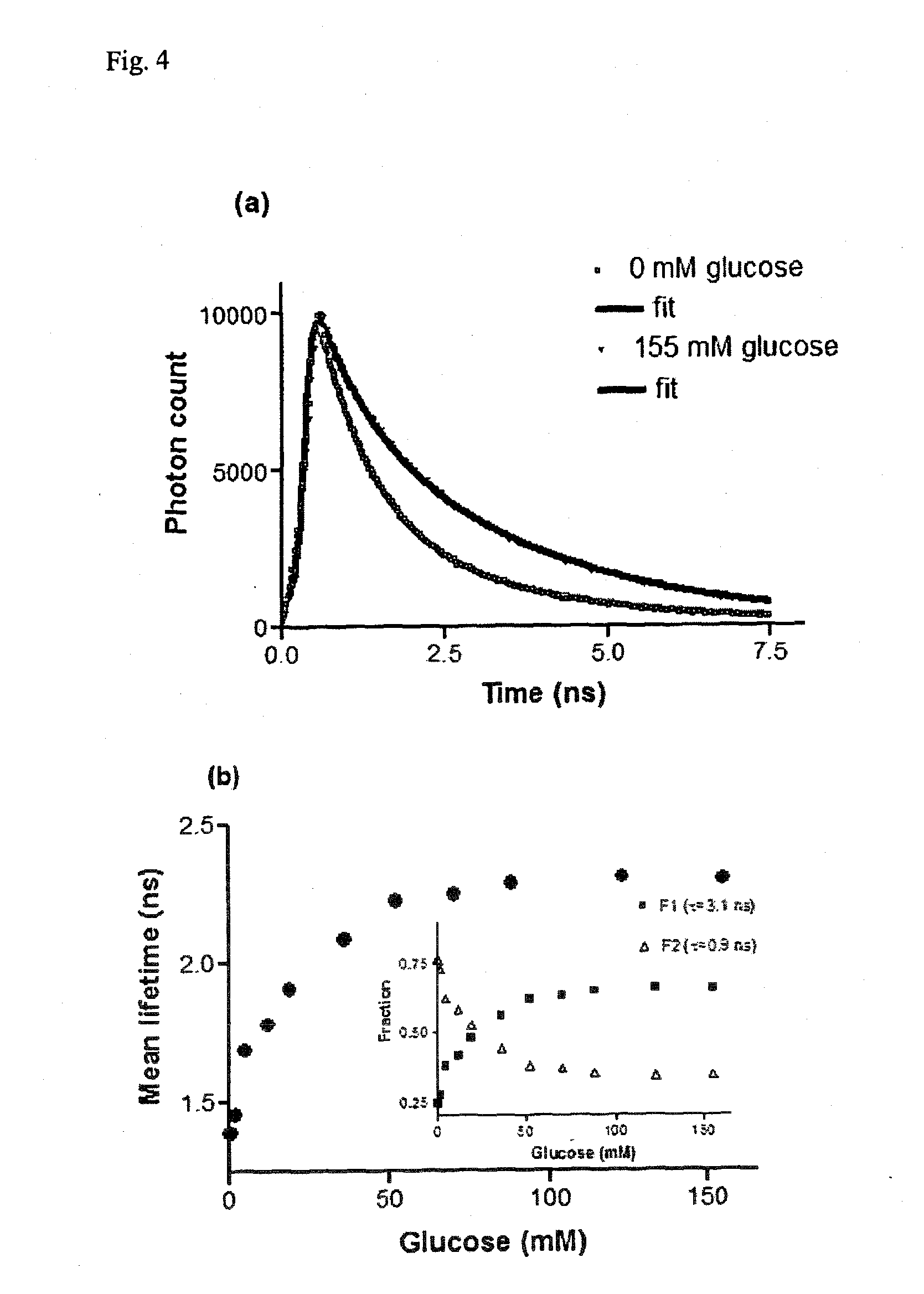
The present invention provides an improved glucose sensor by studying mutants of GLBP and making novel combinations of mutations. The mutant of GLBP covalently attached to a fluorophore has an extended Kd and operating range suitable for clinical use. The sensor works well in serum and is a robust method for developing CGM in vivo. The invention also includes a microcapsule and a fibre optic strand comprising the glucose binding protein, as well as a nucleic acid encoding the glucose binding protein and a method for Ni chelation of polystyrene beads. The mutated glucose binding protein has mutations at positions H152, A213, and L238. The invention provides a recombinant glucose / galactose binding protein that is mutated relative to the wild type sequence at positions H152, A213, and L238. The use of the mutated glucose binding protein for determination of glucose concentration and monitoring is also provided.

Problems solved by technology

Known glucose sensors have suffered from problems and drawbacks regarding their sensitivity and their response, together with their effective operating range.
Known sensors have also suffered from inadequate changes in fluorescence characteristics upon glucose binding.

Method used


Image

  • Glucose sensor
  • Glucose sensor
  • Glucose sensor

Examples


example 1

Construction of Expression Vector pET303-GBP and Purification of Mutants of GBP

[0144]Details of the methodology are given in Khan of al. [25]. In brief, the GBP gene (mgIB) was isolated from the plasmid pTZ18U-mgIB by PCR and ligated into pET303 / CT-His vector using a Rapid DNA ligation kit to form pET303-GBP. For the H152C, H152C / A213R, H152C / L238S and H152C / A213R / L238S mutants, pET303-GBP was used as a template. Site-directed mutagenesis was performed using the Quick-change mutagenesis kit with respective primers for each mutation. DNA sequencing data verified the presence of the desired mutations. A single colony of E. coli BL21(DE3) transformed with the pET303-GBP plasmid containing various mutants was inoculated in LB media containing 100 μg / ml of ampicillin and grown at 37° C. Expression of the proteins was induced by adding isopropyl-2-D-thiogalactopyranoside to a final concentration of 1 mM. Bacterial cells were lysed and the cell extract was clarified by centrifugation. Affi...

example 2

Fluorophore Labelling

[0145]To label GBP mutants with badan, 50 μM protein was dissolved in 5 mM Tris(2-carboxyethyl)phosphine in phosphate-buffered saline (PBS) pH 7.4, and then a 10-fold excess of dye (500 μM) was added and the mixture incubated overnight at 4° C., after which it was purified on a Sephadex G-25 gel-filtration column.

example 3

Serum Preparation

[0146]Venous blood samples from healthy volunteers were collected in Vacuette tubes. The specimens were incubated at room temperature for 4 days to allow blood clotting and glycolysis. The samples were then centrifuged at 3500 g for 20 min and serum removed, pooled and stored until use. The glucose concentrations of serum samples were determined using a hexokinase-based assay (Sigma).


PUM

PropertyMeasurementUnit
Kaaaaaaaaaaa
Kaaaaaaaaaaa
pHaaaaaaaaaa

Abstract

The invention relates to a glucose binding protein comprising amino acid mutations relative to the wild type sequence at the following positions: (i) H 152, (ii) A213; and (iii) L238 wherein the mutation at position H 152 is H152C. The invention further relates to such a glucose binding protein comprising the mutations H152C, A213R and L238S, in particular when linked to an environmentally sensitive dye such as badan.

Description

FIELD OF THE INVENTION[0001]The invention relates to a glucose sensor based on the bacterial glucose / galactose binding protein (GBP).BACKGROUND TO THE INVENTION[0002]Currently available sensors used in clinical practice for continuous glucose monitoring (CGM) in diabetes are subcutaneously implanted needle-type devices that are either amperometric enzyme electrodes, or microdialysis probes which sample interstitial fluid and deliver it to an ex vivo biosensor [1-3]. Both sensor types are based on immobilized glucose oxidase and the electrochemical detection of either hydrogen peroxide or electrons directly coupled to an underlying electrode via a molecular mediator [4]. Whilst evidence for the clinical utility of CGM is now accumulating [5], electrochemical glucose sensors suffer from impaired responses in vivo which necessitates frequent calibration, and contributes to sub-optimal accuracy [6, 7]. The likely reasons for poor CGM performance (in addition to time lags between blood a...

Claims


Application Information

Patent Timeline
Patent Type & Authority Applications(United States)
IPC IPC(8): C07K14/435C07H21/04
CPCC07K17/00C07K17/06G01N33/66G01N33/545G01N33/582G01N33/54313
Inventor PICKUP, JOHNKHAN, FAAIZAH
Owner KINGS COLLEGE LONDON
Learn More