Intra-Culture Perfusion Methods and Applications Thereof

a perfusion method and culture technology, applied in the field of intra-culture perfusion methods and applications thereof, can solve the problems of difficult to grow and maintain such cultures, difficult to deliver and distribute nutrients, drugs and other compounds intra--3d-culture for physiologically closer tissue modeling, etc., and achieve the effect of limiting the loss of cell signaling molecules

Active Publication Date: 2014-04-17
LENA BIOSCI INC
View PDF4 Cites 15 Cited by
  • Summary
  • Abstract
  • Description
  • Claims
  • Application Information

AI Technical Summary

Benefits of technology

[0016]The invention further discloses a perfusion method which limits loss of cell signaling molecules, and provides for a design solution to evacuate air in the domain outside of the culture(s) following culture plating, prior to and during perfusion.
[0017]The invention also answers the need for a perfusion tool which incorporates (1) an intra-culture perfusion method, wherein said method also limits loss of cell signaling molecules, and further enables air evacuation in the domain outside of cultures(s); and (2) the rigid scaffold and / or the synthetic vasculature to deliver nutrients, dissolved gas and test compounds within the interior of 3D cultures consistently, reproducibly, and in high-throughput.

Problems solved by technology

Enormous cost, ethical concerns and increased pressure from regulatory agencies all drive demand for cell-based models and perfusion methodologies that can be used for in vitro screening of both large and small molecule drugs, and to triage toxic and ineffective leads earlier, prior to in vivo studies.
However, it has been difficult finding a cell-based model that mimics function of living tissues, and a corresponding perfusion system which mimics both the function of vasculature and in vivo tissue perfusion for physiologically closer tissue modeling, tissue metabolic sustenance, and the delivery, distribution and testing of compounds within the interior of tissue mimicking cell mass.
However, it has been challenging to grow and maintain such cultures due to diffusive mass transport limitations within their interior, and especially in the case of a common 3D ECM gel culture model or a high-cell-density spheroid model.
Accordingly, without functional vasculature and non-invasive yet efficient intra-3D-culture perfusion, it has been difficult to deliver and distribute nutrients, drugs and other compounds intra-3D-culture for physiologically closer tissue modeling and drug testing in the same.
In standard, diffusion-limited culturing systems in vitro, 3D cultures which model tissues perfused in vivo suffer from the same problems as do slices of said explanted tissue.
Prior art 3D cell culture perfusion tools have also been riddled with problems.
Lack of intra-culture perfusion methods (intra-gel or through a dense mass of cells); cumbersome priming and out-gassing; pressure surges and clogging of miniaturized components, complex setup, frequent user interventions in operation with overall poor culture performance relative to user input and cost have been some of the problems even when throughput was low.
Since prior art flow geometry has rarely been optimized for intra-3D-culture perfusion forcing flow around 3D culture rarely made flow of concentrated solutions pass intra-3D-culture (due to higher culture resistance), and the dominant mode of intra-3D-culture mass transport has remained diffusion-limited.
Accordingly, forcing flow intra-culture (e.g. 3D cell culture gel plug) and forcing it non-invasively has remained a delicate task.
Another frequently overlooked problem in prior art cell culture perfusion, whether with 2D cultures or 3D cultures, is that the vast majority of perfusion tools have been based on unidirectional perfusion.
Although medium recycling has been used to mitigate this effect, relatively high liquid volume to culture volume in a single loop have generally contributed to dilution and delay in bringing these molecules back to cells.

Method used


Image

  • Intra-Culture Perfusion Methods and Applications Thereof
  • Intra-Culture Perfusion Methods and Applications Thereof
  • Intra-Culture Perfusion Methods and Applications Thereof

Examples


example 1

The Anchoring (Hydrophilic, Absorbent) Scaffold Compositions Comprising Hydrophilic and Absorbent Synthetic Vasculature which Extended to all Exterior Surfaces of the Scaffold

[0083]U.S. Provisional Patent Application Ser. No. 61 / 712,943 and the U.S. patent application Ser. No. 13 / 962,403 disclosed production methods and materials which were suitable examples of 3D scaffolds comprising absorbent synthetic vasculature, wherein the vasculature extended to all exterior surfaces of the scaffold. In one embodiment, the entire 3D scaffold was absorbent and made from PVOH fibers. In another embodiment, the scaffold was made of borosilicate glass fibers coated with the absorbent polyvinyl alcohol (PVOH). In a further embodiment, scaffold compositions comprising PVOH fibers and PVOH-coated borosilicate glass fibers were disclosed. In another embodiment, the scaffold was made using absorbent fibrillated cellulose fibers and PVOH-sized borosilicate glass fibers. In yet another embodiment, the s...

example 2

Additional Anchoring, Hydrophilic and Absorbent, Scaffold Compositions Comprising Hydrophilic and Absorbent Synthetic Vasculature which Extended to all Exterior Surfaces of the Rigid Scaffold

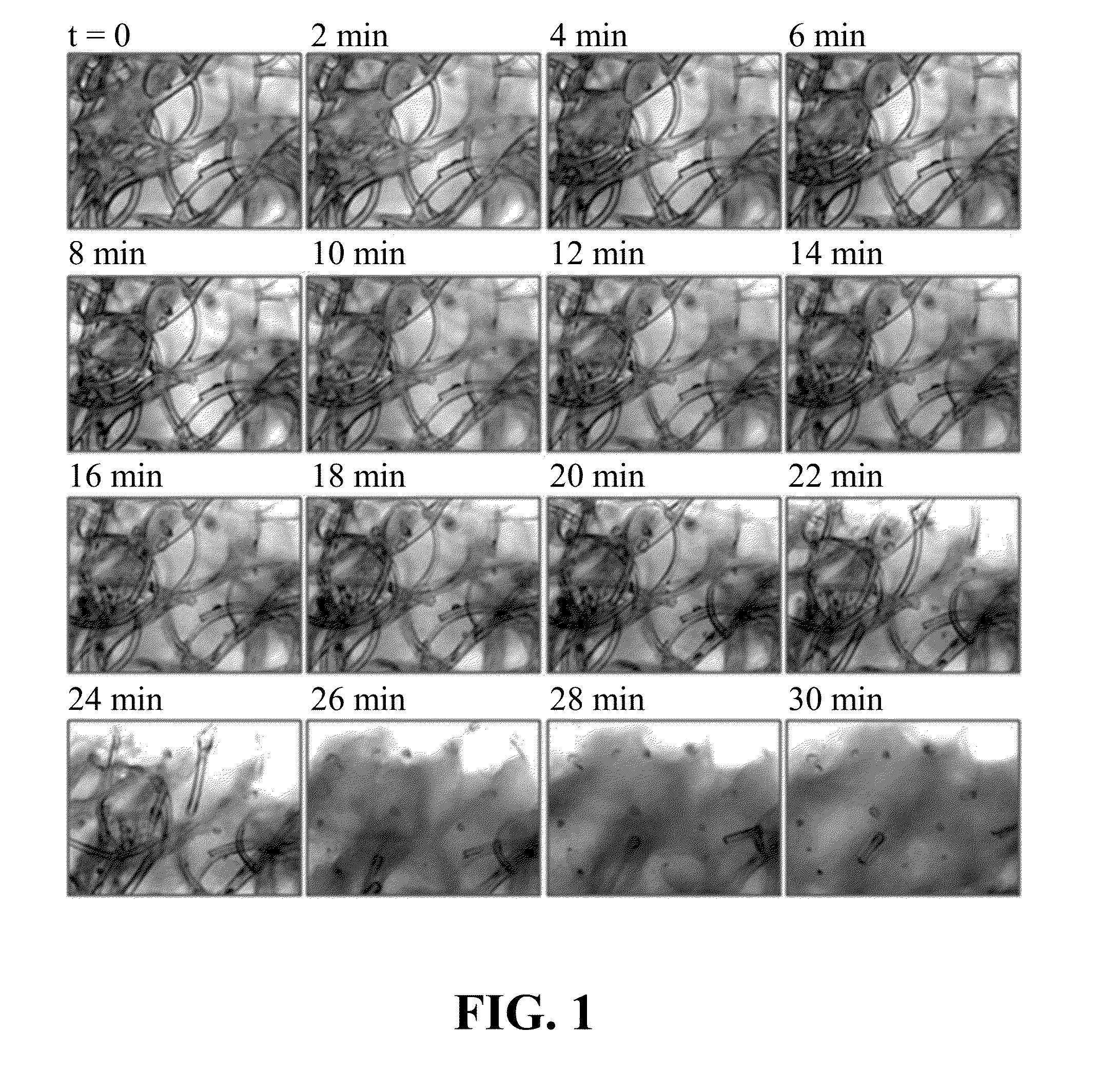
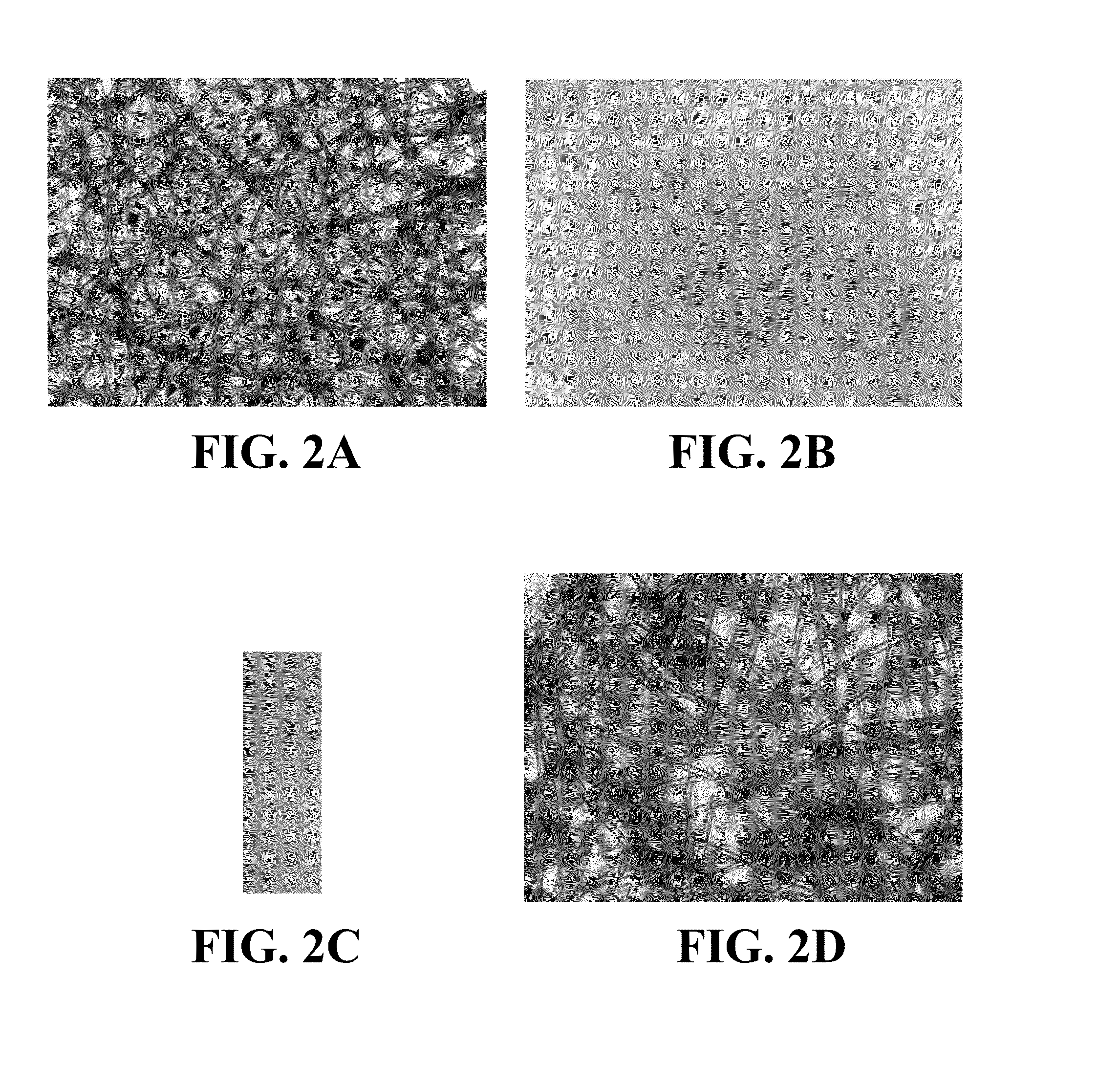
[0088]A material comprising a blend of cellulose and synthetic fibers with PVOH binder was acquired from Pall Corporation, product No. SMCON01 (conjugate pad type 8301). According to the supplier the material thickness was 355.6-444.5 μm, typical basis weight of 50 g / m2, tensile strength of 10.3 lbs in MD, water absorption capacity of 28 μl / cm2 and the average wicking rate of 226 seconds per 3 cm. The material was punched into disks of 9.5 mm in diameter. The measured thickness was approximately 400 μm. FIG. 2A shows a 4× photograph of the material in bright field after staining with a brown dye solution in DI water, followed by 30 minute room temperature drying. At least one type of fiber and / or its coating was absorbent and swollen in the aqueous solution (and stained dark). In wicking tests, ...

example 3

Methods of Controlling the Volume Fraction of Synthetic Vasculature and Capillary-to-Capillary Distances

[0090]The vasculature volume fraction is an important factor in modeling vascularized tissues using 3D cell culture models. Tissues that are perfused in vivo generally have different volume fraction of capillaries within their interior. Accordingly, to model said tissues using 3D cell cultures, a control of the synthetic vasculature volume fraction was desirable. Production methods disclosed herein and in U.S. Provisional Patent Application Ser. No. 61 / 712,943 and in U.S. patent application Ser. No. 13 / 962,403 enabled the control of synthetic vasculature volume fraction by controlling mass fraction of the absorbent component in the final scaffold composition during manufacture, using the exemplary wet-laid process among other disclosed methods, provided for a mixture of absorbent fibers, absorbent fibers and other fibers, absorbent coating on the fibers etc.

[0091]While in most in ...


PUM

PropertyMeasurementUnit
flow rateaaaaaaaaaa
thicknessaaaaaaaaaa
thicknessesaaaaaaaaaa

Abstract

Disclosed are intra-culture perfusion methods applied to standard cell culture disposables using three-dimensional cell culture scaffold and synthetic vasculature compositions in high-throughput screening and high-content screening applications, and assay development.

Description

CROSS REFERENCE TO RELATED APPLICATIONS[0001]This application claims the priority benefit of U.S. Provisional Patent Application Ser. No. 61 / 712,943 filed on Oct. 12, 2012.STATEMENT REGARDING FEDERALLY SPONSORED RESEARCH OR DEVELOPMENT[0002]Some embodiments of this invention were made with government support under agreements 1 R43NS065543 and 5 R43NS065543 awarded by the National Institutes of Health. The U.S. government may have certain rights in this invention.BACKGROUND OF THE INVENTION[0003]To reduce high attrition rates and accelerate discovery and development timelines, the pharmaceutical industry seeks in vitro alternatives to interrogate drug candidates prior to animal, humanized animal, and human studies. Enormous cost, ethical concerns and increased pressure from regulatory agencies all drive demand for cell-based models and perfusion methodologies that can be used for in vitro screening of both large and small molecule drugs, and to triage toxic and ineffective leads earl...

Claims


Application Information

Patent Timeline
Patent Type & Authority Applications(United States)
IPC IPC(8): C12N5/00
CPCC12N5/0068C12N2533/30Y10T29/49826
Inventor VUKASINOVIC, JELENA
Owner LENA BIOSCI INC
Why Eureka
Try Eureka