
Optical regulation of gene expression in the retina

a gene expression and retinal technology, applied in the field of optical regulation of gene expression in the retina, can solve the problems of undesirable persistence of expression, loss of cell division, and inability to persist in dividing cells of gene therapy vectors,

Inactive Publication Date: 2015-04-23
THE BOARD OF TRUSTEES OF THE LELAND STANFORD JUNIOR UNIV
View PDF4 Cites 10 Cited by
  • Summary
  • Abstract
  • Description
  • Claims
  • Application Information

AI Technical Summary

Benefits of technology

The present invention provides methods for modulating gene expression in a subject by using electromagnetic radiation to apply heat to target cells in the subject. This can be done by expressing a transgene in the target cells, which includes a polynucleotide encoding the transgene and a chromophore that can generate heat upon application of electromagnetic radiation. The method can be used to treat genetic diseases, control gene expression, and modulate the immune system. The target cells can include pigmented cells in the retina, iris, and choroid. The method can also involve using a laser to apply the electromagnetic radiation.

Problems solved by technology

Other gene therapy vectors are not able to persist within dividing cells, and are therefore “lost” during cell division.
In some clinical applications, such long-term, persistent expression may be undesirable or have the potential to cause concern, for example, if there is a complete resolution of the disease and continued expression is unnecessary, or if transgene expression leads to adverse events, such as a hypersensitivity reaction or acute or chronic toxicity.
In such cases, continued expression of the transgene could be undesirable or problematic, and it would be desirable to “turn off” or reduce transgene expression, or otherwise “remove” cells containing the transgene.
In other cases, the level of transgene expression is too low resulting in a subtherapeutic dose of the transgene product.
Despite great efforts placed on development of inducible gene expression systems, the developed approaches have been cumbersome and provide an incomplete solution in most clinical settings.
The small molecule agent used for induction may itself have undesirable effects, such as off-target biological effects of rapamycin (e.g., immunosuppression), and off-target effects of steroid hormones, which would be similar to hormone-replacement therapy effects at the levels proposed for the RU-486 system.
Systems that seem to avoid this problem, such as the TetOn system, suffer from other shortcomings that prevent widespread clinical use, such as immune response to the gene product in large animals.
Another shortcoming is that these systems are often “leaky”, leading to a low level of background expression persisting in the “off” state.
Randomizing to switch off a therapy that may provide benefit poses an ethical challenge, and using the switch only “when needed” is problematic analytically.

Method used


Image

  • Optical regulation of gene expression in the retina
  • Optical regulation of gene expression in the retina
  • Optical regulation of gene expression in the retina

Examples


example 1

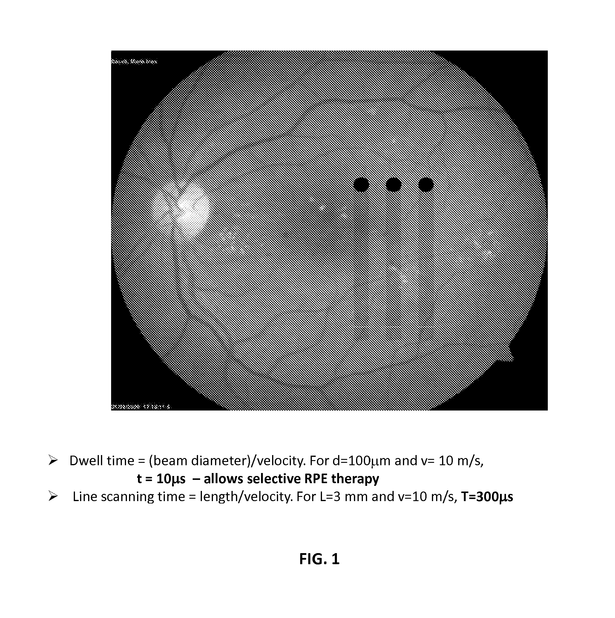
Selectivity of RPE Destruction

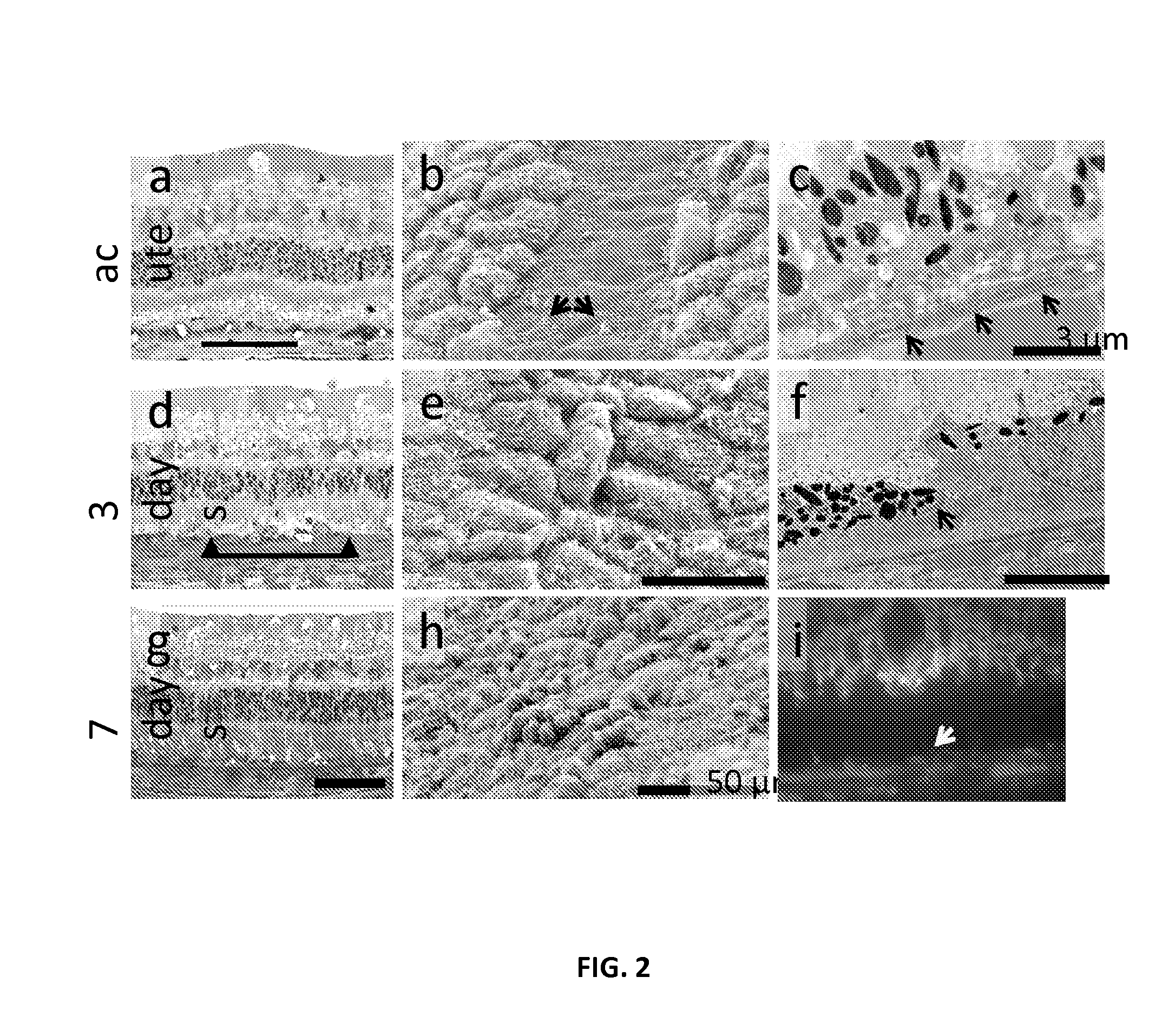
[0199]Pigmented rabbits are transfected using a rAAV2 encoding Green Fluorescent Protein (GFP). Selectivity of RPE destruction was tested with line scanning laser in vivo using fluorescence angiography and spectral domain optical coherence tomography (SD-OCT), and confirmed with light microscopy, transmission (TEM) and scanning electron microscopy (SEM). A typical example of selective destruction of RPE cells produced by scanning laser in a rabbit eye without damage to photoreceptors and Bruch's membrane is shown in FIG. 2. Collapsed RPE cells underneath the intact photoreceptors can be seen in the histological section (FIG. 2a). SEM image shows absence of the RPE cells along the line of laser scanning one day after treatment and evidence of cellular migration by foot processes on the surrounding cells (FIG. 2b). TEM demonstrates RPE disruption with an intact Bruch's membrane underneath. Continuity of the RPE layer is reestablished in three days (FIG. 2...

example 2

Control Expression of GFP in Retina

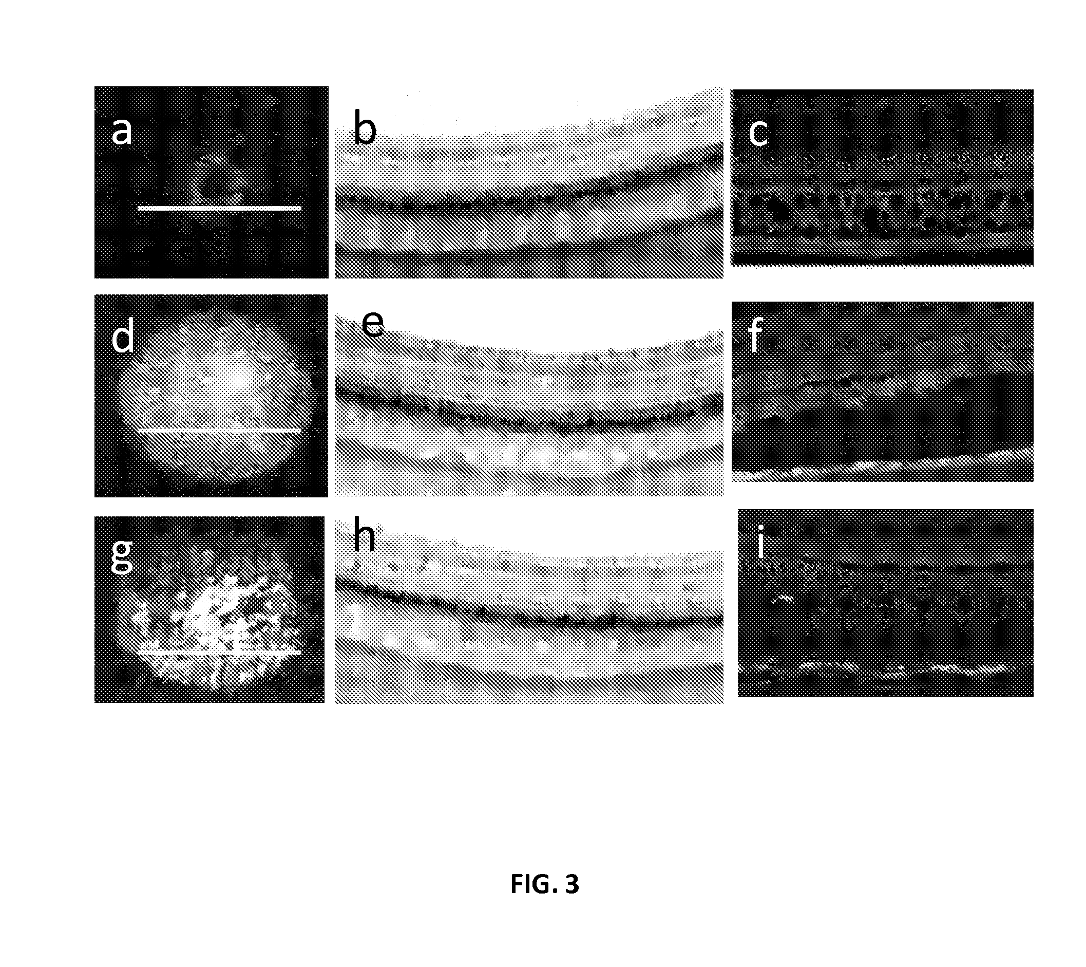
[0201]Each eye received three subretinal injections: one with balanced salt solution (BSS), and two containing an rAAV vector encoding GFP. One bleb was left untreated for control and another was treated by SRT 3 weeks after surgery with a line scanning pattern density of 50% (i.e. laser lines were spaced one line width apart). GFP fluorescence was mapped to quantify the level of GFP expression in transfected retina over time. Possible neurosensory retina damage was monitored in-vivo using high resolution SD-OCT (FIG. 3a-b, d-e, g-h).

[0202]The time course of GFP expression and the effect of SRT on fluorescent signal was plotted in FIG. 4. Both transfected blebs reached maximum fluorescence level at 3 weeks. There was no significant increase in fluorescence signal in the BSS-injected bleb. Fluorescence in the control bleb (transfected, but not treated with laser) remained stable throughout the follow up period. The laser-treated bleb showed a 40% si...

example 3

Up-regulation of GFP in Retina by Application of Laser

[0204]To visualize the expression of heat shock protein, a transgenic mouse expressing luciferase under the control of a heat-shock sensitive promoter (HSP70) were used to visualize laser-induced bioluminescence indicating genetic up-regulation. FIG. 7 illustrates enhanced expression of the heat-shock protein HSP70 visualized by bioluminescent light emission from the treated area in an albino mouse. FIG. 8 illustrates increase of the bioluminescence with laser dose below the RPE damage threshold (40 mW) of a pigmented mouse.


PUM

PropertyMeasurementUnit
beam widthaaaaaaaaaa
poweraaaaaaaaaa
dwell timeaaaaaaaaaa

Abstract

The present invention provides methods and compositions for regulating transgene expression in a subject using heat generated by electro-magnetic radiation in a target cell. The heat is generated by a thermo generator resided in the target cell upon application the electromagnetic radiation. The amount of the heat is controlled by the energy delivered by the electromagnetic radiation.

Description

[0001]This application is a U.S. national phase of PCT / US2013 / 045043, filed Jun. 10, 2013, which application claims priority under 35 USC §119(e) to U.S. Provisional Application No. 61 / 658,316, filed Jun. 11, 2012, each of which is incorporated herein by reference in its entirety.BACKGROUND OF THE INVENTION[0002]Gene therapy generally refers to the use of DNA as a pharmaceutical to treat disease. In general, gene therapy involves replacing a defective gene with a functional copy of that gene. Other forms include directly correcting a mutation, or using DNA that encodes a therapeutic protein or RNA, which may or may not be naturally occurring, but does not necessarily replace a defective copy in an individual, to provide treatment. In gene therapy applications, DNA is generally packaged in a “vector” that provides functional delivery of the therapeutic DNA to target cells in the body.[0003]Among gene therapy vectors in clinical use, some vectors are able to replicate and persist with...

Claims


Application Information

Patent Timeline
Patent Type & Authority Applications(United States)
IPC IPC(8): A61K48/00A61K41/00A61F9/008C12N15/86
CPCA61K48/005C12N15/86A61K41/0057A61F9/008A61F2009/00868C12N2830/002A61F2009/00863A61F2009/00876C12N2750/14143A61K48/0058A61N5/062A61N5/067
Inventor PALANKER, DANIEL V.CHALBERT, JR., THOMAS W.
Owner THE BOARD OF TRUSTEES OF THE LELAND STANFORD JUNIOR UNIV
Learn More