Highly Penetrative Nanocarriers for Treatment of CNS Disease

a nanocarrier and cns disease technology, applied in the field of drug delivery, can solve the problems of limiting the efficacy of gbm, modest improvement in patient survival, poor prognosis, etc., and achieve the effects of improving the penetration depth of locally-delivered drugs, improving survival rate, and improving survival ra

Active Publication Date: 2015-04-30
YALE UNIV
View PDF10 Cites 35 Cited by
  • Summary
  • Abstract
  • Description
  • Claims
  • Application Information

AI Technical Summary

Benefits of technology

[0009]Using fluorescence imaging and positron emission tomography (PET), it was demonstrated that brain-penetrating nanoparticles can be delivered intracranially to large volumes in both rat and pig. Several FDA-approved agents that potently inhibit proliferation and self-renewal of BCSCs were tested. When loaded into brain-penetrating nanoparticles and administered by convection-enhanced delivery (CED), one of these agents, dithiazanine iodide (DI), significantly increased survival in rats bearing BCSC-derived xenografts. Other preferred active agents include Carmustine (BCNU), temozolomide , paclitaxel, and camptothecin. Treatment of brain tumors is improved by 1) enhancing the depth of penetration of locally-delivered therapeutic agents using convection-enhanced delivery (CED), in which agents are infused into the brain under a positive pressure gradient, creating bulk fluid movement in the brain interstitium (Bobo et al., Proc Natl Acad Sci USA 91, 2076-2080 (1994), 2) providing for long-term release of active agents using polymer nanocarriers that are much smaller than conventional particles and still capable of efficient drug loading and controlled release, and 3) delivering agents that are known to be effective against the cells that are most important in tumor recurrence. Reliable methods for making PLGA nanoparticles with these characteristics have been developed that enable direct testing of novel agents that address the complexity of GBM biology.

Problems solved by technology

Despite considerable research efforts, the prognosis for GBM remains poor: median survival with standard-of-care therapy (surgery, systemic chemotherapy with temozolomide, and radiation) is 14.6 months (Stupp et al., N Engl J Med 352, 987-996 (2005)) and five-year survival is 9.8% (Stupp et al., The lancet oncology 10, 459-466 (2009)), with the vast majority of GBMs recurring within 2 cm of the original tumor focus (Hochberg, et al.
Histopathologically, GBM is characterized by its infiltrative nature and cellular heterogeneity, leading to a number of challenges that must be overcome by any presumptive therapy.
However, use of the Gliadel® wafer results in only modest improvements in patient survival, typically two months.
These wafers produce high interstitial drug concentrations in the tissue near the implant, but because drugs move from the implant into the tissue by diffusion—penetration into tissue is limited to approximately 1 mm, which could limit their efficacy (Fung, et al.
Drug developers have long been frustrated by the BBB, which severely limits the types of agents that can be tested for activity in the brain.
Current therapy for glioblastoma multiforma (GBM) is insufficient, with nearly universal recurrence.
Available drug therapies are unsuccessful because they fail to penetrate through the region of the brain containing tumor cells and they fail to kill the cells most responsible for tumor development and therapy resistance, brain cancer stem cells (BCSCs).
But CED of PLGA nanoparticles, which are typically 100-200 nm in diameter, has been limited by the failure of particles to move by convection through the brain interstitial spaces (Sawyer, et al.

Method used


Image

  • Highly Penetrative Nanocarriers for Treatment of CNS Disease
  • Highly Penetrative Nanocarriers for Treatment of CNS Disease
  • Highly Penetrative Nanocarriers for Treatment of CNS Disease

Examples


example 1

Synthesis of Brain-Penetrating PLGA Nanoparticles

[0077]Materials and Methods

[0078]PLGA nanoparticles were synthesized using a single-emulsion, solvent evaporation technique. Dichloromethane (DCM) was chosen initially as the solvent due to its ability to dissolve a wide range of hydrophobic drugs. A partial centrifugation technique was used to produce particles of the desired diameter. Specifically, after solvent evaporation and prior to particle washing, the particle solution was subjected to low-speed centrifugation (8,000 g for 10 min), which caused larger particles to pellet while keeping the smaller particles in the supernatant. The initial pellet contained comparatively large nanoparticles and was removed. Nanoparticles in the supernatant were collected and washed using high-speed centrifugation (100,000 g for 30 min)

[0079]Results

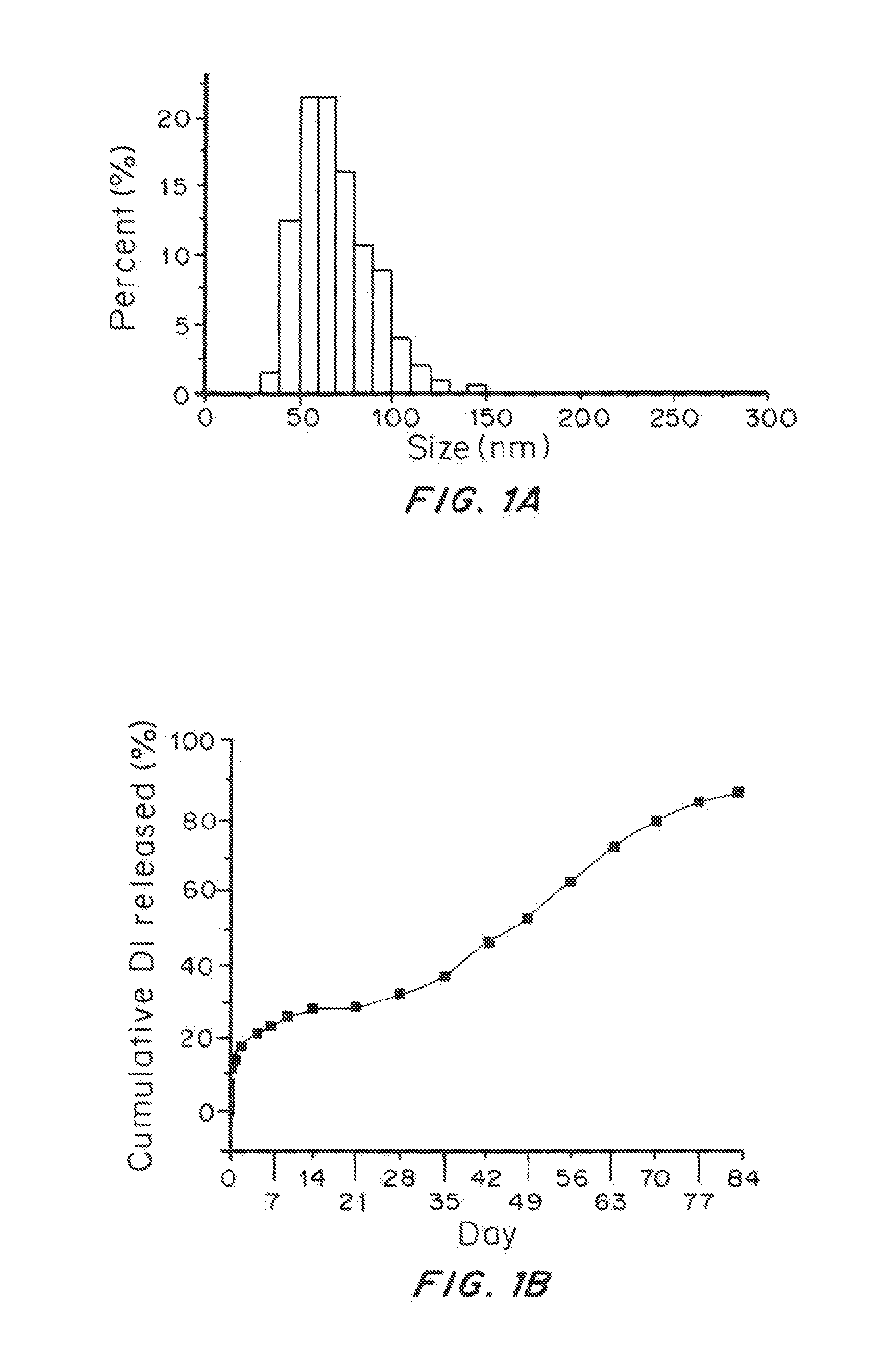
[0080]Scanning electron microscopy (SEM) showed that nanoparticles isolated using this protocol were 74±18 nm in diameter and morphologically spherica...

example 2

Synthesis of Brain Penetrating NPs with Water-Miscible Solvent

[0081]Materials and Methods

[0082]Partially water-miscible organic solvents, such as benzyl alcohol, butyl lactate, and ethyl acetate (EA), allow nanoparticle formulation through an emulsion-diffusion mechanism and are able to produce smaller nanoparticles than water-immiscible solvents such as DCM. TEA was chosen because of its low toxicity. The same method otherwise was used as in Example 1.

[0083]Results

[0084]Nanoparticles synthesized using EA as solvent instead of DCM were 65±16 nm in diameter and morphologically spherical. The yield was improved with EA: 44%±3%.

example 3

Cryoprotection of Brain-Penetrating PLGA Nanoparticles

[0085]Materials and Methods

[0086]Lyophilization is a technique commonly used to stabilize nanoparticles for long-term storage. However, lyophilization can also cause nanoparticles to aggregate, making them difficult to resuspend in an aqueous solution. Furthermore, particle aggregation, if it did occur, could complicate CED infusion and restrict penetration in the brain. To reduce aggregation, the FDA-approved disaccharide trehalose was added as an excipient, at a ratio of 0.5:1 (trehalose:nanoparticles) by mass immediately prior to lyophilization.

[0087]Results

[0088]The addition of trehalose did not alter nanoparticle size, morphology, or yield. SEM images demonstrated that trehalose enhanced the separation of nanoparticles from one another when compared to nanoparticles lyophilized without trehalose. Reconstitution of cryoprotected brain-penetrating nanoparticles resulted in a homogenous solution, while reconstitution of nanopar...


PUM

PropertyMeasurementUnit
diameteraaaaaaaaaa
sizeaaaaaaaaaa
diameteraaaaaaaaaa

Abstract

Brain-penetrating polymeric nanoparticles that can be loaded with drugs and are optimized for intracranial convection-enhanced delivery (CED) have been developed. In the preferred embodiment, these are loaded with FDA-approved compounds, identified through library screening to target brain cancer stem cells (BSCSs). The particles are formed by emulsifying a polymer-drug solution, then removing solvent and centrifuging at a first force to remove the larger particles, then collecting the smaller particles using a second higher force to sediment the smaller particles having a diameter of less than 100 nm, more preferably less than 90 nanometers average diameter, able to penetrate brain interstitial spaces.

Description

STATEMENT REGARDING FEDERALLY SPONSORED RESEARCH OR DEVELOPMENT[0001]This invention was made with Government Support under grant CA149128 awarded to Mark Saltzman by the National Institutes of Health. The Government has certain rights in the invention.FIELD OF THE INVENTION[0002]This application is generally in the field of drug delivery, and more specifically delivery of chemotherapeutics to the brain, especially for the treatment of glioblastoma.BACKGROUND OF THE INVENTION[0003]Of the approximately 40,000 people diagnosed with primary brain tumors in the United States each year, an estimated 15,000 have glioblastoma multiforme (GBM), a WHO grade IV malignant glioma (Mrugala, et al Nat Clin Pract Oncol 5, 476-486 (2008)). Despite considerable research efforts, the prognosis for GBM remains poor: median survival with standard-of-care therapy (surgery, systemic chemotherapy with temozolomide, and radiation) is 14.6 months (Stupp et al., N Engl J Med 352, 987-996 (2005)) and five-year...

Claims


Application Information

Patent Timeline
Patent Type & Authority Applications(United States)
IPC IPC(8): A61K9/51A61K31/428A61K9/00A61K31/337
CPCA61K9/513A61K9/0085A61K31/428A61K31/337A61K9/5153A61K31/17A61K31/4745A61K31/495A61K9/0019A61K9/19
Inventor ZHOU, JIANGBINGPATEL, TORAL R.PIEPMEIER, JOSEPH M.SALTZMAN, WILLIAM MARK
Owner YALE UNIV
Why Eureka
Try Eureka