Extracellular vesicle separation method, colloidal particle and preparation method thereof

a technology of colloidal particles and separation methods, which is applied in the field of separation methods, colloidal particles and preparation methods of extracellular vesicles, can solve the problems of low recovery rate, time saving, and none of them meet the requirements of high recovery rate, time saving, and high purity at the same time. , the effect of high recovery

Pending Publication Date: 2021-07-01
IND TECH RES INST
View PDF0 Cites 0 Cited by
  • Summary
  • Abstract
  • Description
  • Claims
  • Application Information

AI Technical Summary

Benefits of technology

[0006]The disclosure provides an extracellular vesicle separation method, a colloidal particle and a preparation method thereof, in which a separation column is filled with

Problems solved by technology

Although high-purity extracellular vesicles can be obtained, the recovery rate is very low.
In recent years, many extracellular vesicle separation methods or devices, such as polyethylene glycol precipitation method, immunomagnetic bead purif

Method used


Image

  • Extracellular vesicle separation method, colloidal particle and preparation method thereof
  • Extracellular vesicle separation method, colloidal particle and preparation method thereof
  • Extracellular vesicle separation method, colloidal particle and preparation method thereof

Examples


experimental example

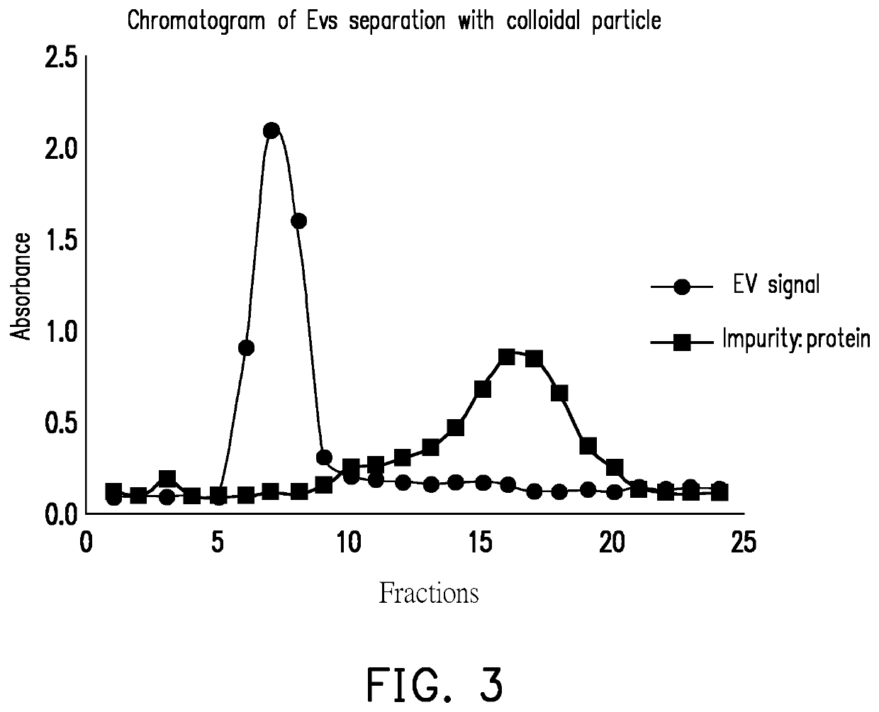
[0039]In order to prove that the colloidal particle of the disclosure effectively separates extracellular vesicles, the following Experimental Examples are particularly provided.

experimental example 1

idal Particle

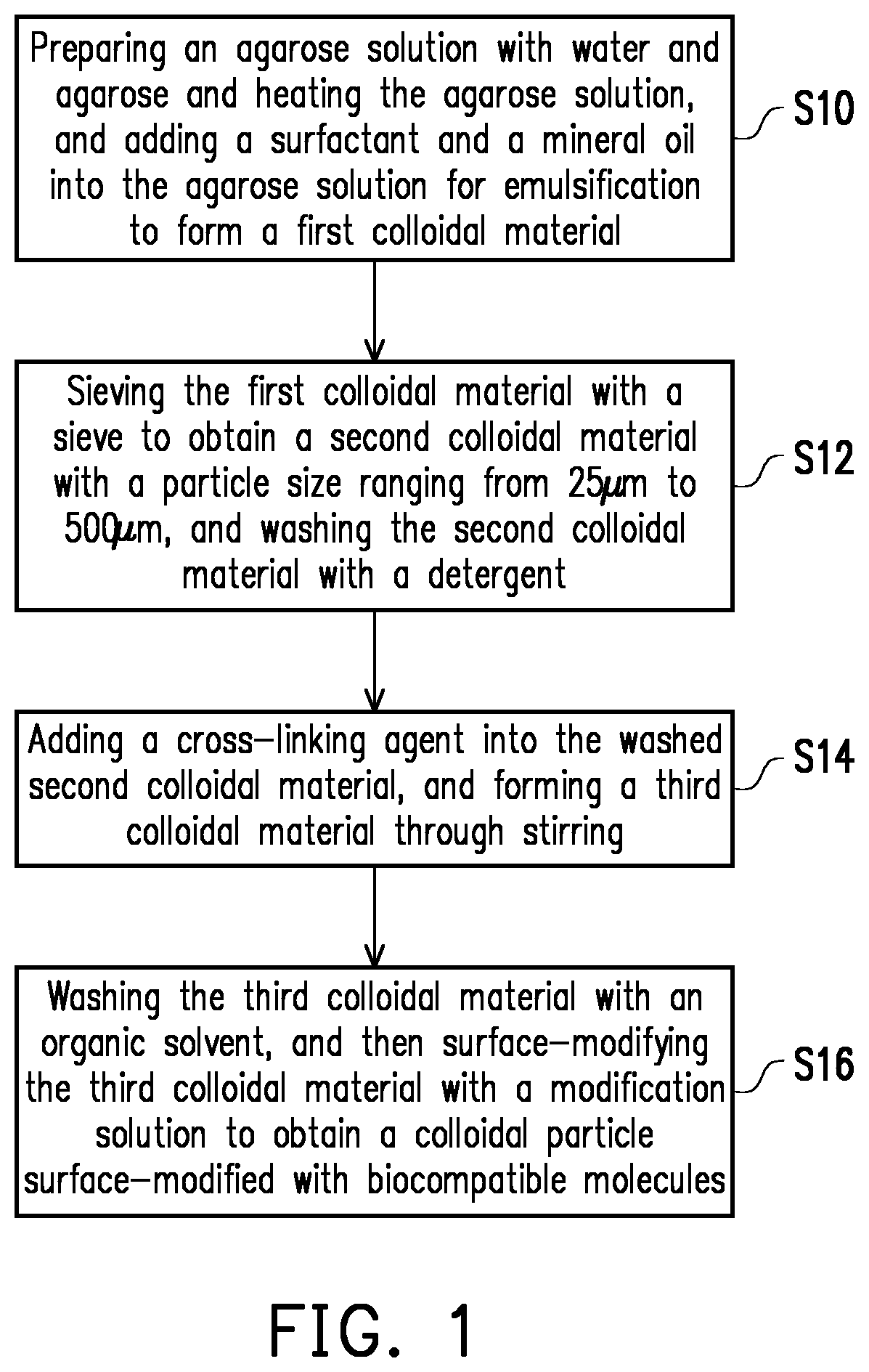
[0040](1) Preparation of an Agarose Particle

[0041]80 ml of water was mixed with 4.8 g of agarose, heated in a microwave oven or an autoclave, and heated at 90° C. for 2 hours for completely dissolved, so as to prepare a 6% agarose solution. Then, 13.5 ml of Span 80, 1.5 ml of Triton X-100, and 85 ml of a mineral oil were mixed evenly and heated to 95° C., then the 6% agarose solution was added thereinto for 30 minutes for emulsification. After the reaction was completed, the reaction temperature was lowered to room temperature. Afterwards, oil stains were washed off with a 1% SDS aqueous solution, and an agarose particle at 74 μm to 250 μm was collected through filtration. The particle was washed with 100 ml of n-hexane and 80 ml of 20% alcohol three times, and then was washed with 80 ml of double distilled water three times. 40 ml of 6% agarose particle (equivalent to the above-mentioned second colloidal material) at 74 μm to 250 μm were obtained.

[0042](2) Cross-Linkin...

experimental example 2

Purification of Cancer Cell Culture Medium

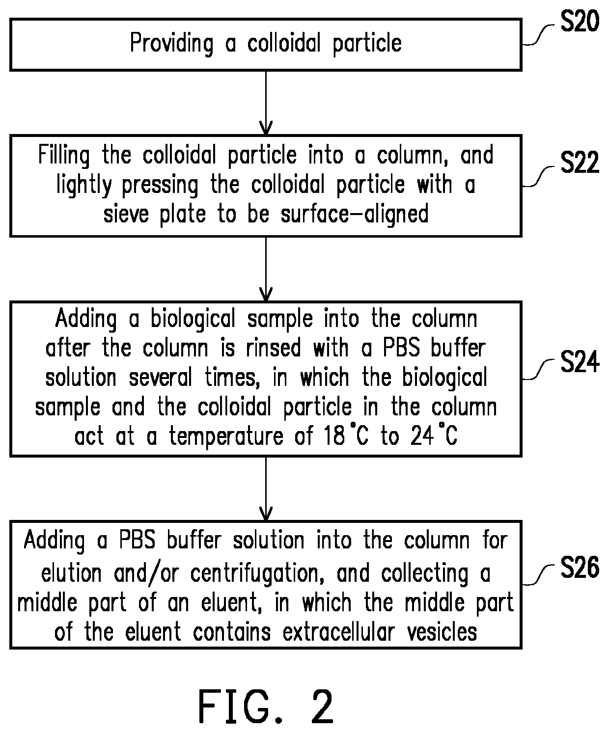
[0046](1) Filling Chromatography Column

[0047]A bottom of a column with a diameter of 8 mm, a length of 70 mm, and a total volume of about 3.5 ml was placed into a sieve plate, and was first rinsed with a PBS buffer solution. Then, the colloidal particle synthesized in Experimental Example 1 was drawn up and filled into the column with a dropper. After the colloidal particle sank to the height (70 mm) to be filled, the surface was aligned using the sieve plate to complete the colloid particle filling, and then the column was eluted with 10 ml of a PBS buffer solution to be balanced.

[0048](2) Extracellular Vesicle Separation

[0049]100 μl of a culture medium of cell line SKBr3 of human breast cancer cells was injected from the top of the column. When the fluid level dropped to or below the sieve plate, the sample injection was completed. The sample was eluted with a PBS buffer solution, and the early, the middle, and the late parts of the eluent...


PUM

PropertyMeasurementUnit
Temperatureaaaaaaaaaa

Abstract

An extracellular vesicle separation method, a colloidal particle, and a preparation method thereof are provided. The colloidal particle is used for extracellular vesicle separation, and includes 2 wt % to 6 wt % of agarose. The colloidal particle has a particle size of 25 μm to 500 μm, and is surface-modified with biocompatible molecules. The biocompatible molecules include sodium carboxymethyl cellulose (CMC), methyl cellulose (MC), glycine, aspartic acid, glutamic acid, bovine serum albumin (BSA), fetal bovine serum (FBS), or a combination thereof.

Description

CROSS-REFERENCE TO RELATED APPLICATION[0001]This application claims the priority benefit of U.S. Provisional Application No. 62 / 954,670, filed on Dec. 30, 2019. The entirety of the above-mentioned patent application is hereby incorporated by reference herein and made a part of this specification.BACKGROUNDTechnical Field[0002]The disclosure relates to a separation method, a colloidal particle and a preparation method thereof, and particularly relates to a separation method, a colloidal particle and a preparation method thereof regarding extracellular vesicles.Description of Related Art[0003]Extracellular vesicles (EVs), a collective term for referring to various vesicles having a membrane structure, have a diameter ranging from 30 nm to 40 nm to 8 μm to 9 μm, are secreted by various types of cells to the surrounding environment, and may be found their present in blood, urine, saliva, and other body fluids. At present, the functionality of the extracellular vesicles has not been comp...

Claims


Application Information

Patent Timeline
IPC IPC(8): B01J13/18B01J13/20B01J13/04B01D15/42B01D15/38B01D15/20
CPCB01J13/185B01J13/20B01J13/04B01D2221/10B01D15/38B01D15/206B01D15/426C08B37/0039C08L5/12B01D15/265
Inventor YEN, YU-FANGCHEN, CHIEN-ANCHEN, CHENG-TAI
Owner IND TECH RES INST
Why Eureka
Try Eureka