A micro physiological model for neuronal and muscular diseases and disorders

a neuronal and muscular disease and micro-physiological model technology, applied in the field of micro-physiological models for neuronal and muscular diseases and disorders, can solve the problems of lack of effectiveness of existing drugs and severely limited the scope of drug development, and achieve the effect of improving a biochemical

Pending Publication Date: 2022-06-02
MASSACHUSETTS INST OF TECH
View PDF0 Cites 0 Cited by
  • Summary
  • Abstract
  • Description
  • Claims
  • Application Information

AI Technical Summary

Benefits of technology

[0022]In some embodiments, the compound improves a biochemical, cellular, molecular, or morphological characteristic of the neuronal cells, muscle bundle, or combination thereof, and may be selected as a candidate ...

Problems solved by technology

However, existing drugs lack effectiveness and only a limited number of drug candidates have been evaluated for the treatment of ALS.
These traditional in vitro culture methods have...

Method used


Image

  • A micro physiological model for neuronal and muscular diseases and disorders
  • A micro physiological model for neuronal and muscular diseases and disorders
  • A micro physiological model for neuronal and muscular diseases and disorders

Examples


example 1

rformance and Characterization of MN Spheroids in Microfluidic Devices

Materials and Methods

[0192]Microfluidic Device Fabrication

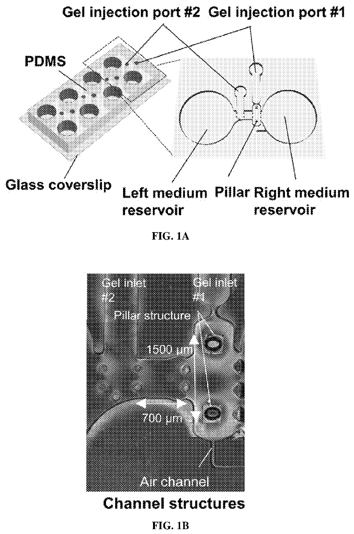
[0193]The mold fabrication process was similar to that previously reported (Uzel et al., Sci. Adv. 2, e1501429 (2016)). Briefly, device structures were designed using Solidworks (Dassault Systémes SolidWorks Corporation), and the patterns were transferred onto a transparency mask using high-resolution printing (FineLine Imaging). Silicon wafers were then fabricated by photolithography using typical SU-8 photoresist techniques. All the mold surfaces were treated with (tridecafluoro-1,1,2,2-tetrahydrooctyl)-1-trichlorosilane for at least 2 hours. For the main microfluidic section, PDMS and curing reagents (Ellsworth Adhesives) were mixed in a 10:1 ratio, poured into the SU-8 mold, and cured at 80° C. for at least 6 hours. The pillar head was then attached to the base of the pillar. The pillar head consisted of 250 μm by 250 μm squares of 25-μm-thin PDMS (obta...

example 2

on of a 3D Motor Unit Model and NMJs in Microfluidic Devices

Materials and Methods

[0211]Formation of 3D Motor Units

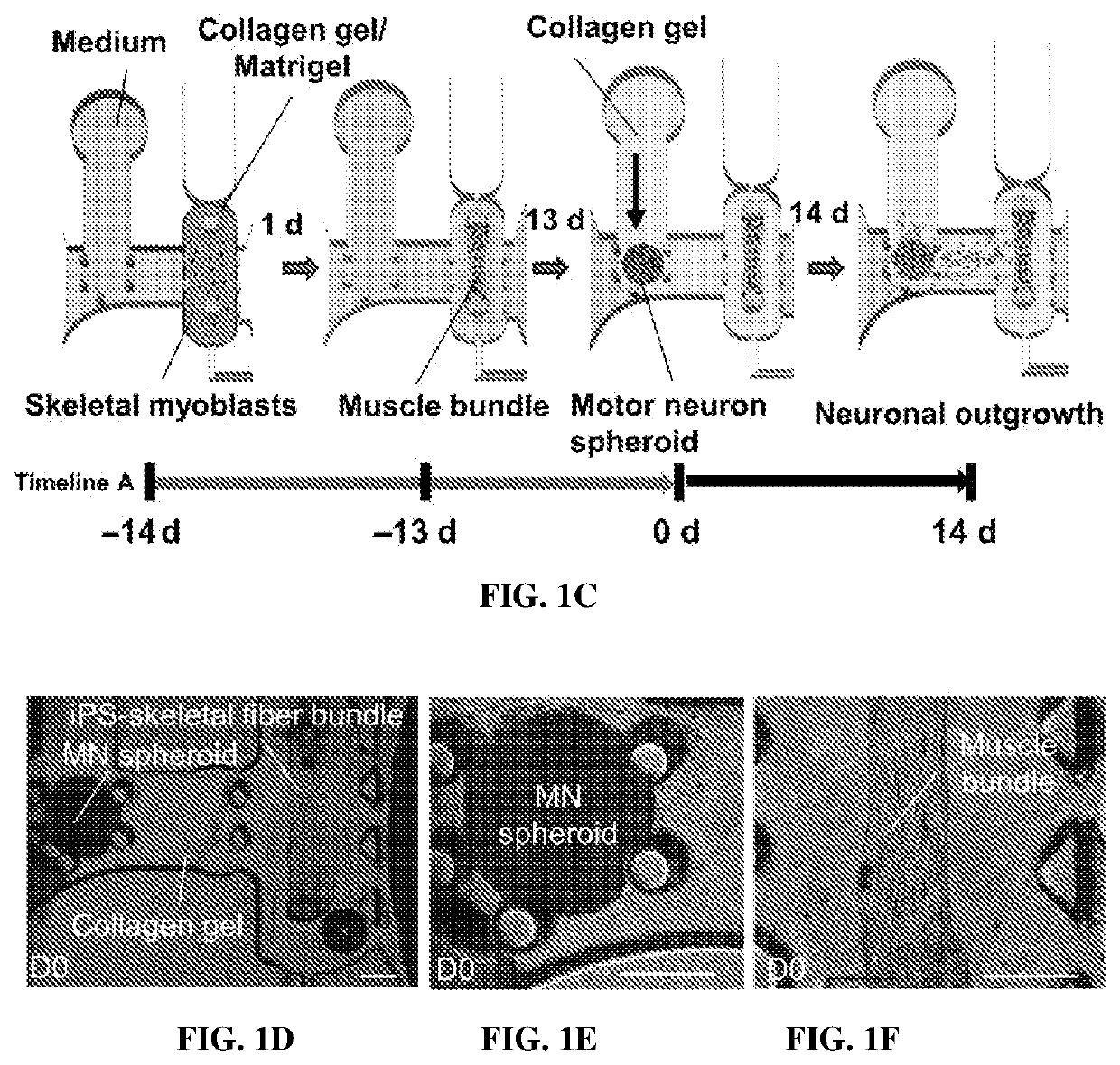
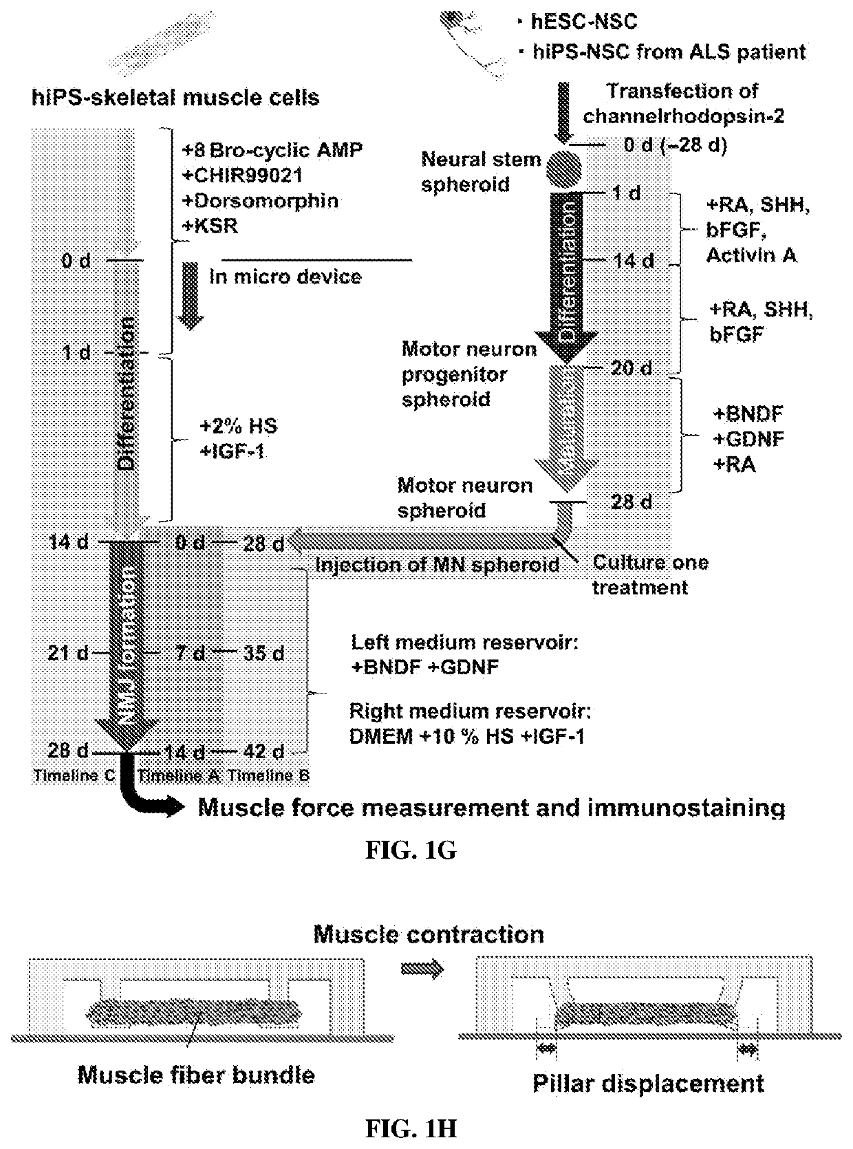
[0212]To avoid single-cell attachment to the glass bottom, the microfluidic device was incubated with 4% Pluronic F-127 (Sigma) in the incubator for 1 hour, rinsed with distilled water, and dried. Human iPSC-skeletal muscle cells were injected into the right compartment of the microfluidic device with a mixture of porcine skin type I collagen gel (Nitta Gelatin, 2.4 mg / ml) and 10% MATRIGEL® (BD Biosciences) via gel injection port no. 1 (FIGS. 1A and 1B). A 3D muscle fiber bundle was formed within 24 hours, with both sides of the muscle attached to the pillar structures. The muscle fiber bundle was then cultured and differentiated using 2% horse serum and IGF-1. After 13 days of differentiation, predifferentiated MN spheroids (normal and ALS) were injected into the left compartment (FIG. 1B) via gel injection port no. 2 with collagen without MATRIGEL®. For the cocultured ...

example 3

icity Induced by Excess Glutamic Acid Treatment Mimics ALS Pathogenesis

[0223]Glutamate (glutamic acid) is widely recognized as one of the major factors contributing to ALS pathogenesis (Blasco et al., Curr. Med. Chem. 21, 3551-3575 (2014)). Prolonged excitation caused by excess glutamate is known to harm neurons (Plaitakis, Ann. Neurol. 28, 3-8 (1990)). Normally, glutamic acid is cleared from neurons via several pathways [e.g., neuron-neuron and neuron-astrocyte junctions (Seifert et al., Nat. Rev. Neurosci. 7, 194-206 (2006))]; however, patients with ALS are unable to remove glutamate and therefore suffer from MN excitotoxicity because of their low threshold of activation, resulting in neuron death. The prevention of MN excitotoxicity is thought to be the mechanism of action of riluzole, one of the symptomatic ALS treatments.

[0224]To mimic this, a high concentration of glutamic acid was added to an established 3D motor unit model. After 7 days of coculture, glutamic acid was added ...


PUM

PropertyMeasurementUnit
Lengthaaaaaaaaaa
Lengthaaaaaaaaaa
Lengthaaaaaaaaaa

Abstract

Microfluidic devices with neuronal cells, muscle cells, and optionally other cell types co-cultured therein are provided. Typically one or more the cells has a mutation that contributes to or causes a neuronal or muscular disease or disorder. For example, in some embodiments, one or more of the cultured cells are derived from a subject with a neuronal or muscular disease or disorder. The microfluidic device can facilitate formation of a 3D motor unit and a neuromuscular junction in vitro, and be used to monitor the molecular, biochemical, cellular, and morphological differences in the formation of such structures by healthy and diseased cells, and for testing compounds, dosages of compounds, dosing regimes, and combinations thereof, that may improve or worsen their formation. An exemplary combination drug therapy identified in this way is also provided.

Description

STATEMENT REGARDING FEDERALLY SPONSORED RESEARCH[0001]This invention was made with government support under Grant CBET-0939511 awarded by the National Science Foundation. The government has certain rights in the invention.FIELD OF THE INVENTION[0002]The field of the invention generally relates to devices, systems, compositions, and methods for monitoring the development of heathy and diseased neuronal motor units and neuromuscular junctions, and testing the effects compounds thereon.BACKGROUND OF THE INVENTION[0003]Amyotrophic lateral sclerosis (ALS), also known as Lou Gehrig's disease, is a neurodegenerative disease in which motor neuron (MN) loss in both the spinal cord and motor cortex causes progressive paralysis, muscle atrophy, and death (Ling et al., Neuron 79, 416-438 (2013), Hardiman et al., Nat. Rev. Neurol. 7, 639-649 (2011)). The U.S. Centers for Disease Control and Prevention estimates that 12,000 to 15,000 people in the United States have ALS (Mehta et al., Surveill. S...

Claims


Application Information

Patent Timeline
IPC IPC(8): C12M3/06C12M1/34C12M1/36C12N5/0793C12M1/26G01N33/50A61K45/06C12M1/12C12M1/00A61K31/436A61K31/4706A61K31/496
CPCC12M23/16C12M41/40C12M41/48C12N5/0619C12M33/04A61K31/496A61K45/06C12M23/04C12M23/34A61K31/436A61K31/4706G01N33/5058G01N33/5061B01L2300/0663B01L2300/0609B01L3/502761B01L2200/0663B01L3/5085
Inventor KAMM, ROGER DALEOSAKI, TATSUYA
Owner MASSACHUSETTS INST OF TECH
Why Eureka
Try Eureka