Cell motility and chemotaxis test device and methods of using same

Inactive Publication Date: 2006-01-03
SURFACE LOGIX INC
View PDF7 Cites 23 Cited by
  • Summary
  • Abstract
  • Description
  • Claims
  • Application Information

AI Technical Summary

Benefits of technology

[0033]The present invention provide a method of monitoring haptotaxis. The method includes providing a device for monitoring haptotaxis having a housing defining a chamber. The chamber includes a first well region including at least one first well, the first well region configured to receive a test agent therein and further including biomolecules immobilized therein; a second well region including at least one second well, the second well region configured to receive a sample comprising cells therein and further being horizontally offset with respect to the first well region in a tes

Problems solved by technology

Conversely, lack of capillary production can contribute to tissue death in cardiac muscle after, for example, a heart attack.
Assays employing transwells require a labor-intensive protocol that is not readily adaptable to high-throughput screening and processing.
Because of the configuration of a transwell system, it is difficult to integrate with existing robotic liquid handling systems and automatic image acquisition systems.
Therefore, much of the screening and processing, such as counting cells, is done manually which is a time-consuming, tedious, and expensive process.
Specifically, due to the time and expense associated with examining an entire filter, only randomly selected representative areas may be counted.
Transwell-based assays have intrinsic limitations imposed by the thin membranes utilized in transwell systems.
If a cell chemotaxis assay requires the chemotactic gradient to be generated over a long distance (>100-200 μm) and to be stable over at least two hours, currently available transwell assays cannot be satisfactorily performed.
Notwithstanding the above, perhaps the most significant disadvantage of transwells is the lack of real-time observation of chemotaxis and chemoinvasion.
In particular, the changes in cell morphology during chemotaxis cannot be observed in real-time with the use of transwells.
Consequently, once a cell is observed it can no longer be reintroduced into the assay or studied at subsequent periods of exposure to a test agent.
In light of the multiple samples required for each test, in addition to the positive and negative controls required to obtain reliable data, a single chemotaxis assay is can require dozens of filters, each of which needs to be individually examined and counted-an onerous and time-consuming task.
Because of the size

Method used


Image

  • Cell motility and chemotaxis test device and methods of using same
  • Cell motility and chemotaxis test device and methods of using same
  • Cell motility and chemotaxis test device and methods of using same

Examples


example 1

Procedure for Fabrication of Chemoinvasion Device

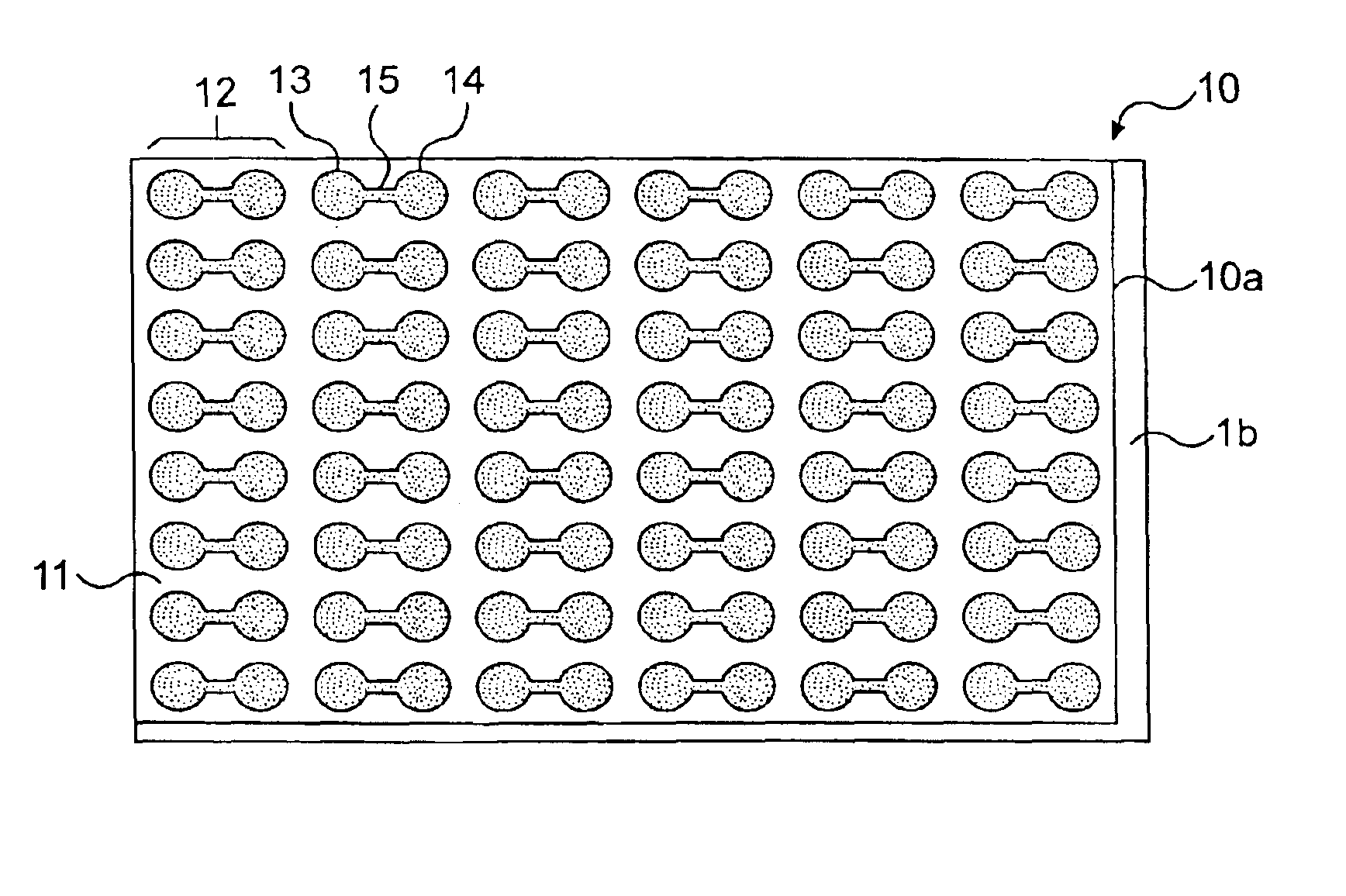
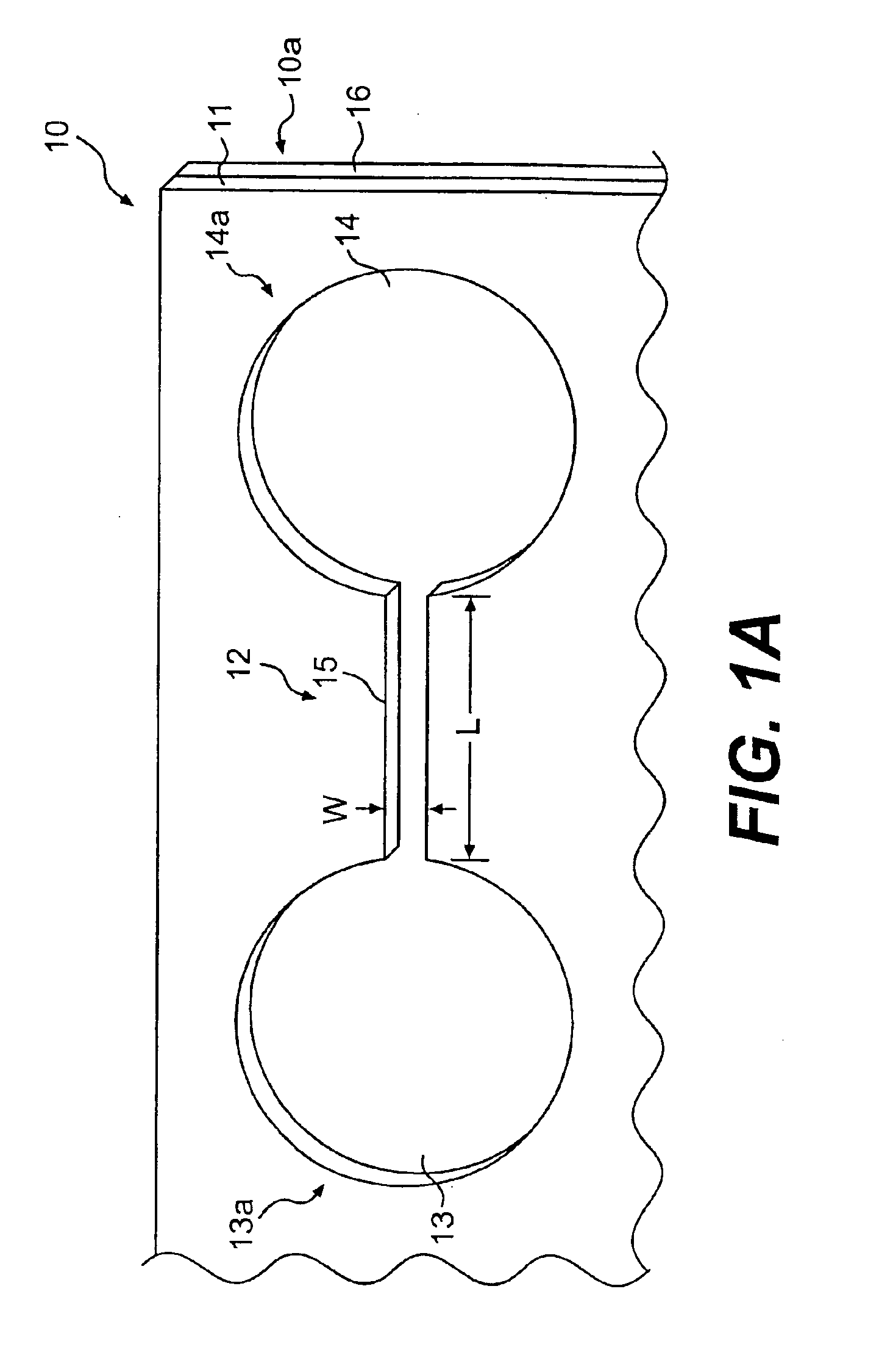
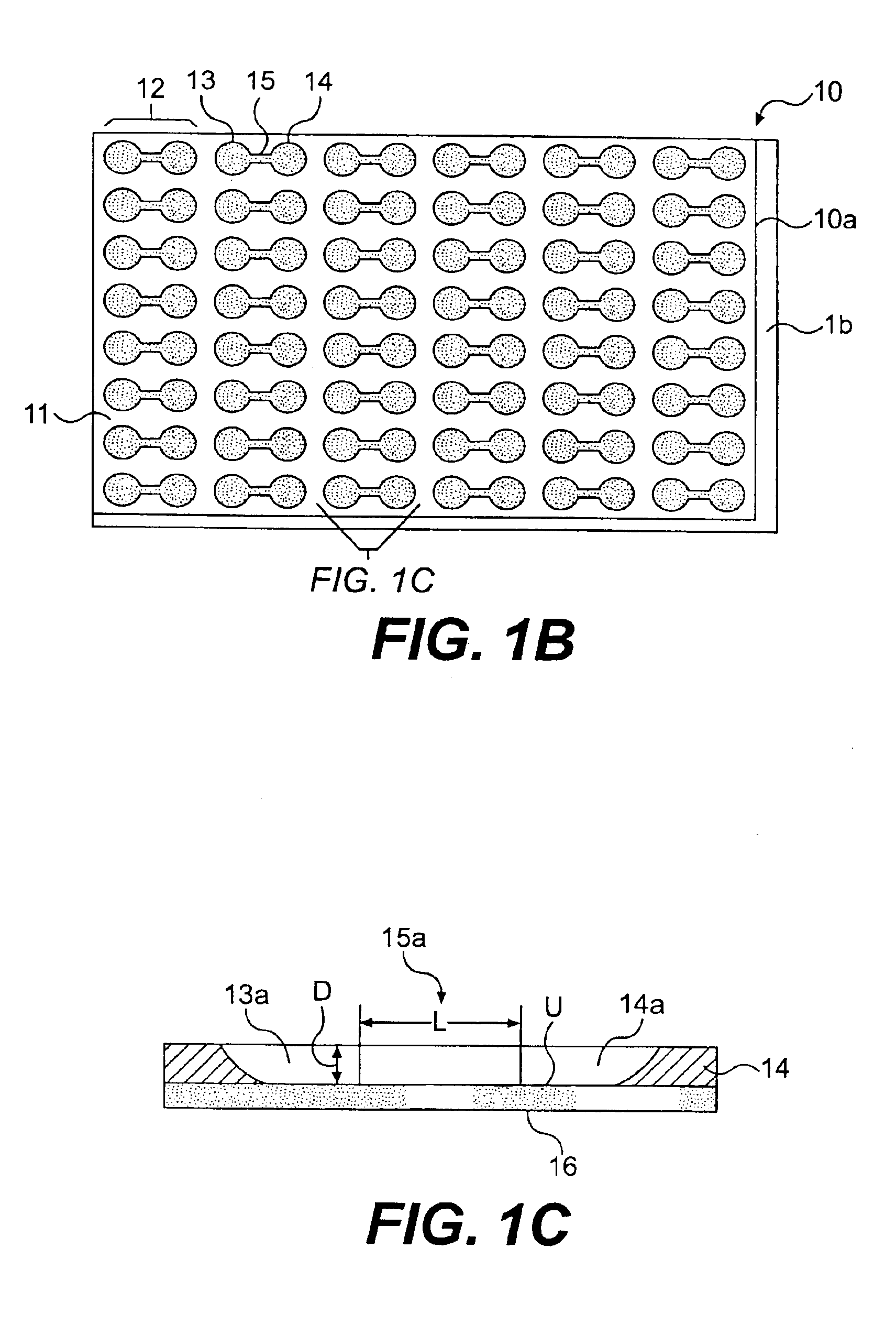
[0145]A silicon wafer (6 inches) is spin coated with photoresist (SU8-50) at 2000 rpm for 45 seconds. After baking the wafer on a hot plate at 115° C. for 10 minutes, the wafer is allowed to cool to room temperature. A mask aligner (EVG620) is used to expose the photoresist film through a photomask. Exposure of 45 seconds is followed by another hard bake at 115° C. for 10 minutes. The silicon wafer is allowed to cool to room temperature for over 30 minutes. The uncrosslinked photoresist is removed using propylene glycol methyl ether acetate (PGMEA). The wafer is dried under a stream of nitrogen, and the patterned photoresist is ready for subsequent processing.

[0146]In one embodiment, the patterned photoresist is spin-coated with another layer of SU8-100 at 1500 rpm for 45 seconds. A mask aligner is used to selectively expose macrofeatures (i.e. wells) of the top member but not expose channel regions connecting the wells and other area...

example 2

Cell Chemoinvasion Assay

Placement of Cells and Test Agent in Chamber

[0148]The first and second wells of a chamber of a top member are filled with phosphate buffered saline solution, PBS. The bottom of the second well may be treated with fibronectin (1 mg / ml) or other extracellular matrix protein for 30 minutes, followed by washing twice with PBS. After aspirating PBS, astrocytoma cells (U87-MG) are plated in 50 μl of freshly warmed medium in the second well (25,000 cells per well of a 24-well plate, in volume of 50 μl of solution per well). The cells deposit through the second well of the chamber, and attach to the bottom of the second well.

[0149]Cells are left to attach and spread in the second well overnight in a 37° C. incubator. At the start of the experiment, the cell medium is exchanged for fresh serum-free medium. 10 μg of bFGF (basic fibroblast growth factor) per ml of medium is added to the first well of each chamber.

Image Acquisition and Data Analysis

[0150]Digital Images a...

example 3

Cell Chemoinvasion Inhibition Assay Using Solution Gradient

Placement of Cells and Test Agent in Chambers

[0151]With respect to three chambers, the wells of each chamber of a top member are filled with PBS. The bottom of the second wells may be treated with fibronectin (1 mg / ml) or other extracellular matrix protein for 30 minutes, followed by washing twice with PBS. After aspirating PBS, U87-MG cells are plated in 50 μl of freshly warmed medium in the second wells (10,000 cells per well of a 24-well plate, in volume of 50 μl of medium per well). The cells deposit through the second wells of each chamber, and adhere to the bottom of the second wells.

[0152]Cells are left to attach and spread in the second wells overnight in a 37° C. incubator. At the start of the experiment, the cell medium is exchanged for fresh serum-free medium or 1% serum. 1 μg of bFGF (basic fibroblast growth factor) per ml of medium is added to the first wells of the chamber. A solution gradient is allowed to for...


PUM


Abstract

The present invention discloses a device for monitoring chemotaxis or chemoinvasion including a housing comprising: a support member and a top member, the top member mounted to the support member by being placed in substantially fluid-tight conformal contact with the support member. The support member and the top member are configured such that they together define a discrete chamber adapted to allow a monitoring of chemotaxis or chemoinvasion therein. The discrete chamber includes a first well region including at least one first well, the at least one first well configured to received a test agent therein; a second well region including at least one second well, the at least one second well configured to receive a sample comprising cells therein; and a channel region including at least one channel connecting the first well region and the second well region with one another. The second well region is preferably horizontally offset with respect to the first well region in a test orientation of the device.

Description

RELATED APPLICATIONS[0001]This application is a continuation-in-part of U.S. application Ser. No. 10 / 097,351 filed Mar. 15, 2002; U.S. application Ser. No. 10 / 097,329 filed Mar. 15,2002; U.S. application Ser. No. 10 / 097,302 filed Mar. 15, 2002; U.S. application Ser. No. 10 / 097,304 filed Mar. 15, 2002 now U.S. Pat. No. 6,818,403; U.S. application Ser. No. 10 / 097,322 filed Mar. 15, 2002 now U.S. Pat. No. 6,811,968; U.S. application Ser. No. 10 / 097,306 filed Mar. 15, 2002, all of which are herein incorporated by reference in their entirety. This application also claims priority to both Provisional U.S. Application No. 60 / 363,354, filed on Mar. 12, 2002, and Provisional U.S. Application No. 60 / 373,783, filed on Apr. 24, 2002, all of which are herein incorporated reference in their entirety.TECHNICAL FIELD[0002]The present invention relates generally to device for monitoring cell motility and chemotaxis.BACKGROUND ART[0003]Test devices, such as those used in chemotaxis, haptotaxis and ch...

Claims


Application Information

Patent Timeline
IPC IPC(8): C12N5/08B01D57/02B01L3/00C12M1/34C12Q1/20
CPCB01L3/5025B01L2400/086B01L2300/0829
Inventor KIM, ENOCHKIRK, GREGORYBROWN, MATTHEWOSTUNI, EMANUELE
Owner SURFACE LOGIX INC
Why Eureka
Try Eureka