

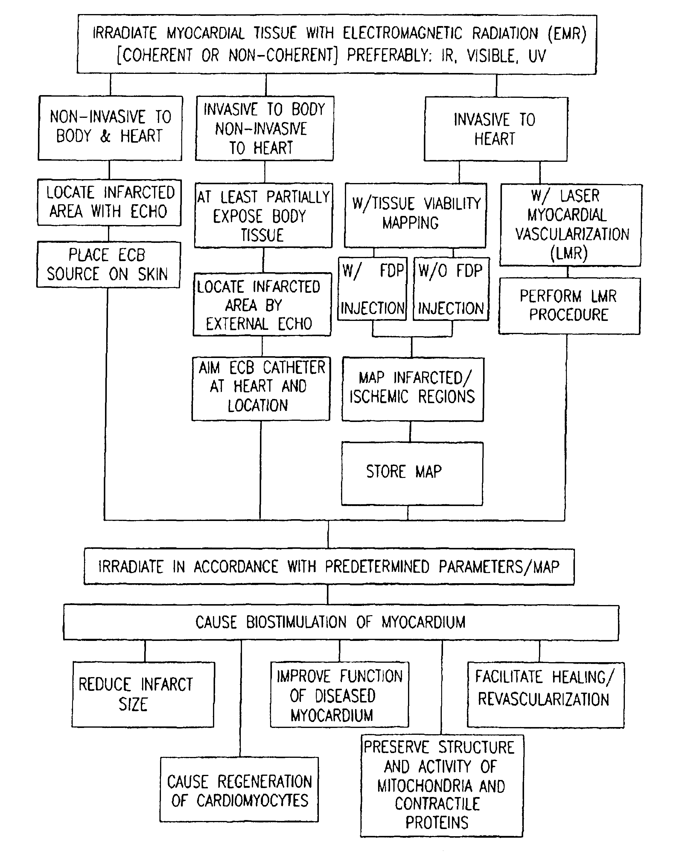
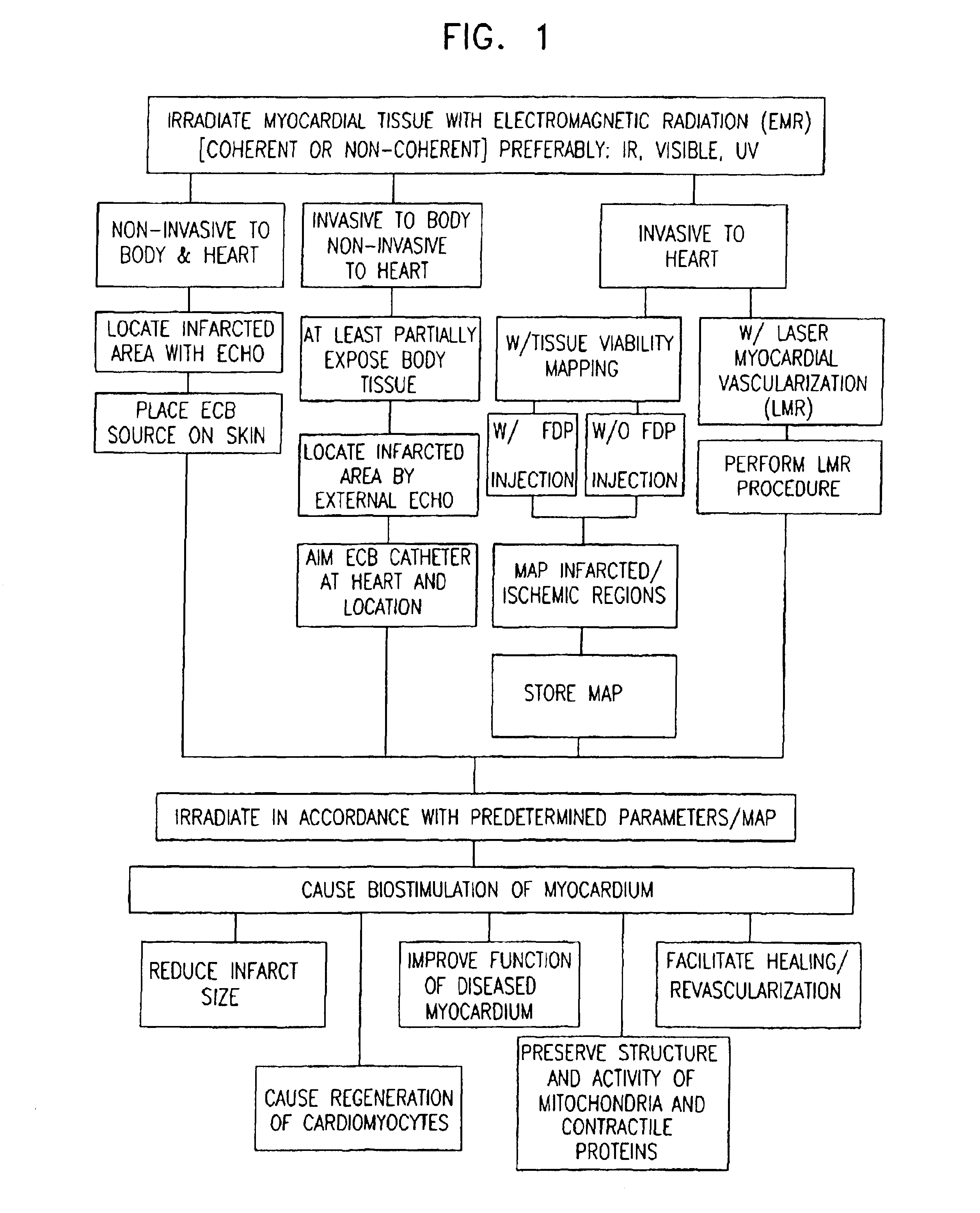
Apparatus for providing electromagnetic biostimulation of tissue using optics and echo imaging
a tissue biostimulation and electromagnetic technology, applied in the field of cardiac treatment, can solve the problems of increasing increasing the cell mass and not the number of cells, and high cost of procedures, so as to improve the overall viability of the implant, improve the survival rate of the implant, and improve the overall viability
- Summary
- Abstract
- Description
- Claims
- Application Information
AI Technical Summary
Benefits of technology
Problems solved by technology
Method used
Image

Examples
experiment i
[0112
[0113]Forty-nine male Sprague-Dawley ether-anesthetized rats, 6-8 weeks old (220-260 g body weight) were used for the experiments. Two-cm incisions to the left of and parallel to the sternum were made; the fifth and sixth ribs were separated with a clamp, and the hearts were exteriorized by applying pressure to the lateral aspects of the thoracic cage. The left coronary arteries were then occluded 2-3 mm from their origins with single sutures. The chests were closed and the rats allowed to recover. This procedure induces myocardial infarction (MI) and chronic ischemia.
[0114]Twenty-three rats were irradiated immediately following surgery using a diode laser (wavelength 780 nm; 38 mW). The irradiation was performed by placing and gently pressing a laser source tip onto the exposed intercostal muscles approximately in a perpendicular direction to the lateral wall of the left ventricle. The laser source was moved at a constant velocity for 2 min to cover the chest area from the ste...
experiment ii
[0121
[0122]Heart infarction was induced in 22 rats as described in Experiment I. Ten rats served as control and 12 rats underwent a different laser irradiation from that in Experiment I. The laser used was a diode laser with power of 47 mW and wavelength of 830 nm and irradiation was performed as described in Experiment I, but for 5 min immediately following induction of infarction. On the third and fourth day post-infarction, the rats underwent a further two sessions of laser irradiation under the same conditions as above. The rats were sacrificed after 21 days and the infarct size calculated as in Experiment I. The infarct size of control rats was 31±7%, whereas in the laser irradiated rats it comprised only 5.5±1.2% [significantly (p<0.001) lower than control]. The infarct size of the laser irradiated rats was also significantly smaller (p<0.05) than that of the laser irradiated rats in Experiment I.
[0123]From the results of Experiment II, it is seen that in various wavelengths (...
experiment iii
[0124
[0125]Heart attacks were induced in another group of 21 rats as described in Experiment I. Ten rats served as control and 11 rats underwent the laser treatment as described in Experiment I. After 21 days post induction of heart attack, the hearts were removed and connected to a Langendorff apparatus in an artificial physiological solution. They were then injected with TTC (triphenyltetrazolium chloride) through the coronary arteries. This solution stains “healthy” uninjured heart cells with red and injured scar tissue in the heart with a pale yellow color. The hearts were then fixed in neutral buffered formalin and sectioned transversely. Quantitative morphometric measurements and statistics were performed as in Experiment I.
[0126]The infarct size (injured pale yellow area as percent of total area of the left ventricle) was 32±5% in the control group which was significantly (P<0.01) higher than laser irradiated group which comprised 8±3%.
PUM
 Abstract
Description
Claims
Application Information