
Cross-linked heparin coatings and methods

Inactive Publication Date: 2008-12-23
BIOSURFACE ENG TECH
View PDF43 Cites 25 Cited by
  • Summary
  • Abstract
  • Description
  • Claims
  • Application Information

AI Technical Summary

Benefits of technology

[0033]A primary advantage of the present invention is that it provides for coating contacting surfaces of medical devices of complex geometries and surfaces with a durable and low-cost coating that promotes the desired biological or therapeutic effect.

Problems solved by technology

Although vascular grafts constructed using such material are generally clinically successful, there is a tendency for small bore vascular grafts to undergo thrombosis.
A particular disadvantage of adsorbable heparins is that they generally have a short resident time in vivo and are easily leached over a period of hours.
Such chemistries are technically challenging and expensive, with some medical devices are not amenable to the chemistry.
Medical devices made of polytetrafluoroethylene are particularly difficult to adapt to direct heparin conjugation strategies.
Adsorption strategies suffer from the fact that the adsorbed materials are not covalently conjugated and are frequently quickly desorbed from the surface.
This coating is durable, but the method is cumbersome and requires a large number of steps.
In this method the use of multiple layers is cumbersome.
Both U.S. Pat. No. 5,650,234 and U.S. Pat. No. 6,099,562 used glow-discharge methods which are limited to coating “line-of-sight” surfaces, and are not generally applicable to devices with complex geometries, such as tubes or matrices.
The use of a viscous coating results in a thick coating and makes penetration of matrices difficult.
Furthermore, coatings of tubes are prone to variations in thickness along the length of the tubes due to wicking during drying.
The method does not teach a way of allowing the heparin to interact with the surface to prolong resident time either by covalent conjugation to the surface or by adsorption methods.
It can be seen that the foregoing methods do not provide a simple and durable heparin coating, preferably a cross-linked heparin coating, which can be applied by means of simple chemistry steps, and particularly applied to microporous structures such as vascular grafts made from ePTFE.

Method used


Image

  • Cross-linked heparin coatings and methods
  • Cross-linked heparin coatings and methods
  • Cross-linked heparin coatings and methods

Examples


example 1

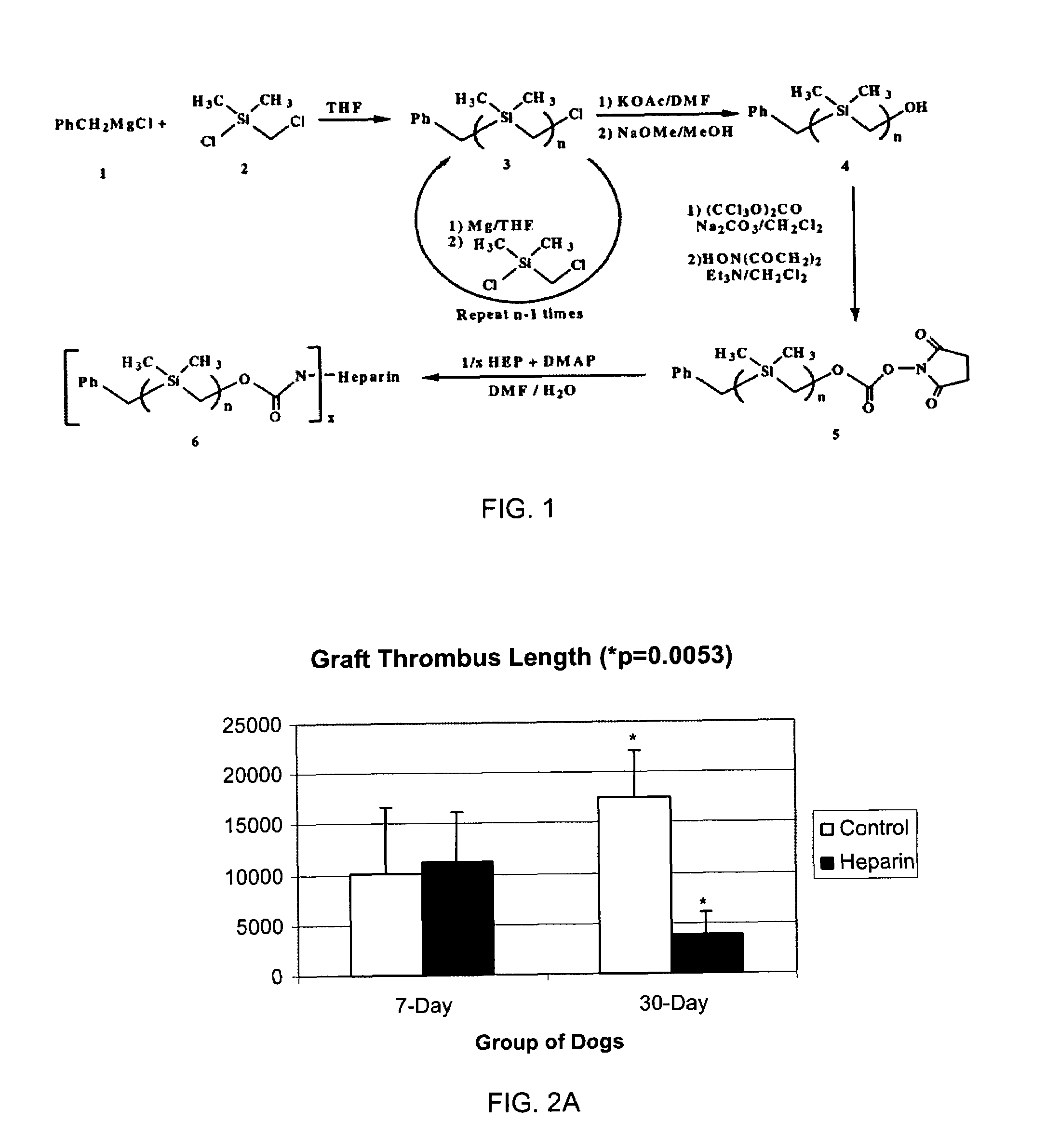
[0077]FIG. 1 depicts a scheme for preparation of silyl-heparin. Treatment of benzylmagnesium chloride 1 with chloro(chloromethyl)-dimethysilane 2 gave benzyl(chloromethyl)dimethysilane 3 (n=1). 3 then underwent a repetitive reaction resulting in chain elongation. For chain elongation, treatment of 3n with magnesium gave the Grignard Reagent which was, in turn, treated with chloro-(chloromethyl)dimethylsilane 2 to give the homologous silyl compound 3 n+1. This Grignard reaction was repeated as needed to obtain the desired chain length for the silyl compound. At the desired chain length, 3 (or 3n) was treated with potassium acetate, followed by trans-esterification of the corresponding acetate with basified methanol to give the alcohol 4. The alcohol 4, when treated with triphosgene, gave the corresponding chloroformate, which on treatment with N-hydroxysuccinimide gave the N-hydroxysuccinimidyl carbonate 5. Heparin was conjugated to 5 in 1:1 DMF / H2O, where DMF is dimethyl formamide, ...

example 2

[0078]CARBOFLO® vascular graft material made of ePTFE with carbon impregnation of approximately 25-30% of the luminal wall by coextrusion (4 mm diameter) was cut to lengths of 8 cm. The grafts were immersed in acetone for 30 minutes at 37° C. until all air bubbles had disappeared and the graft “cleared” indicative of complete wetting. The grafts were transferred to an acetonitrile solution containing 0.34 mg / mL of bis-benzotriazole carbonate (polyethylene glycol) (molecular weight approximately 3.4 K daltons). The grafts were incubated in this solution for 30 minutes at room temperature. The grafts were then transferred to an aqueous solution containing 60% acetonitrile and 1.5% silyl-heparin of Example 1 and allowed to incubate for 1 hour at room temperature. The grafts were then rinsed in four serial changes of acetonitrile using a 15 minute incubation at each rinse. The grafts were then air-dried at 56° C. for at least 2 hours. The presence of heparin on the grafts was confirmed ...

example 3

[0079]CARBOFLO® vascular graft material (4 mm diameter) was cut to lengths of 8 cm. The grafts were immersed in acetone for 30 minutes at 37° C. until all air bubbles had disappeared and the graft “cleared” indicative of complete wetting. The grafts were then transferred to an aqueous solution containing 60% acetonitrile and 1.5% silyl-heparin of Example 1 and allowed to incubate for 30 minutes at room temperature. The grafts were transferred to freshly prepared aqueous solution containing 60% acetonitrile and 0.34 mg / mL of bis-benzotriazole carbonate (polyethylene glycol) (molecular weight approximately 3.4 K daltons). The grafts were incubated in this solution for 1 hour at room temperature. The grafts were then rinsed in four serial changes of acetonitrile using 15 minute incubation at each rinse. The grafts were then air-dried at 56° C. for at least 2 hours. The presence of heparin on the grafts was confirmed by staining with 0.01% aqueous dimethylmethylene blue and by use of a ...


PUM

PropertyMeasurementUnit
Fractionaaaaaaaaaa
Fractionaaaaaaaaaa
Fractionaaaaaaaaaa

Abstract

A thromboresistant coating for a medical device, method of coating and coated medical device, the coating including an in situ cross-linked co-polymer of a cross-linkable biomolecule, preferably an adsorbable biomolecule such as a heparin activity biomolecule with at least one prosthetic hydrophobic unit, and a multifunctional crosslinking agent, such as a bis-variant of polyethylene glycol, polyethylene oxide, or polyethylene glycol, wherein the crosslinking is by means of covalent complexation through at least two functional groups of the multifunctional crosslinking agent.

Description

[0001]This application claims the benefit of the filing of U.S. Provisional patent Application Ser. No. 60 / 432,504, entitled “Cross-Linked Heparin Coatings and Methods”, filed on Dec. 10, 2002, and the specification thereof is incorporated herein by reference.BACKGROUND OF THE INVENTION[0002]1. Field of the Invention (Technical Field)[0003]The present invention relates to methods of making covalently cross-linked coatings for medical devices, and particularly cross-linked complex carbohydrate molecules such as heparin and related molecules, coatings made by such methods and medical devices including such coatings, particularly coated vascular graft devices.[0004]2. Description of Related Art[0005]Note that the following discussion refers to a number of publications by author(s) and year of publication, and that due to recent publication dates certain publications are not to be considered as prior art vis-a-vis the present invention. Discussion of such publications herein is given fo...

Claims


Application Information

Patent Timeline
IPC IPC(8): B05D3/00A61K31/727C08B37/10A61L15/28
CPCA61K31/727A61L27/34A61L27/54A61L33/0029C08B37/0075C08L5/10C08L71/02A61L2300/236A61L2300/42A61L2300/606Y10T428/31663
Inventor ZAMORA, PAUL O.
Owner BIOSURFACE ENG TECH
Features
  • Generate Ideas
  • Intellectual Property
  • Life Sciences
  • Materials
  • Tech Scout
  • Unparalleled Data Quality
  • Higher Quality Content
  • 60% Fewer Hallucinations
Learn More