Method of determining toxicity with three dimensional structures

a three-dimensional structure and toxicity technology, applied in the field of tissue engineering, drug discovery and drug development, can solve the problems of inability to develop candidate drugs, failure to progress out of pre-clinical development of significant number of drug candidates, and inability to achieve the effect of achieving the effect of reducing the risk of cancer, and reducing the number of patients

Active Publication Date: 2010-03-02
CHARLES STARK DRAPER LABORATORY +1
View PDF65 Cites 28 Cited by
  • Summary
  • Abstract
  • Description
  • Claims
  • Application Information

AI Technical Summary

Problems solved by technology

Thus, a significant number of drug candidates in pre-clinical development fail to progress out of this stage due to unacceptable levels of toxicity in test systems.
Failure in development of candidate drugs often occurs when an unacceptable level of toxicity develops.
However, patient responses to drugs are typically more complex and less predictable than responses in the healthy subjects.
While technological advances in cell, molecular, and biochemical assays have made significant strides, a number of problems still exist.
First, in vitro assays using purified or recombinant enzymes and cell cultures provide the first step in determining pharmacologic and toxicologic parameters to be used thereafter in animal models, but are often too simplistic to account for the multifactorial events that occur during drug metabolism in a human system or human organ.
Second, data obtained in animal models cannot be reliably extrapolated to human systems.
Third, many drugs used to treat chronic diseases such as HIV infection or Alzheimer's disease necessitate dosing regimens that are applied over long periods of time, and in some cases, over the lifetime of an individual.
The high attrition rate of drug candidates is a major economic deterrent in the pharmaceutical industry, as drug failure may be identified only after great time and expense are invested.
These failures can be attributed, in part, to a lack of effective pre-clinical models and assay systems.
In addition, many serious diseases (e.g., hepatitis C) lack reliable animal model systems, a problem that has severely handicapped the drug discovery process.

Method used


Image

  • Method of determining toxicity with three dimensional structures
  • Method of determining toxicity with three dimensional structures
  • Method of determining toxicity with three dimensional structures

Examples


example 1

Micromachining of Template to Tissue Engineer Branched Vascularized Channels for Liver Fabrication

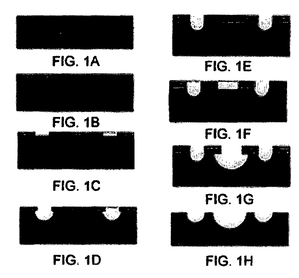
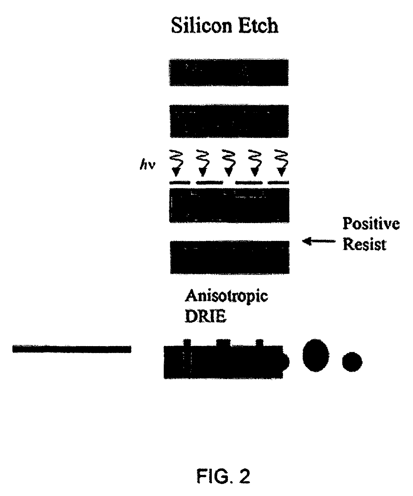
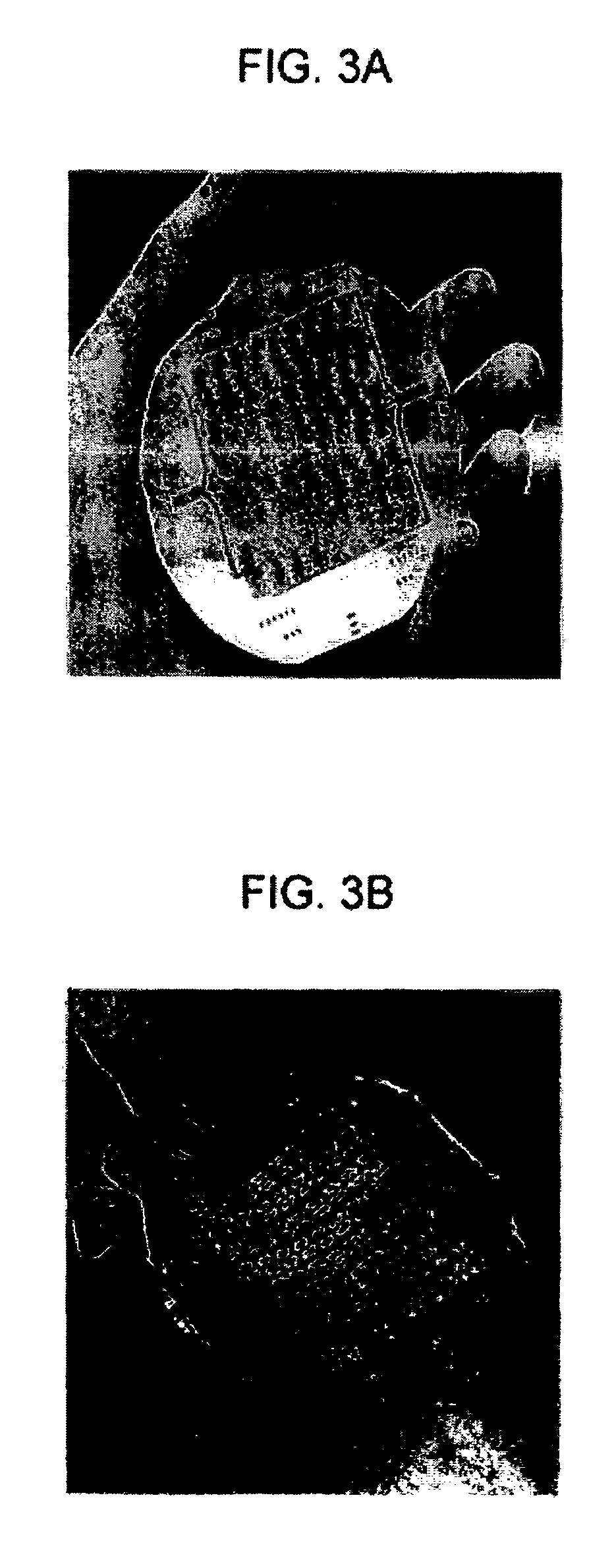
[0344]Micromachining technologies were used on silicon and pyrex surfaces to generate complete vascular systems that can be integrated with engineered tissue before implantation. Trench patterns reminiscent of branched architecture of vascular and capillary networks were etched using standard photolithographic techniques onto silicon and pyrex surfaces to serve as templates. Hepatocytes and endothelial cells were cultured and subsequently lifted as single-cell monolayers from these two dimensional molds. Both cell types were viable and proliferative on these surfaces. In addition, hepatocytes maintained albumin production.

Materials and Methods

Micromachining Techniques

[0345]Templates for the formation of sheets of living vascularized tissue were fabricated utilizing micromachining technology. For the present work, a single level etch was utilized to transfer a vascular network pattern in...

example 2

Endothelialized Microvascular Networks Grown on Micromachined Pyrex® Templates for Tissue Engineering of Vital Organs

[0371]This Example shows the design, modeling, and experimental / computational testing of the endothelialized microvascular matrices grown on micromachined Pyrex® templates.

[0372]Patterns of microvascular networks were etched using microfabrication technologies on Pyrex® wafers. The pattern consisted of 10 generations of bifurcations from a single inflow channel of width 3 mm down to channels of width of 30 μm.

[0373]The channels were then collected to a single outflow. All channels were etched to the same depth of 30 μm. The Pyrex® wafer was sealed to a flat silicone rubber sheet of the same size. Endothelial cells harvested from rat lung were successfully seeded and expanded under continuous flow conditions in this microvascular network. Red blood cells harvested from rat were heparinized and perfused into the endothelialized channels, and successfully collected at th...

example 3

Microfluidics Device for Tissue Engineering Microvasculature

Endothelial Cell Culture

[0377]In this Example, the fabrication of the microfluidic mold, in vitro seeding, and extended cell culture in the mold is demonstrated. Capillary networks were fabricated in biocompatible PDMS, sterilized, coated with cell adhesion molecules, and seeded with cells. Cell-containing molds were then connected to a closed-loop bioreactor for long-term culture. Continuous-flow culture of endothelial cells for up to 4 weeks without occlusion or contamination was achieved.

[0378]Traditional soft lithography microfluidics were used as a prototype matrix. These cell-containing microfluidics are capable of supporting long-term culture in vitro, because in vitro expansion of cells prior to implantation can take several weeks. The prototype matrix is designed to supply sufficient oxygen and nutrients and to remove excretory products while avoiding large shear stresses. The matrix is useful for long-term microfl...


PUM

PropertyMeasurementUnit
incubation timeaaaaaaaaaa
temperaturesaaaaaaaaaa
pHaaaaaaaaaa

Abstract

A method of determining toxicity of a test agent in a tissue including A) incubating a test agent within a three-dimensional tissue engineered structure comprising a first mold or polymer scaffold, a semi-permeable membrane, and a second mold or polymer scaffold, wherein the semi-permeable membrane is disposed between the first and second molds or polymer scaffolds, and wherein the first mold or polymer scaffold has microchannels comprising vessels that bifurcate, and B) obtaining information from step A) to assess toxicity.

Description

RELATED APPLICATIONS[0001]This is a continuation-in-part application of International Application Serial Number PCT / US04 / 01098, filed Jan. 16, 2004, which claims priority to U.S. Ser. No. 60 / 440,539, filed on Jan. 16, 2003, the contents each of which are hereby incorporated herein by reference.[0002]Reference is made herein to U.S. Ser. No. 10 / 187,247, filed Jun. 28, 2002, which claims priority to U.S. Ser. No. 60 / 367,675, filed Mar. 25, 2002, and which is a CIP of 09 / 560,480, filed Apr. 28, 2000, now U.S. Pat. No. 6,455,311, which claims priority to U.S. Ser. No. 60 / 165,329, filed Nov. 12, 1999 and to U.S. Ser. No. 60 / 131,930, filed Apr. 30, 1999, the contents each of which are expressly incorporated herein by reference. Reference is also made herein to U.S. Ser. No. 10 / 038,891, filed Jan. 2, 2002, which claims priority to U.S. Ser. No. 60 / 259,283 filed Jan. 2, 2001. Reference is also made herein to International Appln. No. PCT / US03 / 29880, which claims priority to U.S. Ser. No. 60 / ...

Claims


Application Information

Patent Timeline
Patent Type & Authority Patents(United States)
IPC IPC(8): C12Q1/24A01N1/00C12N5/00C12QC12Q1/00C12Q1/26C12Q1/34C12Q1/42C12Q1/44C12Q1/48G01N33/50G01N33/53
CPCC12Q1/00C12Q1/18C12Q1/26C12Q1/34C12Q1/42C12Q1/44C12Q1/48G01N33/5038G01N33/5088B33Y80/00
Inventor VACANTI, JOSEPH P.RUBIN, ROBERTCHEUNG, WINGBORENSTEIN, JEFFREY T.
Owner CHARLES STARK DRAPER LABORATORY
Why Eureka
Try Eureka