
Microporation of tissue for delivery of bioactive agents

a bioactive agent and tissue technology, applied in the field of bioactive agent transmembrane delivery to an organism, can solve the problems of limited microporation of plants, long lag time, and control of drug delivery and irreversible skin damage, so as to improve the permeability of said selected site, enhance the transmembrane flux rate, and reduce the barrier

Inactive Publication Date: 2010-07-20
ALTEA THERAPEUTIC CORP +1
View PDF144 Cites 51 Cited by
  • Summary
  • Abstract
  • Description
  • Claims
  • Application Information

AI Technical Summary

Benefits of technology

This patent describes a method for increasing the rate at which a permeant (a substance that can pass through a membrane) enters an organism. The method involves enhancing the permeability of the membrane at a specific site on the organism by using sonic energy to create a micropore in the membrane. An enhancer (a substance that can increase the flux rate of the permeant) is then applied to the site to further enhance the transmembrane flux rate of the permeant. The enhancer can be sonic energy, electroporation, iontophoresis, chemical enhancers, mechanical force, or a magnetic field. The method can also involve combining multiple enhancers or using different methods of enhancing the membrane permeability. Overall, this invention provides a way to increase the rate at which permeants enter an organism and improve the efficiency of transmembrane flux.

Problems solved by technology

Microporation of plants has been previously limited to select applications in individual cells in laboratory settings.
Iontophoresis involves the application of an external electric field and topical delivery of an ionized form of drug or an un-ionized drug carried with the water flux associated with ion transport (electro-osmosis): While permeation enhancement with iontophoresis has been effective, control of drug delivery and irreversible skin damage are problems associated with the technique.
However, the method taught by Stanley et al. results in an extremely long lag time, ranging from 2 to 24 hours depending on the chemical enhancer composition used, before detectable amounts of glucose can be detected diffusing through human skin (heat-separated epidermis) in vitro.
Thus, Stanley et al. clearly does not teach a method for transporting blood glucose or other analytes non-invasively through the skin in a manner that allows for rapid monitoring, as is required for blood glucose monitoring of diabetic patients and for many other body analytes such as blood electrolytes.
While the use of sonic energy for drug delivery is known, results have been largely disappointing in that enhancement of permeability has been relatively low.
There is no consensus on the efficacy of sonic energy for increasing drug flux across the skin.
Systems in which rodent skin were employed showed the most promising results, whereas systems in which human skin was employed have generally shown disappointing results.
It is well known to those skilled in the art that rodent skin is much more permeable than human skin, and consequently the above results do not teach one skilled in the art how to effectively utilize sonophoresis as applied to transdermal delivery and / or monitoring through human skin.
Jacques et al. is restricted to use of few wavelengths of light and expensive lasers.
Unfortunately, ‘tape-stripping’ leaves a open sore which takes weeks to heal, and for this, as well as other reasons, is not considered as an acceptable practice for enhancing transcutaneous transport in wide applications.
However, due to the prohibitively high costs associated with these light sources, there have been no commercial products developed based on this concept.

Method used


Image

  • Microporation of tissue for delivery of bioactive agents
  • Microporation of tissue for delivery of bioactive agents
  • Microporation of tissue for delivery of bioactive agents

Examples


example 1

[0175]In this example, skin samples were prepared as follows. Epidermal membrane was separated from human cadaver whole skin by the heat-separation method of Klingman and Christopher, 88 Arch. Dermatol. 702 (1963), involving the exposure of the full thickness skin to a temperature of 60° C. for 60 seconds, after which time the stratum corneum and part of the epidermis (epidermal membrane) were gently peeled from the dermis.

example 2

[0176]Heat separated stratum corneum samples prepared according to the procedure of Example 1 were cut into 1 cm2 sections. These small samples were than attached to a glass cover slide by placing them on the slide and applying an pressure sensitive adhesive backed disk with a 6 mm hole in the center over the skin sample. The samples were then ready for experimental testing. In some instances the skin samples were hydrated by allowing them to soak for several hours in a neutral buffered phosphate solution or pure water.

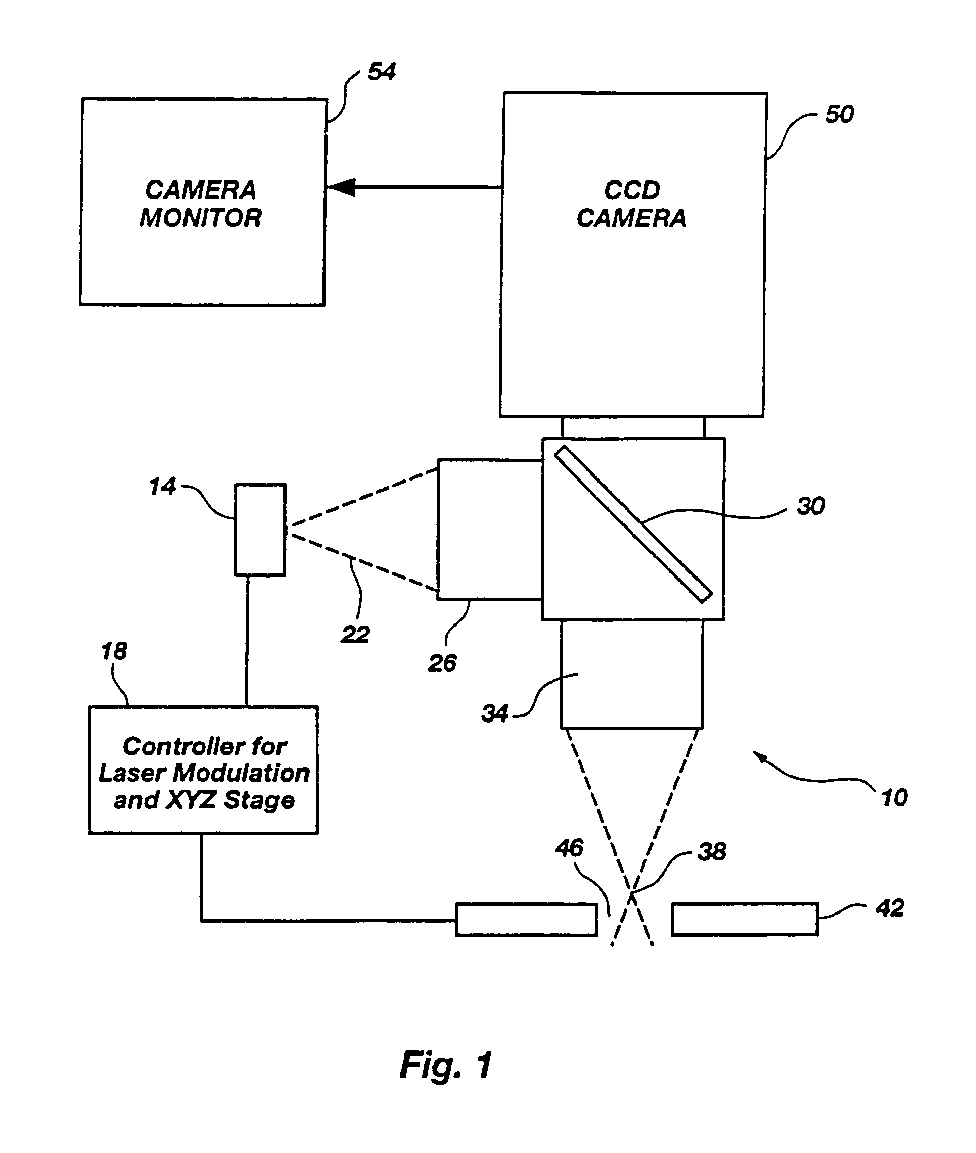
[0177]As a test of these untreated skin samples, the outputs of several different infrared laser diodes, emitting at roughly 810, 905, 1480 and 1550 nanometers were applied to the sample. The delivery optics were designed to produce a focal waist 25 μm across with a final objective have a numerical aperture of 0.4. The total power delivered to the focal point was measured to be between 50 and 200 milliwatts for the 810 and 1480 nm laser diodes, which were capable of o...

example 3

[0179]To evaluate the potential sensation to a living subject when illuminated with optical energy under the conditions of Example 2, six volunteers were used and the output of each laser source was applied to their fingertips, forearms, and the backs of their hands. In the cases of the 810, 905 and 1550 nm lasers, the subject was unable to sense when the laser was turned on or off. In the case of the 1480 nm laser, there was a some sensation during the illumination by the 1480 nm laser operating at 70 mW CW, and a short while later a tiny blister was formed under the skin due to the absorption of the 1480 nm radiation by one of the water absorption bands. Apparently the amount of energy absorbed was sufficient to induce the formation of the blister, but was not enough to cause the ablative removal of the stratum corneum. Also, the absorption of the 1480 nm light occurred predominantly in the deeper, fully hydrated (85% to 90% water content) tissues of the epidermis and dermis, not ...


PUM


Abstract

A method of enhancing the permeability of a biological membrane, including the skin or mucosa of an animal or the outer layer of a plant to a permeant is described utilizing microporation of selected depth and optionally one or more of sonic, electromagnetic, mechanical and thermal energy and a chemical enhancer. Microporation is accomplished to form a micropore of selected depth in the biological membrane and the porated site is contacted with the permeant. Additional permeation enhancement measures may be applied to the site to enhance both the flux rate of the permeant into the organism through the micropores as well as into targeted tissues within the organism.

Description

[0001]This application is a continuation application of Ser. No. 10 / 284,408, filed Oct. 31, 2002, which is a continuation of Ser. No. 09 / 331,124, filed Aug. 12, 1999, now U.S. Pat. No. 6,527,716, which is a 371 of PCT / US97 / 24127 filed Dec. 30, 1997, which claims priority to Ser. No. 08 / 778,415 filed Dec. 31, 1996, now abandoned, the entire contents of which are hereby incorporated.BACKGROUND OF THE INVENTION[0002]This invention relates generally to the field of transmembrane delivery of drugs or bioactive molecules to an organism. More particularly, this invention relates to a minimally invasive to non-invasive method of increasing the permeability of the skin, mucosal membrane or outer layer of a plant through microporation of this biological membrane, which can be combined with sonic, electromagnetic, and thermal energy, chemical permeation enhancers, pressure, and the like for selectively enhancing flux rate of bioactive molecules into the organism and, once in the organism, into...

Claims


Application Information

Patent Timeline
Patent Type & Authority Patents(United States)
IPC IPC(8): A61M5/00A61B1/313A61B17/00A61B18/04A61B18/20A61K41/00A61M37/00A61N1/32
CPCA61B1/313A61B5/14514A61B18/04A61K41/0047A61M37/0092A61N1/325A61N1/327A61B5/150129A61B18/10A61B18/203A61B2017/00026A61B2017/00172A61B2017/00765A61B2018/00452A61B2018/00577A61B2018/00642A61B2018/00791A61M2037/0007A61N1/0412A61N1/0428A61N1/0476A61N2007/0034A61B5/150022A61B5/150076A61B5/150083A61B5/150091A61B5/150099A61B5/150175A61B5/15136
Inventor EPPSTEIN, JONATHAN A.
Owner ALTEA THERAPEUTIC CORP
Learn More