Non-invasive vagal nerve stimulation to treat disorders

a vagal nerve and non-invasive technology, applied in the field of non-invasive vagal nerve stimulation to treat disorders, can solve the problems of inability to fully utilize vagal nerves, inability to effectively stimulate vagal nerves, etc., to achieve the effect of reducing the level of inhibitory neurotransmitter, reducing inactivity, and heightening activity in areas

Active Publication Date: 2014-09-23
ELECTROCORE
View PDF4 Cites 73 Cited by
  • Summary
  • Abstract
  • Description
  • Claims
  • Application Information

AI Technical Summary

Benefits of technology

[0032]One advantage of the present invention is that the electrical impulses described above are sufficient to generate an electric field at the vagus nerve above a threshold for generating action potentials within fibers of the vagus nerve responsible for activating neural pathways causing release of inhibitory neurotransmitters within a brain of the patient. In a preferred embodiment, the inhibitory neurotransmitters are one or more of the following: GABA, serotonin and / or norepinephrine.
[0033]The applicant has discovered that many seemingly disparate disorders may, in fact, have common causes that manifest into different symptoms or disorders in different individuals. Specifically, the applicant has discovered that in certain individuals who may suffer from disorders, such as depression, asthma, COPD, migraine, cluster headache, anxiety, fibromyalgia, epilepsy and the like, certain areas of the brain are prone to periodic or continuous excessive excitatory neurotransmitter levels. These periodic excessive excitatory neurotransmitter levels can be caused by certain “triggers”, such as noxious substances entering the lungs that cause airway reactivity or other triggers, such as chocolate or seafood that can cause migraines in certain individuals. In other cases, the patient may have pathologically high excitatory neurotransmitter levels on a continuous basis without any particular trigger. One example, of an excitatory neurotransmitter is glutamate, which is known to be associated with migraines.
[0034]The excessive excitatory neurotransmitter levels in a patient's brain can be caused by inaccurate signals from the body transmitted through the vagus nerve or other nerves or these excessive levels can be caused by the brain overreacting to normal signals coming from the body. In some cases, these excessive excitatory neurotransmitter levels may be caused by inappropriate inactivity in the production and / or release of inhibitory neurotransmitters, such as GABA, serotonin and / or norepinephrine. A reduced level of these inhibitory neurotransmitter levels can result in excessive levels of the excitatory neurotransmitters that they are meant to balance.
[0035]The present invention seeks to address this inbalance in neurotransmitters in the brain that can result in many of the disorders mentioned above. Specifically, afferent nerve fibers in the vagus nerve are stimulated with the devices, signals and methods described above to heighten activity in areas of the brain (e.g., the periaqueducatal gray, locus ceruleus and / or raphe nuclei) resulting in the release of inhibitory neurotransmitter levels, such as GABA, norephinephrine and / or serotonin). For example, as discussed above, heightened activity in the locus coeruleus will result in a release of norephinephrine or an increase the release of norephinephrine. This release of inhibitory neurotransmitters suppresses the excessive excitatory neurotransmitters and creates balance in the brain such that the brain either does not overreact to certain stimuli and / or to modulate the pathologically high level of excitatory neurotransmitters.
[0036]Another of the key advantages with the present invention is that the electrical field is above the threshold for generating action potentials within A and B fibers of the vagus nerve but below the threshold for the C fibers. Thus, the A and B fibers are selectively stimulated without stimulating the C fibers of the vagus nerve. The method of the present invention allows for selective stimulation of nerves responsible for activating neural pathways that will cause the release of inhibitory neurotransmitters within the brain to treat a variety of disorders in a patient. At the same time, the stimulation has substantially no effect on heart rate or blood pressure and it will not cause bronchoconstriction.
[0037]In a preferred embodiment, the electric field at the vagus nerve is between about 10 to 600 V / m and more preferably less than 100 V / m. The electrical field gradient is preferably greater than 2 V / m / mm. The electrical impulses are substantially constrained from modulating the nerves between the outer skin surface of the patient and the vagus nerve. The electric field is preferably not sufficient to produce substantial movement of the skeletal muscles of the patient.

Problems solved by technology

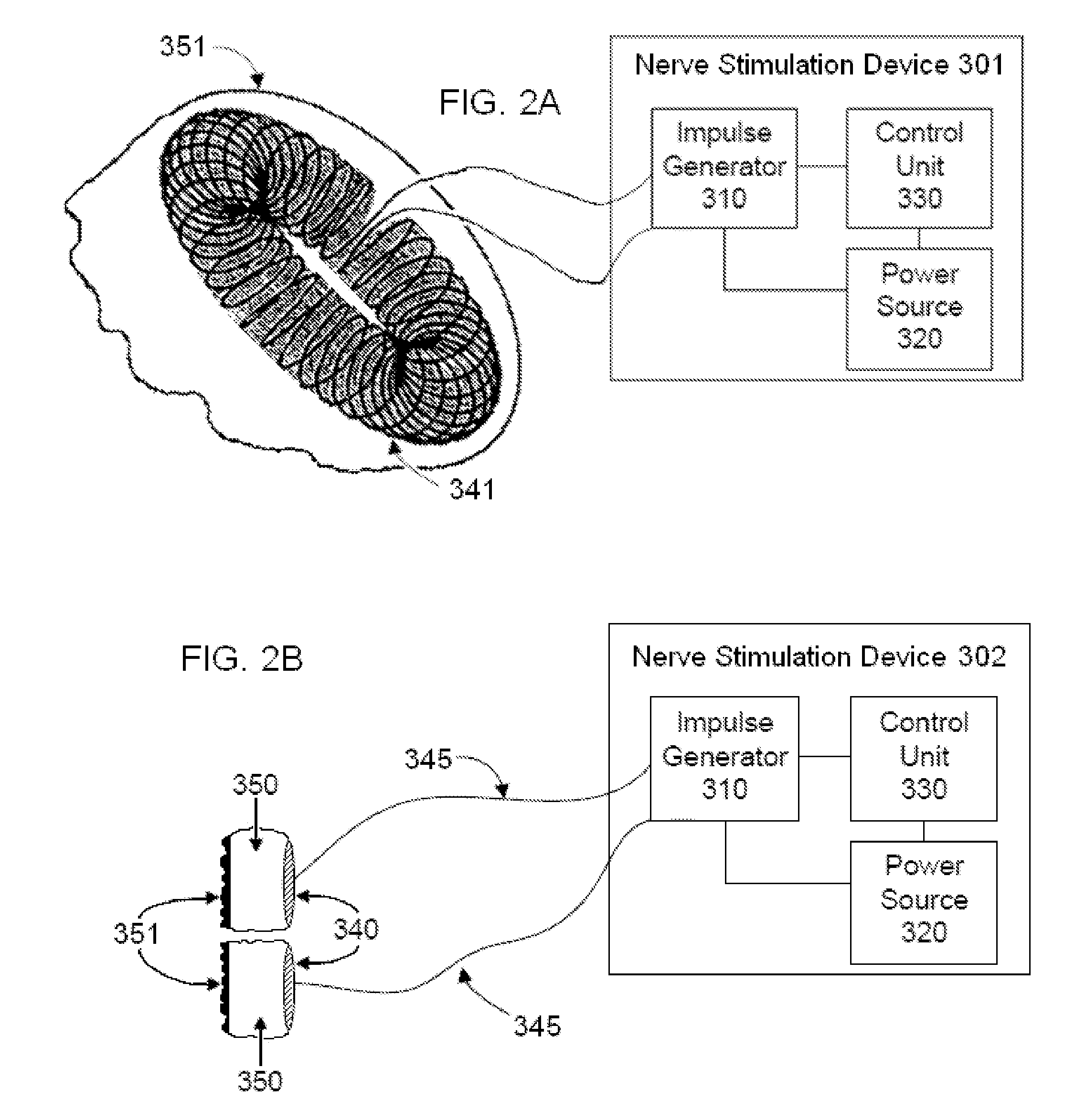
The electrical circuits for magnetic stimulators are generally complex and expensive and use a high current impulse generator that may produce discharge currents of 5,000 amps or more, which is passed through the stimulator coil to produce a magnetic pulse.
Non-invasive procedures are generally painless and may be performed without the dangers and costs of surgery.
Smooth muscle surrounding the airways goes into spasm, resulting in constriction of airways.
Over time, this inflammation can lead to scarring of the airways and a further reduction in airflow.
This inflammation leads to the airways becoming more irritable, which may cause an increase in coughing and increased susceptibility to asthma episodes.
Second, airway hyper-responsiveness resulting from local and central neural reflex stimulation and by mediators of inflammation can increase bronchoconstriction.
The combination can be lethal.
For some patients, however, these medications, and even the bronchodilators are insufficient to stop the constriction of their bronchial passages.
Minute amounts of allergens may cause a life-threatening anaphylactic reaction.
Anaphylactic shock usually results in death in minutes if untreated.
It is a life-threatening medical emergency because of rapid constriction of the airway, resulting in brain damage through oxygen deprivation.
In addition, anaphylactic shock can be brought on by exercise.
Yet all such reactions are mediated by a series of hypersensitivity responses that result in uncontrollable airway occlusion driven by smooth muscle constriction, and dramatic hypotension that leads to shock.
Cardiovascular failure, multiple organ ischemia, and asphyxiation are the most dangerous consequences of anaphylaxis.
Clinical treatment typically includes administration of antihistamines (which inhibit the effects of histamine at histamine receptors, but which are usually not sufficient in anaphylaxis), and high doses of intravenous corticosteroids.
Tragically, many of these patients are fully aware of the severity of their condition, but nevertheless die while struggling in vain to manage the attack medically.
Many of these fatal incidents occur in hospitals or in ambulances, in the presence of highly trained medical personnel who are powerless to break the cycle of inflammation and bronchoconstriction (and life-threatening hypotension in the case of anaphylaxis) affecting their patient.
Unfortunately, prompt medical attention for anaphylactic shock and asthma are not always available.
For example, epinephrine is not always available for immediate injection.
Even in cases where medication and attention is available, life-saving measures are often frustrated because of the nature of the symptoms.
Constriction of the airways frustrates resuscitation efforts, and intubation may be impossible because of swelling of tissues.
This cardiovascular stress can result in tachycardia, heart attacks and strokes.
COPD is a progressive disease that makes it increasingly difficult for the patient to breathe.
COPD can cause coughing that produces large amounts of mucus, wheezing, shortness of breath, chest tightness and other symptoms.
In COPD, there is abnormally low air flow within the bronchial airways for a variety of reasons, including loss of elasticity in the airways and / or air sacs, inflammation and / or destruction of the walls between many of the air sacs and overproduction of mucus within the airways.
In emphysema, the walls between many of the air sacs are damaged, causing them to lose their shape and become floppy.
This damage can also destroy the walls of the air sacs, leading to fewer and larger air sacs instead of many small ones.
This causes the lining to thicken and form thick mucus, making it difficult to breathe.
Many of these patients also experience periodic episodes of acute airway reactivity (i.e., acute exacerbations), wherein the smooth muscle surrounding the airways goes into spasm, resulting in further constriction and inflammation of the airways.
Frequent acute exacerbations of COPD cause lung function to deteriorate quickly, and patients never recover to the condition they were in before the last exacerbation.
As with asthma, current medical management of these acute exacerbations is often insufficient [Dick D. BRIGGS Jr.
However, many patients do not respond to those treatments, or they experience unwanted side effects.
If the pharmacological treatment does not stop or prevent the wheezing, the surgery may be deferred, but this is not always practical or possible in view of the need for surgery.
For some patients, those agents may not be effective or are otherwise contraindicated, and the bronchospasm may continue even after the surgery is completed.
Unlike cardiac arrhythmias, which can be treated chronically with pacemaker technology, or in emergent situations with defibrillators (implantable and external), there is no commercially available medical equipment that can chronically reduce the baseline sensitivity of the smooth muscle tissue in the airways, to reduce the predisposition to asthma attacks, to reduce the symptoms of COPD or to break the cycle of bronchial constriction associated with an acute asthma attack or anaphylaxis.
Although energy has been applied previously to patients in such a way as to bring about bronchodilation, those investigations involve methods that are invasive.
The vagus nerve innervates the heart, which raises additional concerns that even if VNS could be used to dilate bronchi, such vagus nerve stimulation could trigger cardiac or circulatory problems, including bradycardia, hypotension, and arrhythmia, particularly if the right vagus nerve is stimulated [SPUCK S, Tronnier V, Orosz I, Schönweiler R, Sepehrnia A, Nowak G, Sperner J. Operative and technical complications of vagus nerve stimulator implantation.

Method used


Image

  • Non-invasive vagal nerve stimulation to treat disorders
  • Non-invasive vagal nerve stimulation to treat disorders
  • Non-invasive vagal nerve stimulation to treat disorders

Examples


Embodiment Construction

[0066]Once air is inhaled through the mouth or nose, it travels through the trachea and a progressively bifurcating system of bronchi (containing cartilage) and bronchioles (which contain little or no cartilage), until it finally reaches the alveoli, where the gas exchange of carbon dioxide and oxygen takes place. Through constriction or relaxation of smooth muscle within their walls, the bronchioles change diameter to either reduce or increase air flow. The bronchioles between the fourth and eighth bifurcation are thought to be most important in that regard. Normally, an increase in diameter (bronchodilation) to increase air flow is stimulated by circulating epinephrine (adrenaline) or sympathetic nerve fibers or so-called iNANC nerve fibers, and a decrease in diameter (bronchoconstriction) is stimulated by parasympathetic cholinergic nerve fibers, histamine, cold air, and chemical irritants. Reflexes have evolved to regulate the caliber of bronchioles, in which afferent nerves sen...


PUM


Abstract

Devices, systems and methods are disclosed for treating a variety of diseases and disorders that are primarily or at least partially driven by an imbalance in neurotransmitters in the brain, such as asthma, COPD, depression, anxiety, epilepsy, fibromyalgia, and the like. The invention involves the use of an energy source comprising magnetic and / or electrical energy that is transmitted non-invasively to, or in close proximity to, a selected nerve to temporarily stimulate, block and / or modulate the signals in the selected nerve such that neural pathways are activated to release inhibitory neurotransmitters in the patient's brain.

Description

CROSS REFERENCE TO RELATED APPLICATIONS[0001]This application is a continuation-in-part of U.S. patent application Ser. No. 13 / 222,087 filed Aug. 31, 2011, which is a continuation-in-part of U.S. patent application Ser. No. 13 / 183,765 filed Jul. 15, 2011 which claims the benefit of priority of U.S. Provisional Patent Application No. 61 / 488,208 filed May 20, 2011 and is a continuation-in-part to U.S. patent application Ser. No. 13 / 183,721 filed Jul. 15, 2011, which claims the benefit of priority of U.S. Provisional Patent Application No. 61 / 487,439 filed May 18, 2011 and is a continuation-in-part of U.S. patent application Ser. No. 13 / 109,250 filed May 17, 2011, which claims the benefit of priority of U.S. Provisional Patent Application No. 61 / 471,405 filed Apr. 4, 2011 and is a continuation-in-part of U.S. patent application Ser. No. 13 / 075,746 filed Mar. 30, 2011, which claims the benefit of priority of U.S. provisional patent application 61 / 451,259 filed Mar. 10, 2011 and is a con...

Claims


Application Information

Patent Timeline
Patent Type & Authority Patents(United States)
IPC IPC(8): A61N1/00A61B5/00A61N1/36A61N1/40A61N2/00A61N2/02
CPCA61N1/3601A61N1/40A61N2/006A61B5/4836A61N2/008A61N2/02A61N1/36014A61N1/36034
Inventor SIMON, BRUCEERRICO, JOSEPH P.RAFFLE, JOHN T.
Owner ELECTROCORE
Why Eureka
Try Eureka