Image guided intraoperative simultaneous several ports microbeam radiation therapy with microfocus X-ray tubes

a microfocus x-ray tube and imaging guided technology, applied in the field of imaging guided intraoperative simultaneous several ports microfocus radiation therapy with microfocus x-ray tube, can solve the problems of not being able to achieve synchrotron radiation dose rate, not being able to achieve a present medical accelerator or an orthovoltage tube, and not being able to achieve a present medical accelerator

Inactive Publication Date: 2014-12-23
SAHADEVAN VELAYUDHAN
View PDF46 Cites 26 Cited by
  • Summary
  • Abstract
  • Description
  • Claims
  • Application Information

AI Technical Summary

Problems solved by technology

However, its dose rate is nowhere near to that of synchrotron radiation.
It is not achievable with a present medical accelerator or from an orthovoltage tube.
It is not achievable with a present medical accelerator.
Hence the increased spacing of the microbeam do not minimize dose to the tumor.
Such high dose and dose rate radiation therapy is not possible with present medical accelerators.
Its radiobiological effectiveness for tumor cure and control is much inferior as compared to that of synchrotron radiation.
It definitely cannot provide anywhere closer to the radiobiological effectiveness of synchrotron radiation with dose rate of 20,000 Gy/sec in treating a tumor.
The other major disadvantage of the X-ray tube based microbeam radiation as taught in U.S. Pat. No. 7,643,610 is its 0.8 mm sized large penumbra (18).
Using the 1 mm wide microbeam as in U.S. Pat. No. 7,643,610 (19) is a significant hazard as it increases the normal tissue damage and necrosis.
However, synchrotrons are not readily available to most of the cancer treatment centers in the world.
Hence its inferior RBE and poor tumor cure and control.
It definitely cannot provide anywhere close to the radiobiological effectiveness of synchrotron radiation.
It results in tumor recurrence.
However, it does not represent the additive isodose of simultaneous beams.
To treat a second patient, a second source is used, hence it is relatively expensive to maintain.
Because of the potential high dose to the skin, to the chest wall and the ribs for patients with small breast, this system cannot be used for accelerated partial breast irradiation (APBI) for such patients.
Even for those patients who are eligible for APBI with this system, there is the potential danger of balloon deflation and deformity.
It can cause skin fibrosis, rib fracture and breast deformity.
Furthermore, its voltage is more easily controlled and it is very compact.
This causes another week long waiting period for this third surgery.
If additional boost radiation to the tumor bed is also given it will also take additional time.
Because of the poor dose rate of the orthovoltage X-ray beam, 0.17 to 0.83 Gy/sec, it does not offer the same radiobiological advantages of stereotactic microbeam radiosurgery with synchrotron radiation with the dose rate of 20,000 Gy/sec.
The tumor recurrence is primarily due to conventional radiation therapy's inability to sterilize cancer stem cells.
The “differentiated” cancer cells might be sterilized with conventional radiation therapy but the residual radioresistant cancer stem cells could cause tumor recurrence.
The low dose rate conventional radiation therapy is less effective for sterilization of the cancer stem cell.
However

Method used


Image

  • Image guided intraoperative simultaneous several ports microbeam radiation therapy with microfocus X-ray tubes
  • Image guided intraoperative simultaneous several ports microbeam radiation therapy with microfocus X-ray tubes
  • Image guided intraoperative simultaneous several ports microbeam radiation therapy with microfocus X-ray tubes

Examples


Embodiment Construction

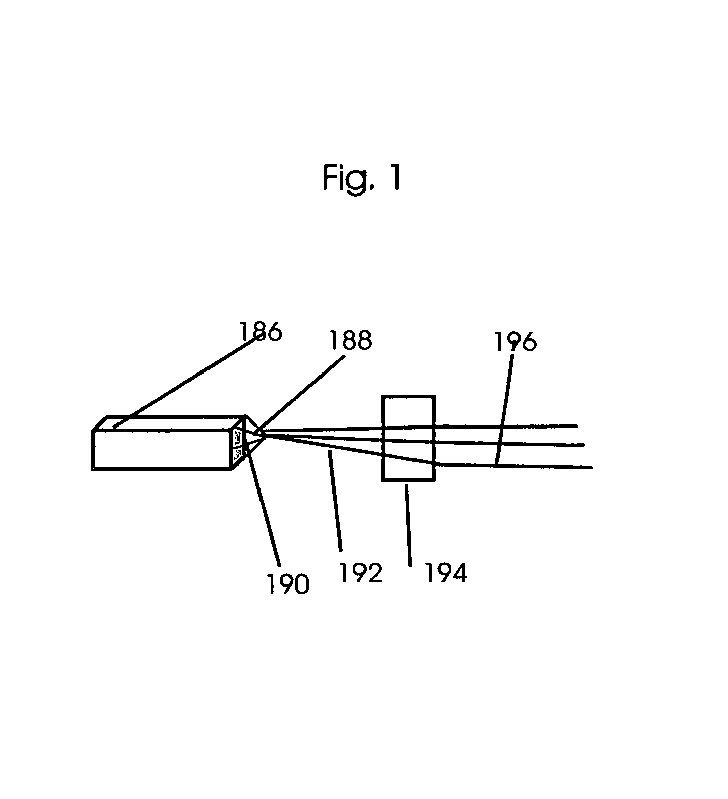
[0245]FIG. 1 illustrates a commercially available micro focus x-ray tube 186 that is equipped with analyzer crystal 190 that filters the bremsstrahlung polychromatic x-ray beam 188 to mostly monochromatic. These commercially available x-ray tubes are equipped with adjustable focal spots ranging from as low as 5 to 50 μm. The 5 μm sized focal spot is very close to the laser produced x-ray's focal spot. They are available with targets ranging from chromium, copper, molybdenum, tungsten etc. The fully packaged commercial tubes have remote control and software controlled operational capabilities. They meet all the radiation safety precautions including instructions and warnings signs on safe operation that is displayed on its display panel.

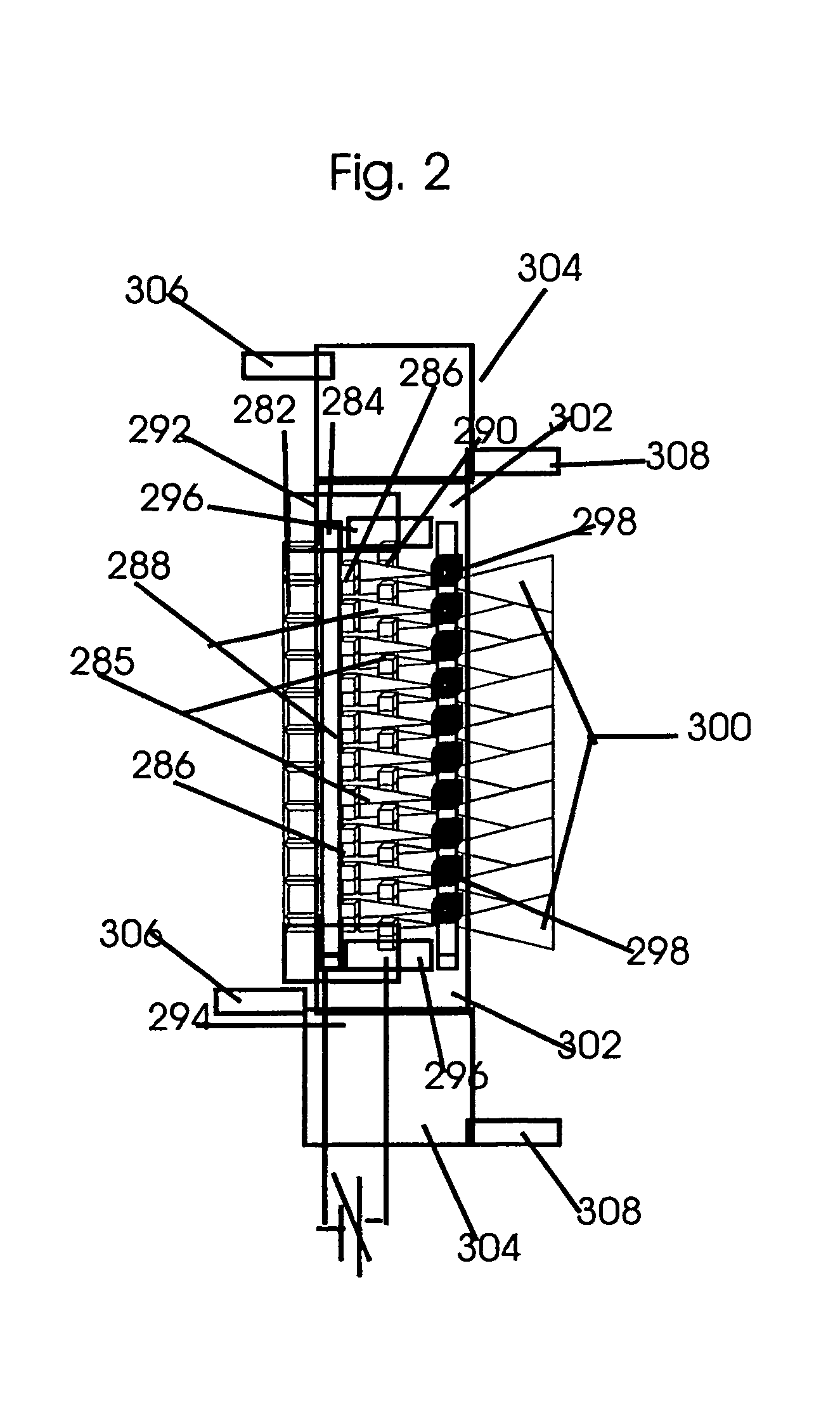
[0246]FIG. 2 is a detailed illustration of the basic structures of a CNT based single set, 10 simultaneous converging beams X-ray tube. The miniaturized tiny CNT based field emission cathode 288 is constructed with the metal-oxide-semiconductor field-...


PUM


Abstract

A microbeam intraoperative, 10-50 kilovoltage radiation therapy system with multiple simultaneous microfocus X-ray sources and method for image guided, microsecond duration, 100 to 1,000 Gy microbeam radiation therapies with dose rate ranging from 1,000 to 20,000 Gy per second and RBE closer to high LET with minimal toxicity to normal tissue is provided. Dismountable coronal, sagital and transverse half-circle gantry sections are assembled on to a surgical table for mounting the microfocus X-ray sources for intraoperative radiosurgery. A stereotactic breast core biopsy system is adapted for combined breast biopsy, positron emission tomography and computerized tomography with phase-contrast imaging. Cosmetic whole breast preserving electronic brachytherapy is combined with stereotactic core biopsy. The low dose valley regions of the intersecting microbeams at the tumor site are filled with scattered and characteristic radiation from high Z-element that is bound or implanted into the tumor.

Description

FIELD OF INVENTION[0001]X-ray beam therapy, class 378, 424, 530FEDERALLY SPONSORED RESEARCH[0002]None;SEQUENCE LISTING[0003]Table of Contents attachedBACKGROUND OF THE INVENTION[0004]Synchrotron high dose rate microbeams with dose rate exceeding 20 Gy / sec is curative even for the most radiation resistant tumors like the glioblastoma multiforme (Slatkin and Dilimanian). This invention is aimed at similar high dose rate radiation therapy but with much more affordable and easily manageable microbeams from microfocus X-ray tubes that are configured to provide multiple simultaneous interlaced microbeams for simultaneous several ports irradiation. Their additive high dose rate is close to that of high flux synchrotron radiation.[0005]Generally, after the diagnosis of malignant diseases it is treated by surgery, radiation therapy and chemotherapy. There are also an increasing number of benign diseases that are treated with radiation and chemotherapy. The methods of high dose rate radiation...

Claims


Application Information

Patent Timeline
IPC IPC(8): A61N5/00A61B17/52
CPCA61N5/1027A61B10/0233A61N5/1084A61N2005/1022A61N2005/1052A61N2005/1054A61N2005/1055A61N2005/1091
Inventor SAHADEVAN, VELAYUDHAN
Owner SAHADEVAN VELAYUDHAN
Why Eureka
Try Eureka