Single nanoparticle having a nanogap between a core material and a shell material, and preparation method thereof

a nanoparticle and core material technology, applied in the field of single nanoparticles, can solve the problems of fluorescent dyes, inconvenient long-term storage or experiment, difficult handling, etc., and achieve the effects of high reproducibility, uniform thickness, and large surface area

Active Publication Date: 2016-11-01
KOREA RES INST OF CHEM TECH +1
View PDF24 Cites 1 Cited by
  • Summary
  • Abstract
  • Description
  • Claims
  • Application Information

AI Technical Summary

Benefits of technology

[0063]The nanostructure of nanogap particle has a large surface area and provides the nanogap of high reproducibility and uniform thickness. Accordingly, the entire surface of the core can be used for enhancing the SERS, and the location of the dye also can be positioned precisely inside the nanogap. Furthermore, in actual use, it can be synthesized simply with high synthetic yield. Therefore, very high signal amplification effect is shown, and the problem of signal reproducibility and quantification, which is the crucial prerequisite to the commercialization, can be overcome due to high reproducibility.

Problems solved by technology

However, it is not easy to handle because of the harmful radiation and the radiation of some isotopes has short half-life instead of large emission energy, causing inconvenience in long-term storage or experiment.
However, the fluorescent dyes have limitations in that the fluorescent dyes are connected by deformation of the part which relatively little affects the activity through structure activity relationship, incapable of direct substitution of the elements of the active ligand as radioactive isotopes.
In addition, these fluorescent markers emit weaker intensity of fluorescence over time (photobleaching) and have a very narrow wavelength range of activation light and a wide wavelength range of emission light leading to the disadvantage of interference between different fluorophores.
Also, the number of available fluorophores is extremely limited.
However, they have disadvantages of high toxicity and difficulty of mass production.
In addition, the number of available quantum dots, although theoretically variable, is highly restricted in practice.
However, due to the low signal intensity, it did not reach a level of practical use despite long research period.
However, there has been little progress in the preparation methods for single-molecule SERS active substrates based on salt induced aggregation of silver (Ag) nanoparticles with the Raman active molecule (eg, Rhodamine 6G) since the initial study.
However, in the previous SERS detection methods for the various analytes, the substrate and / or colloidal metal particles, such as aggregated silver nanoparticles, coated on the supporter were typically used, sometimes yielding SERS detection with increased sensitivity by 106 to 108 times, without being able to detect single-molecule of small analytes such as nucleotides.
However, despite the advantages of SERS, the mechanism of SERS phenomenon are not only not fully understood, the preparation and control of well-defined nanostructures are also difficult, as well as many unsolved problems exist in terms of reproducibility and reliability arising from the changes in enhancement efficiency depending on the wavelength of the light used to measure the spectrum, and the polarization direction remains an unsolved problem for the application of the SERS phenomenon including the development and commercialization of nanobiosensors.
However, the characteristics of the physical structure and distance range from nanoparticles, where enhanced sensitivity is achieved, of hot spots, and spatial correlation between the analytes to enhance the sensitivity and aggregate of nanoparticles have not been presented.
In addition, the aggregated nanoparticles are inherently unstable in solution, and give an adverse effect on the reproducibility of the detection of single-particle analyte.
However, if surface-enhanced Raman scattering (SERS) is to be obtained using these structures, quantification of the signal, reproducibility of the results, ease and simplicity of synthesis, cost, and stability of the probe still remain the problems.
In other words, if two or more nanoparticles are combined by a nanogap, the amplified optical signal detection is detectable, but simplicity of material synthesis, stability, reproducibility of the signal and quantification cannot be secured.
Meanwhile, although synthesis and assembly of various nanostructures for DNA have been studied in-depth, there have been very few researches on other roles of DNA.

Method used


Image

  • Single nanoparticle having a nanogap between a core material and a shell material, and preparation method thereof
  • Single nanoparticle having a nanogap between a core material and a shell material, and preparation method thereof
  • Single nanoparticle having a nanogap between a core material and a shell material, and preparation method thereof

Examples


example 2

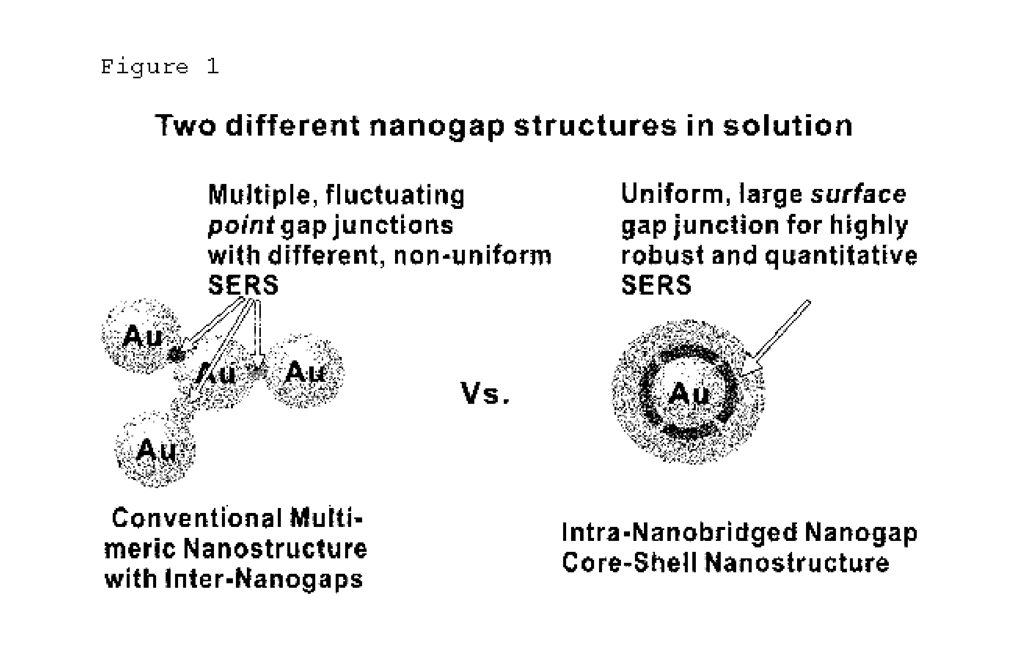
Fem Calculation of Gold Nanoparticle and Core-Shell Particle Surrounded by Nanogap without Bridge and Silica

[0103]In order to understand relation between Au—NNP and electromagnetic wave, FEM (3D finite-element-method was applied to the calculation (Wustholz, K. L. et al. Structure-activity relationships in gold nanoparticle dimers and trimers for surface-enhanced Raman spectroscopy. J. Am. Chem. Soc. 132, 10903-10910 (2010)), and the results were compared with Au—Au core-shell nanoparticle surrounded by silica (FIG. 9). In every calculation, four intra-nanobridges were assumed to be formed between Au core and Au shell. Radius of core is 20 nm, nanobridge is cylindrical shape of 2.5 nm×1.2 nm, size of gap or thickness of silica is 1.2 nm, and thickness of shell is 11 nm. Linearly polarized plane wave incident along the x-axis was used for plasmon excitation. The intensity of EM enhancement is represented in FIG. 9a, which indicates that EM enhancement is located intensively on the in...

example 3

Preparation of Nanoparticle with Modified Location of Raman Dye

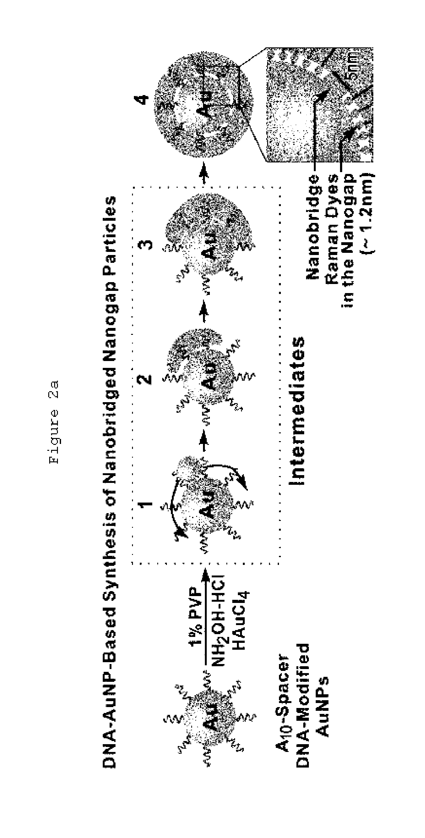
[0104]DNA strand was used for forming platform for Raman dye modification as well as forming intra-nanogap.

[0105]Three different kinds of reduced thiolated oligonucleotides with modified location of dye (ROXgap (760 μL, 4.3 μM): 3′-HS—(CH2)3-(ROX)-A10-PEG18-AAACTCTTTGCGCAC-5′(i.e., 3′-HS—(CH2)3-(ROX)-SEQ ID No.1-PEG18-SEQ ID No.2-5′), ROXshell (131 μL, 24.9 μM): 3′-HS—(CH2)3-A10-PEG18-(ROX)-AAACTCTTTGCGCAC-5′ (i.e., 3′-HS—(CH2)3-SEQ ID No.1-PEG18-(ROX)-SEQ ID No.2-5′) and ROXouter (456 μL, 7.1 μM), 3′-HS—(CH2)3-A10-PEG18-AAACTCTTTGCGCAC-(ROX)-5′ (i.e., 3′-HS—(CH2)3-SEQ ID No.1-PEG18-SEQ ID No.2-(ROX)-5′)) was mixed with and reacted to citrate-gold nanoparticles (1 ml, 1.0 nM) for 20 minutes at room temperature, respectively. In order to obtain as final phosphate concentration of 10 mM (pH 7.4), the resultant solution was adjusted with 100 mM phosphate buffer (for ROXgap, ROXshell and ROXouter, 176 μL, 113 μL and 146 μL a...

example 4

Preparation of Nanoparticle with Adjusted Amount of Dye

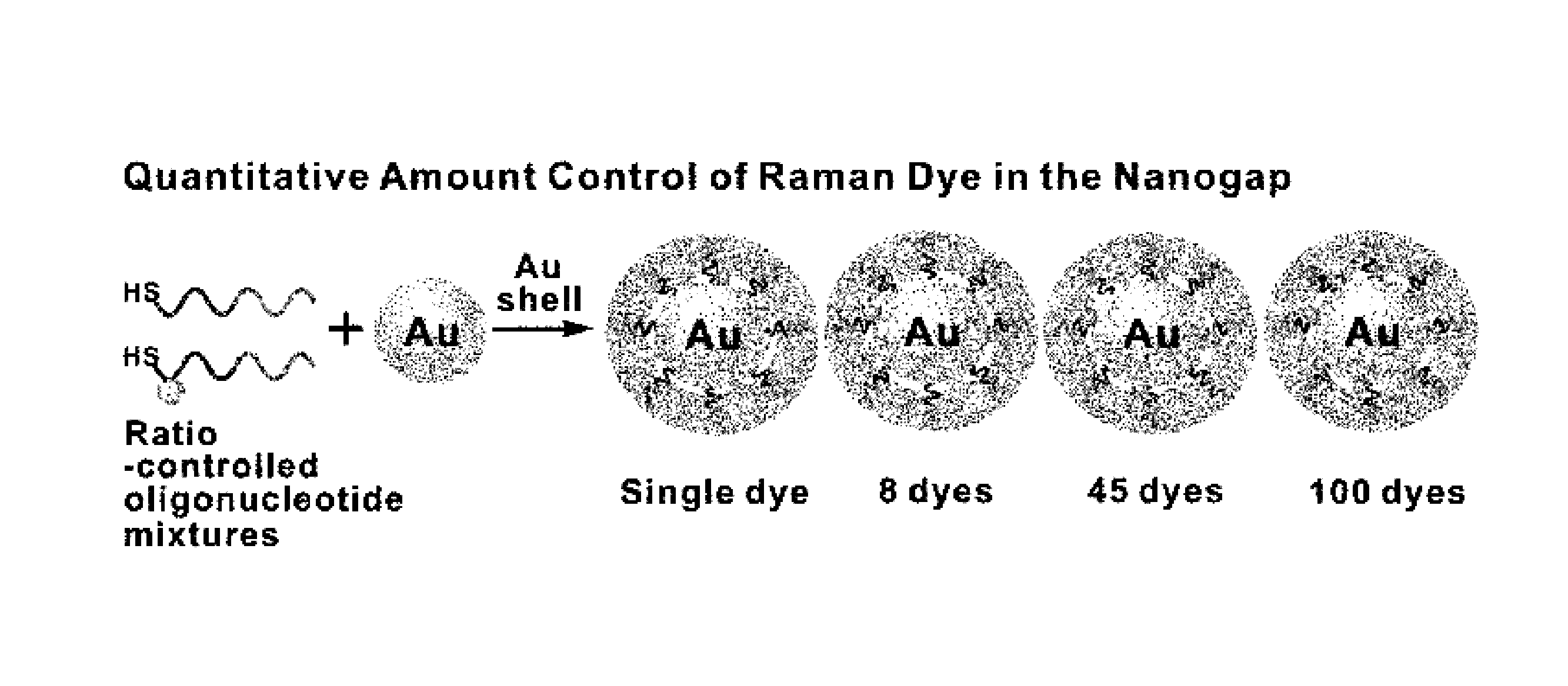
[0111]The number of Raman dyes in the intra-nanogap was adjusted as follows, characteristics were identified accordingly and the whole process was schematically shown in FIG. 11a.

[0112]It is known that if poly A spacer is used in the condition of 0.3 M PBS, the number of oligonucleotide loading on 20 nm gold nanoparticle can be approximately 100 according to the size of nanoparticle and DNA loading characteristic of DNA spacer (S. J. Hurst, A. K. R. Lytton-Jean, C. A. Mirkin, Anal. Chem. 78, 8313 (2006)). Hereupon, the mixtures of surface protecting sequence and ROXgap-modified sequence (surface protecting sequence: 3′-HS—(CH2)3-A10-PEG18-AAACTCTTTGCGCAC-5′ (i.e., 3′-HS—(CH2)3-SEQ ID No.1-PEG18-SEQ ID No.2-5′), ROXgap-modified sequence: 3′-HS—(CH2)3-(ROX)-A10-PEG18-AAACTCTTTGCGCAC-5′) (i.e., 3′-HS—(CH2)3-(ROX)-SEQ ID No.1-PEG18-SEQ ID No.2-5′) of four different kinds of ratio (99:1 (259 μL, 12.6 μM: 2.4 μL, 13.8 μM), 90:10 (235...


PUM

PropertyMeasurementUnit
diameteraaaaaaaaaa
thicknessaaaaaaaaaa
diameteraaaaaaaaaa

Abstract

The present invention is to provide a nanoparticle, which can be used effectively for Raman analysis based on very high amplification effect of electromagnetic signal by plasomonic coupling of nanogap formation inside thereof and high reproducibility, and which includes core and surrounding shell with nanogap formation between the same and the method of synthesis thereof. The present invention is also to provide the method for detecting the analyte using the above nanoparticle and the analyte detection kit including the above nanoparticle.

Description

TECHNICAL FIELD[0001]The present invention relates to a single nanoparticle, which has extremely high amplifying capability of electromagnetic signal by plasomonic coupling of the nanogap formed between core material and shell material and which shows the homogeneous signal intensity and quantitative signal contrasted with concentration of particle caused by homogeneous distribution and quantitative control of signal substance on the surface of core material, and preparation method thereof.BACKGROUND ART[0002]Highly accurate detection of single molecules from biological sample and other samples can be widely used in medical diagnostics, pathology, toxicology, environmental sampling, chemical analysis, and many other areas, and nanoparticles and chemicals labeled with specific substances have been used in researches for metabolism, distribution and coupling of small amounts of synthetic substances and bio-molecules in biochemistry for last a few years. Typically, there are methods us...

Claims


Application Information

Patent Timeline
Patent Type & Authority Patents(United States)
IPC IPC(8): C12M1/36A61K49/00G01N33/543G01N21/65
CPCG01N21/658A61K49/0002G01N33/54346G01N2021/653
Inventor SUH, YUNG DOUGNAM, JWA MINLIM, DONG KWONJEON, KI SEOK
Owner KOREA RES INST OF CHEM TECH
Why Eureka
Try Eureka