
Modified rat accellular spinal cord bracket material and preparation method thereof

A scaffold material and decellularization technology, which is applied in the field of tissue engineering biomaterials, can solve the problems of high cytotoxicity, poor structural stability, and stiff texture of materials, and achieve the effects of broad application prospects, high porosity, and low cost

Inactive Publication Date: 2013-04-03
THE SECOND AFFILIATED HOSPITAL ARMY MEDICAL UNIV
View PDF3 Cites 7 Cited by
  • Summary
  • Abstract
  • Description
  • Claims
  • Application Information

AI Technical Summary

Problems solved by technology

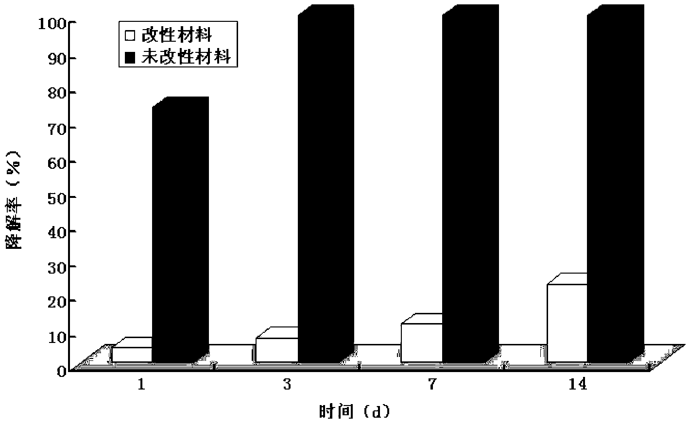
[0005] Previous studies have also found that after decellularization, the spinal cord has poor structural stability, is easy to degrade and collapses quickly, and it is difficult to meet the requirements for biological scaffold materials for spinal cord repair and reconstruction. Appropriate methods need to be used to improve the biological properties of decellularized spinal cord biologically derived materials. characteristics to improve its resistance to enzymatic degradation and maintain its good three-dimensional structure for a longer period of time
Traditional chemical cross-linking agents, such as formaldehyde, glutaraldehyde (glutaraldehyde, GA), acetaldehyde and epoxy compounds, have been widely used in the modification of biological materials, but the materials after treatment have high cytotoxicity, stiff texture and Disadvantages such as long-term degeneration limit its further application (Schoen FJ, Harasaki H, Kim KM, et al. Biomaterial-associated calcification: pathology, mechanisms, and strategies for prevention. J Biomed Mater Res. 1988 Apr; ):11-36.)

Method used


Image

  • Modified rat accellular spinal cord bracket material and preparation method thereof
  • Modified rat accellular spinal cord bracket material and preparation method thereof
  • Modified rat accellular spinal cord bracket material and preparation method thereof

Examples


Embodiment Construction

[0048] 1. Preparation of modified rat acellular spinal cord scaffold material

[0049] (1) Laboratory animals and equipment

[0050] Whole rat spinal cord frozen at -70°C (purchased from Experimental Animal Center of Daping Hospital, Third Military Medical University), sodium pentobarbital (P3761, Sigma), sodium chloride (51024060, Sinopharm Group), TritonX-100 (ST795) , Biyuntian), Sodium Deoxycholate (D001601, Amresco), Genipin (6902-77-8, Linchuan Zhixin Biological Technology Co., Ltd.), Glutaraldehyde (111-30-8, Shanghai Ziyi Reagent Factory ), phosphate buffer solution (PBS) (AR0030, Boster), surgical instruments (Shanghai Medical Instrument Factory), ultrapure water purifier (Millipore, USA), freeze dryer (Thermo Savant Modulyo D, USA), desktop Constant temperature oscillator (THZ-22, Jiangsu Taicang Experimental Equipment Factory), refrigerator (Midea), -70℃ deep low temperature refrigerator (Sanyo, Japan).

[0051] (2) Method

[0052] Preparation of modified rat acellular sp...


PUM


Abstract

The invention relates to a modified rat accellular spinal cord bracket material and a preparation method thereof. With the preparation method, the spinal cord accellular technology is optimized; chemical crosslinking treatment is improved; a material prepared by the method has a relatively complete three-dimensional mesh structure, structure stability and relatively high enzymolysis resistance; on the basis of removing cells and myelin sheath components in a spinal cord, the material effectively retains extracellular matrix and has highly-simulated three-dimensional mesh structure and high porosity; the immunogenicity is weak; the cell compatibility, the biological compatibility and the biological safety are high; equipotential transplantation and dissection transplantation are conveniently realized in in-vivo transplantation; and the modified rat accellular spinal cord bracket material is suitable for reparation, reconstruction and research of the spinal cord.

Description

Technical field [0001] The invention relates to tissue engineering biomaterials, in particular to acellular spinal cord bioderived materials used for spinal cord injury repair research and a preparation method. Background technique [0002] In order to construct a tissue engineering scaffold that meets the anatomical and physiological needs of the spinal cord, scholars have conducted extensive explorations on biological scaffold materials. The commonly used spinal cord bioscaffold materials are mostly biodegradable natural or synthetic polymer biomaterials (Jain A, Kim YT, McKeon RJ, et al. In situ gelling hydrogels for conformal repair of spinal cord defects and local delivery of BDNF after spinal cord injury. Biomaterials,2006,27(3):497-504.He L,Zhang Y,Zeng C,et al.Manufacture ofPLGA multiple-channel conduits with precise hierarchical pore architectures and invitro / vivo evaluation for spinal cord injury.Tissue Eng Part C Methods.2009Jun;15(2):243-255.), such as collagen, gela...

Claims


Application Information

Patent Timeline
IPC IPC(8): A61L27/36
Inventor 蒋涛任先军阴洪
Owner THE SECOND AFFILIATED HOSPITAL ARMY MEDICAL UNIV
Learn More