Cellularization biological liver stent with anticoagulation property and preparation method of cellularization biological liver stent

A decellularization and bio-scaffold technology, which is applied in the fields of clinical medicine, biomedicine and regenerative medicine, can solve the problems of high preparation cost, poor blood compatibility and fast release speed, and achieve strong anticoagulant performance and anticoagulant effect Uniform, slow release effect

Active Publication Date: 2015-08-19
WEST CHINA HOSPITAL SICHUAN UNIV
View PDF4 Cites 11 Cited by
  • Summary
  • Abstract
  • Description
  • Claims
  • Application Information

AI Technical Summary

Problems solved by technology

However, purely decellularized decellularized liver bioscaffolds have poor blood compatibility and are prone to thrombosis in clinical treatment.
[0004] The hemocompatibility of the decellularized liver bioscaffold can be improved by perfusion anticoagulant modification of the decellularized liver scaffold. Strong, uniform anticoagulant effect of each part of the scaffold, and a long anticoagulant time of decellularized scaffold material is very difficult, the existing modification methods are complicated, the preparation cost is high, and the anticoagulant effect is not ideal
[0005] For example, the patent application with publication number 101850136 discloses a method for modifying porcine liver decellularized scaffold material layer by layer self-assembly to improve anticoagulant performance, but it requires eight alternate perfusions with two solutions and one washing solution. It is easy to mix air bubbles in the perfusion solution during the alternate perfusion process, especially for large organs (such as pig liver, pig kidney) at high flow rate perfusion, it is easy to mix air bubbles in the solution alternate process, resulting in failure of the perfusion modification process; at the same time, the modified The heparin content can only reach 12 μg / mg dry weight of tissue at most, and the anticoagulant effect is not good, and the modified heparin and the material are combined by electrostatic force, the combination is not firm enough, and the release speed is fast, and the release amount reaches more than 50% in 24 hours, which cannot long-term anticoagulation
[0006] In addition, Chen-Chi Tsai et al. "Effects of heparin immobilization on the surface characteristics of a biological tissue"fixed with a naturally occurring crosslinking agent(genipin):an in vitro study", Biomaterials 22(2001) 523-533, Sun Jiu et al. , "Study on the anticoagulant performance of heparin end-binding modified porcine liver decellularized scaffold materials", Biomedical Engineering and Clinical, May 2014, Volume 18, No. 3, Wu Qiong et al. "Heparin layer-by-layer self-assembly modified porcine liver decellularized scaffolds Research on Anticoagulant Properties of Materials", Beijing: China Science and Technology Papers Online, 2014-3-19 and many other documents provide methods for heparinized liver slices, specifically soaking liver slices in heparin solution for modification, However, because the three-dimensional structure and vascular network of decellularized liver scaffolds are very complex, simple soaking cannot achieve the goal at all.

Method used


Image

  • Cellularization biological liver stent with anticoagulation property and preparation method of cellularization biological liver stent
  • Cellularization biological liver stent with anticoagulation property and preparation method of cellularization biological liver stent
  • Cellularization biological liver stent with anticoagulation property and preparation method of cellularization biological liver stent

Examples


Embodiment 1

[0040] Example 1 Preparation of decellularized liver bioscaffold with anticoagulant properties of the present invention

[0041] 1. Experimental method

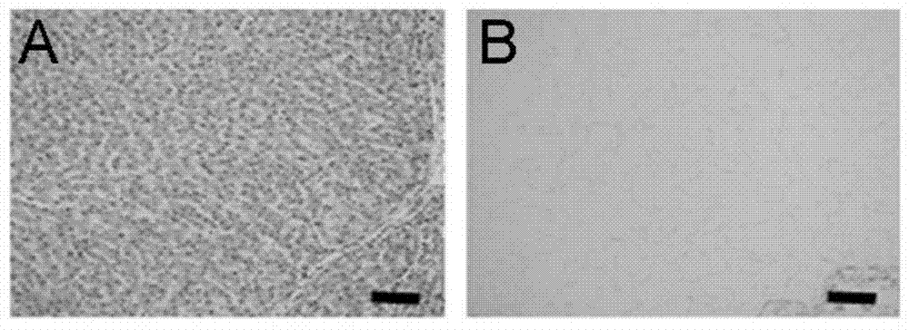
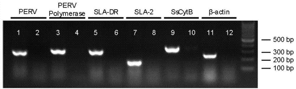
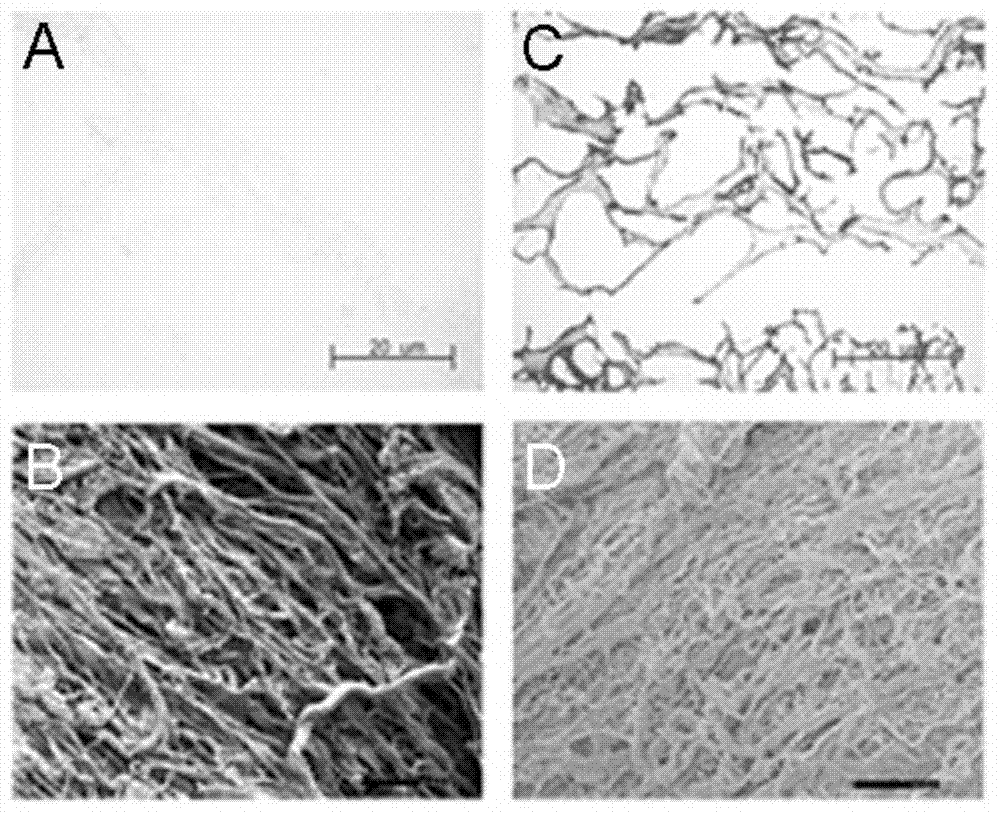
[0042] (1) Obtaining decellularized porcine liver bioscaffold

[0043] The decellularized liver bioscaffold of the present invention can be obtained by using the isolated porcine liver as a raw material and processed by a conventional decellularization method. For example, it can be prepared according to the following method:

[0044] Take out the liver from a Bama pig with a weight of 15-25 kg, and perfuse it with 0.1% ethylenediamine tetraacetic acid (EDTA) until the blood in the liver is completely drained, and obtain the isolated pig liver. save.

[0045] The isolated porcine liver was taken, thawed at 4°C, and perfused with 1% Triton X-100 solution at a flow rate of 200 mL / min for 3 h, and 1% sodium dodecylsulfate (SDS) solution at a flow rate of 200 mL / min for 6 h. Then lavage with 1% Triton X-100 solution and deioni...

Embodiment 2

[0072] Example 2 Preparation of decellularized liver bioscaffold with anticoagulant properties of the present invention

[0073] 1. Experimental method

[0074] (1) Preparation of decellularized porcine liver bioscaffold: According to the method in Example 1, a decellularized porcine liver bioscaffold was prepared.

[0075] (2) Anticoagulant modification

[0076] a. Anticoagulant modification

[0077] Preparation of heparin sodium perfusion solution: prepare a heparin sodium aqueous solution with a concentration of 3.3 g / L at 0° C., and adjust the pH value to 2.7. Then add sodium nitrite to a final concentration of 33mg / L, stir at 0°C for 2 hours, and adjust the pH value to 7.0. Then add NaBH 3 The final concentrations of CN and NaCl were 10 mg / L and 0.15 mol / L, respectively, and the pH was adjusted to 3.5.

[0078] At room temperature, the prepared heparin sodium perfusion solution was poured into the decellularized porcine liver scaffold, and the perfusion was circulate...

Embodiment 3

[0089] Example 3 Preparation of decellularized liver bioscaffold with anticoagulant properties of the present invention

[0090] 1. Experimental method

[0091] (1) Preparation of decellularized porcine liver bioscaffold: According to the method in Example 1, a decellularized porcine liver bioscaffold was prepared.

[0092] (2) Anticoagulant modification

[0093] a. Anticoagulant modification

[0094]Preparation of heparin sodium perfusion solution: prepare a heparin sodium aqueous solution with a concentration of 2 g / L at 0°C, and adjust the pH value to 2.7. Then add sodium nitrite to a final concentration of 33mg / L, stir at 0°C for 2 hours, and adjust the pH value to 7.0. Then add NaBH 3 The final concentrations of CN and NaCl were 10 mg / L and 0.15 mol / L, respectively, and the pH was adjusted to 3.5.

[0095] At room temperature, the prepared heparin sodium perfusion solution was poured into the decellularized porcine liver scaffold, and the perfusion was recirculated f...


PUM

PropertyMeasurementUnit
concentrationaaaaaaaaaa

Abstract

The invention provides a cellularization biological liver stent with an anticoagulation property and a preparation method of the cellularization biological liver stent. The preparation method comprises the following steps: (a) pouring a solution containing heparin and/or heparinate into the cellularization biological liver stent, wherein the pouring speed is 80mL/min-150mL/min, and the pouring time is 5-12 hours; and (b) carrying out sterilization. The prepared cellularization biological liver stent is strong in anticoagulation property and good in cytocompatibility and plays a very important support role in the survival, differentiation and proliferation of cells planted on the cellularization biological liver stent, and a convenient and cheap source and the preparation method are provided for the construction of a heterologous tissue-engineered biological anti-coagulation stent with a clinical application value.

Description

technical field [0001] The invention belongs to the fields of clinical medicine, biomedicine and regenerative medicine, and specifically relates to a decellularized liver biological scaffold with anticoagulant performance and a preparation method thereof. Background technique [0002] The liver is a very important organ of the human body. It is known as the "detoxification factory and metabolic chemical factory" of the human body. However, when it is severely damaged by various factors such as viruses, alcohol, and drugs, it will cause massive necrosis of liver cells and lead to liver failure. . So far, liver transplantation, which is limited by the shortage of donor organs and complicated surgical procedures, is the only clinically effective way to cure liver failure. Tissue engineered liver, as a potential alternative treatment for liver transplantation, is gradually gaining attention. Among them, the quality of the liver scaffold material is one of the core elements in ...

Claims


Application Information

Patent Timeline
Patent Type & Authority Applications(China)
IPC IPC(8): A61L27/36A61L33/10
Inventor 包骥步宏王宇嘉吴琼
Owner WEST CHINA HOSPITAL SICHUAN UNIV
Why Eureka
Try Eureka