Amphipathic self-assembly ultra-short peptide nano hemostatic material

A hemostatic material and ultra-short peptide technology, applied in the field of nano-medical materials, can solve the problems of increased synthesis cost, low mechanical strength of hydrogel, etc., and achieve the effects of low synthesis cost, abundant species, and easy control of synthesis conditions.

Active Publication Date: 2015-11-25
CHINA UNIV OF PETROLEUM (EAST CHINA)
View PDF5 Cites 13 Cited by
  • Summary
  • Abstract
  • Description
  • Claims
  • Application Information

AI Technical Summary

Problems solved by technology

(US5670483; WO2005/014615A2; ZhongliLuo, ShunkangWang, ShuguangZhang, Fabrication of self-assemblyd-formpeptidenanofiberscaffoldd-EAK16forrapidhemostasis, Biomaterials, 32(8), 2013-2020) currently disclosed in the prior art can self-assemble to form nanoscale hydrogel hemostasis Self-assembled short peptides have the characteristics of full charge matching in structure, and the number of amino acids is relatively large (such as RADA and EAK contain 16 amino acids), whic

Method used


Image

  • Amphipathic self-assembly ultra-short peptide nano hemostatic material
  • Amphipathic self-assembly ultra-short peptide nano hemostatic material
  • Amphipathic self-assembly ultra-short peptide nano hemostatic material

Examples


Embodiment 1

[0054] Amphiphilic self-assembled ultrashort peptide of the present invention, its amino acid sequence general formula is I 3 -X-Q-G-K (1-2) , as in the above formula, X is selected from Val, the number of K is 1, the N-terminal of the polypeptide is acetylated, and the C-terminal is aminated, and its amino acid sequence is as follows:

[0055] Acetylated Ac-IIIVQGK-NH2; (abbreviated as IIIVQGK)

[0056] The preparation method of the above-mentioned amphiphilic short peptide (amphiphilic short peptide sample 1):

[0057] Step 1: Distillation of dichloromethane (DMF) and dimethylformamide (DCM) solvents

[0058] Distill the purchased DMF solution under reduced pressure at 60°C, remove about 10mL of the liquid before and after distillation, and obtain pure DMF solvent; carry out vacuum distillation on the purchased DCM, first add a small amount of CaH 2 Water-absorbing agent, suspended steaming for 2-3 hours, and then distilled at atmospheric pressure to obtain pure DCM solve...

Embodiment 2

[0079] Determination of the secondary structure of the amphiphilic short peptide sample prepared by the present invention

[0080] Taking the amphiphilic short peptide sample IIILQGK as an example, its secondary structure in HEPES buffer was detected by circular dichroism spectrometer. The specific measurement method is as follows:

[0081] Step 1: Preparation of HEPES buffer:

[0082] Dissolve 0.05958g of HEPES powder in 1L of ultrapure water, and adjust the pH to 7.4 with 0.4M NaOH solution.

[0083] Dissolve 6.6 mg of the amphiphilic short peptide sample IIILQGK powder in 1 mL of HEPES solution, so that the final concentration of the peptide is 8 mM, add the peptide solution to a quartz sample cell with an optical path of 0.1 mm, and scan at a wavelength of 190 nm to 250 nm. The wavelength interval is 0.1nm, the response time is 1s, all experiments are carried out at room temperature, the measurement results are as follows image 3 shown.

[0084] It can be seen from th...

Embodiment 3

[0086] Self-assembly morphology detection of amphiphilic short peptide samples IIILQGK, IIIVQGK in HEPES buffer (AFM, TEM)

[0087] The specific method is as follows:

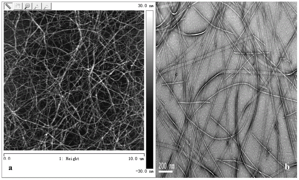
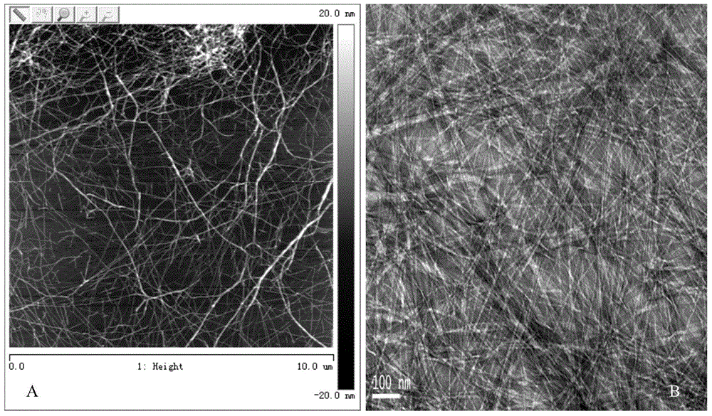
[0088] AFM scanning: Take 10 μL of prepared peptide sample dropwise on the surface of a clean mica sheet, let it absorb for 10 seconds, and then blow dry the sample with high-purity nitrogen gas. The scanning is completed under the AFM microscope in tapping mode, and the height map and phase of the sample are obtained. In the figure, the scanning angle is 0°, and the scanning rate is 1~1.5Hz. In this experiment, an RTESP silicon probe (Veeco, Santa Barbara, CA) was used, with a tip radius of ~10 nm, a vibration arm length of 125 μm, and an elastic coefficient of 42 N / m. The same sample was scanned 5 times at different positions, and the results showed that IIILQGK and IIIVQGK self-assembled into fiber structures in HEPES solution ( figure 2 a and Figure 4 a)

[0089] TEM: Take a drop of polypeptide soluti...


PUM

PropertyMeasurementUnit
Mechanical strengthaaaaaaaaaa

Abstract

The invention aims to provide amphipathic self-assembly ultra-short peptide. The amino acid sequence general formula of the peptide is I3-X-Q-G-K(1-2). According to the amphipathic self-assembly ultra-short peptide, the content of amino acid is small, synthesis cost is low, and synthesis conditions can be controlled easily; high-strength in-situ hydrogel can be formed at the moment the ultra-short peptide makes contact with blood, so that a fast hemostatic function is achieved; meanwhile, the series short peptide is low in toxicity, good in biocompatibility and capable of not causing a non-specific immunity reaction of lymphocyte, thereby being an ideal nano hemostatic material. By the application of the amphipathic self-assembly ultra-short peptide, the variety of existing nano medicine materials can be enriched, and it is of great significance to the life and health of people.

Description

technical field [0001] The invention belongs to the technical field of nano medical materials, in particular to an amphiphilic self-assembled ultrashort peptide nano hemostatic material. Background technique [0002] Due to trauma, surgery, etc., there will be a lot of bleeding. If the bleeding is not stopped in time, it may cause infection and aggravate the trauma. Therefore, controlling bleeding is an important step in first aid and trauma treatment. Currently, local hemostatic materials mainly include zeolites, polysaccharides such as chitosan, and biomaterials such as gelatin. Zeolites have good effect and low cost, but they have exothermic reaction and the particles are not easy to remove; polysaccharides have good biocompatibility but have requirements on the size and shape of the wound; biomaterials have good absorbability, which is beneficial to wound healing and does not leave Scars, its disadvantage is high cost, slow hemostatic speed (a kind of rapid hemostatic p...

Claims


Application Information

Patent Timeline
IPC IPC(8): C07K7/06A61L15/32A61L15/42
Inventor 陈翠霞徐海张宇白景琨王景新吕建仁
Owner CHINA UNIV OF PETROLEUM (EAST CHINA)
Why Eureka
Try Eureka