
A biodegradable nano yarn inhibiting fibrosis and its preparation and application

A technology of nano yarn and fibrillation, which is applied in the direction of fiber chemical characteristics, cellulose/protein conjugated artificial filament, tissue regeneration, etc., can solve the problems of hypertrophic scars and insufficient functionalization, and achieve good repair and regeneration, good Good mechanical properties and biocompatibility

Active Publication Date: 2019-06-25
深圳市迈捷生命科学有限公司
View PDF1 Cites 0 Cited by
  • Summary
  • Abstract
  • Description
  • Claims
  • Application Information

AI Technical Summary

Problems solved by technology

[0006] The technical problem to be solved by the present invention is to provide a biodegradable nano yarn that inhibits fibrosis and its preparation and application. The problem of underfunctionalization of

Method used

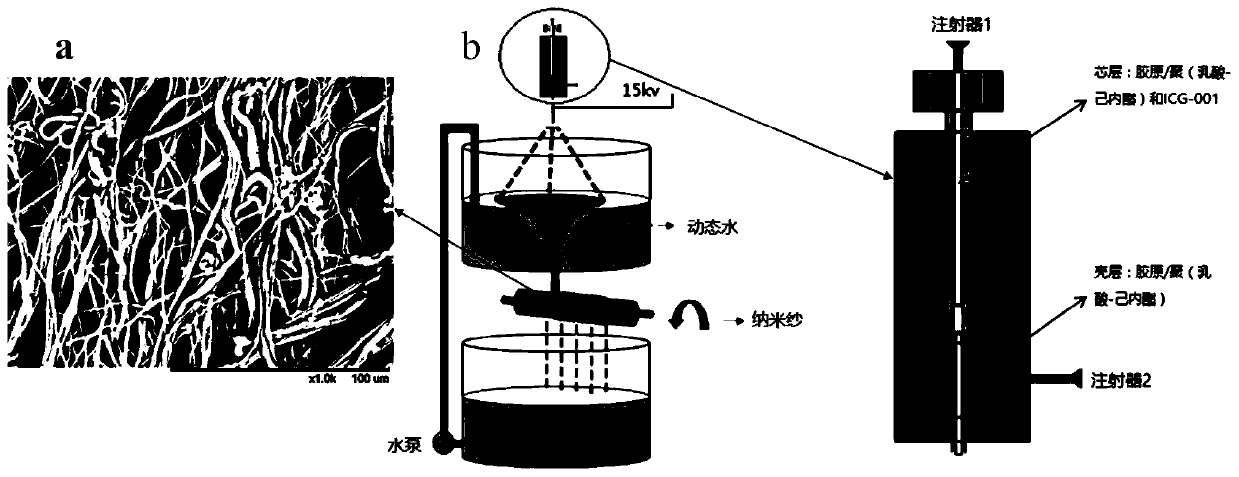

Image

  • A biodegradable nano yarn inhibiting fibrosis and its preparation and application
  • A biodegradable nano yarn inhibiting fibrosis and its preparation and application
  • A biodegradable nano yarn inhibiting fibrosis and its preparation and application

Examples


Embodiment 1

[0033] Dissolve 10 mL of a mixture of collagen I and poly(lactide-caprolactone) (the mass ratio of type I collagen to poly(lactide-caprolactone) is 25:75) in hexafluoroisopropanol solution (mass Concentration 11%), electric stirring at 25°C for 10 hours (rotating speed 550r / min), as a shell layer, and finally ultrasonic vibration for 20 minutes.

[0034] An appropriate amount of ICG-001 drug powder was dissolved in DMSO solution, and the concentration of anti-fibrotic drug ICG-001 was 600 μmol / mL, as the nuclear layer solution.

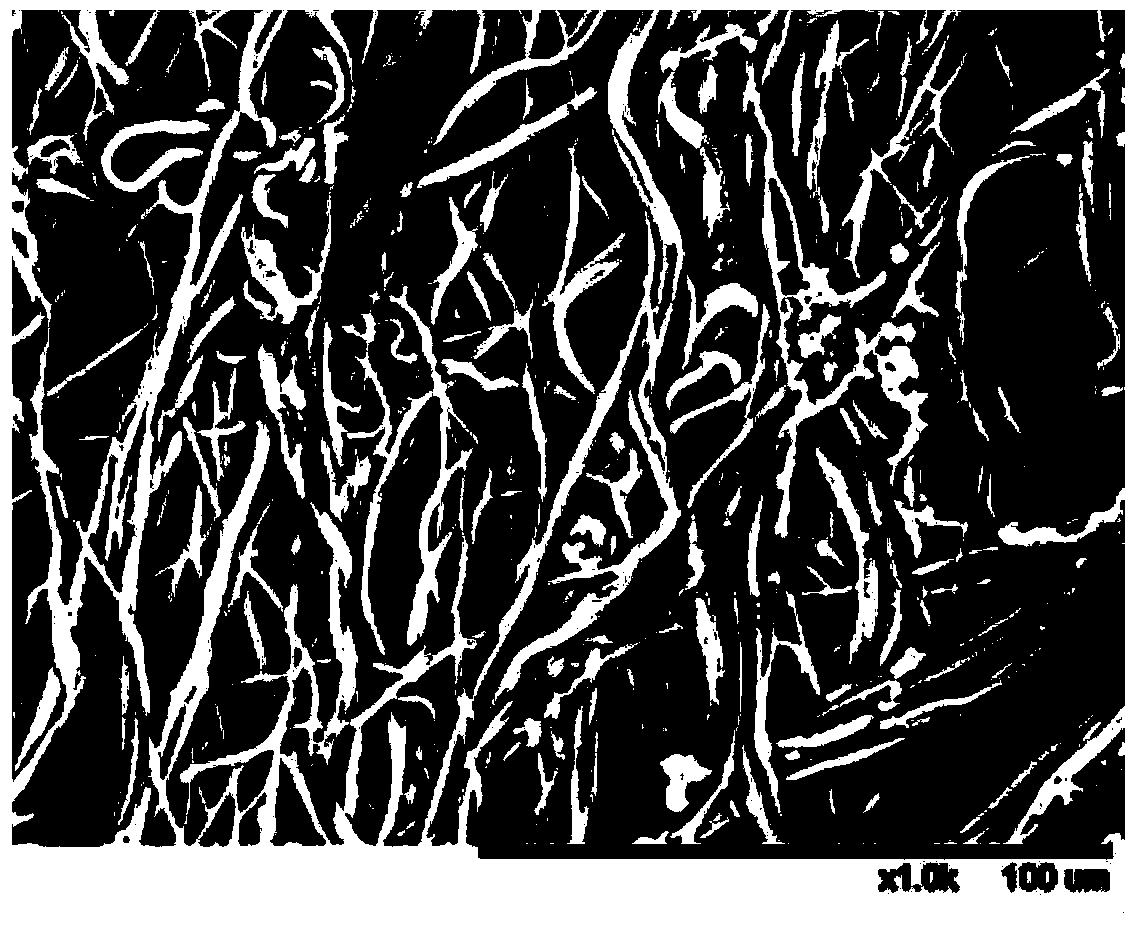
[0035] At a temperature of 25°C, a relative humidity of 50%, a sheath spinning flow rate of 1.0mL / h, a core spinning flow rate of 0.1mL / h, and a spinning voltage of 15kV, according to figure 1 The shown device performs the preparation of drug-loaded nanospun yarns.

[0036] Finally, the prepared drug-loaded nano yarns were frozen at -80°C overnight and then dried in a freeze-drying oven. By freeze-adsorption-sublimation method, they were freeze-dried...

Embodiment 2

[0039] Cytotoxicity test of drug-loaded nano-yarns

[0040](1) Inoculate the fibroblasts of dogs in the logarithmic growth phase into a 96-well plate, ensuring that the number of cells incubated for 2 days is 6000 / well, and the number of cells incubated for 4 days and 7 days is 4000 / well and respectively. 3000 pcs / hole;

[0041] (2) The inoculated cells were adhered to the wall for 6 hours in an incubator, and then the prepared drug-loaded nano yarn and the non-drug-loaded nano yarn as a control were placed in the medium to soak in the medium for allow;

[0042] (3) After incubation for 1, 3, 5, and 7 days, the cells were subjected to MTT determination;

Embodiment 3

[0044] Animal experiment of drug-loaded nano yarn in repairing defect urethra in urethral tissue engineering

[0045] (1) 6 male New Zealand rabbits with almost the same age and physical condition were used as model animals for animal experiments, and a group of 3 rabbits were artificially excised a 2 cm defect in the urethra of the rabbits, respectively, and the drug-loaded nano yarn and the non-drug-loaded The nano yarn implanted in the defect.

[0046] (2) In order to avoid wound infection within 2 weeks after operation, the rabbits were continuously injected with antibiotics. After 3 months, the rabbit's urethra was positioned and contrasted.


PUM

PropertyMeasurementUnit
pore sizeaaaaaaaaaa
porosityaaaaaaaaaa

Abstract

The invention relates to a biodegradable nano yarn capable of inhibiting fibrosis, and preparation and application thereof. The biodegradable nano yarn is prepared from the following raw materials in parts by weight: 100 parts of I-type collagen / poly(lactic acid-caprolactone) composite solution and 100 parts of anti-fibrosis drug solution. The preparation method comprises the following steps: using the I-type collagen / poly(lactic acid-caprolactone) composite solution as a shell solution; and using the anti-fibrosis drug solution as a core solution, spinning, freezing over night, and carrying out freeze-drying. The nano yarn can be used as a biodegradable and absorbable biological material scaffold, and can be used for resisting fibrosis in the urethra tissue engineering restoration to prevent postoperative urethral restenosis and urethral submucoasl excessive fibrosis. The preparation method is simple and efficient, and is low in price. The material has the advantages of favorable mechanical properties, obvious anti-fibrosis effect, favorable biodegradability, favorable biocompatibility, and reasonable release rate and time.

Description

technical field [0001] The invention belongs to the field of drug-loaded nanomaterials and their preparation and application, in particular to a biodegradable nano yarn that inhibits fibrosis and its preparation and application. Background technique [0002] The hypertrophic scar caused by cell fibrosis in the postoperative wound is a major problem for surgeons. Excessive fibrosis of cells will not only affect the patient's wound recovery, but may also lead to the failure of the operation and secondary damage to the patient. Taking urological surgery as an example, more than 70% of patients who have undergone urethral repair surgery have to undergo secondary surgery due to excessive fibrosis of wound cells leading to urethral restenosis. As the main effector cells in wound healing, fibroblasts can synthesize and secrete collagen matrix in the early stage of injury to promote healing. However, in the later stage of wound healing, the overexpression of collagen causes a larg...

Claims


Application Information

Patent Timeline
Patent Type & Authority Patents(China)
IPC IPC(8): A61L27/24A61L27/18A61L27/54A61L27/58D01F8/02D01F8/14D01F1/10
CPCA61L27/18A61L27/24A61L27/54A61L27/58A61L2430/22D01F1/10D01F8/02D01F8/14C08L67/04
Inventor 莫秀梅郭煦然张楷乐
Owner 深圳市迈捷生命科学有限公司
Learn More