Preparation method of graphene biosensor for specific protein detection

A biosensor, protein detection technology, applied in the measurement of phase influence characteristics, etc., can solve the problems of user and enterprise burden, high cost of use, and high price of SPR instruments.

Inactive Publication Date: 2017-05-31
NANKAI UNIV
View PDF3 Cites 2 Cited by
  • Summary
  • Abstract
  • Description
  • Claims
  • Application Information

AI Technical Summary

Problems solved by technology

However, SPR instruments are expensive, and because of the expensive chips that need to be replaced frequently, the subsequent use costs become more expensive, which brings serious burdens to users and enterprises

Method used


Image

  • Preparation method of graphene biosensor for specific protein detection
  • Preparation method of graphene biosensor for specific protein detection
  • Preparation method of graphene biosensor for specific protein detection

Examples


Embodiment 1

[0046] The preparation of graphene, its specific technical scheme is as follows:

[0047] 1) Cleaning and processing of quartz slices:

[0048]The cleaning of the quartz substrate is specifically as follows: the quartz substrate is ultrasonically cleaned with acetone, isopropanol, and ultrapure water respectively, and dried with nitrogen. The treatment of the quartz substrate is as follows: the quartz substrate is cleaned with oxygen plasma, the power of the machine is: 150W; the flow rate of oxygen used is: 300-400ml / min; the processing time: 1 minute.

[0049] 2) Preparation of graphene:

[0050] The graphene oxide aqueous solution of 4mg / ml is spin-coated to the quartz substrate surface that cleans and handles, spin-coats 3 times, then it is put into tube furnace, in the mixed gas of argon and hydrogen (95% argon gas, 5% hydrogen) atmosphere, 800 degrees of thermal annealing for 1 hour, to obtain a graphene film with a uniform thickness of ~8nm; figure 1 is the result of...

Embodiment 2

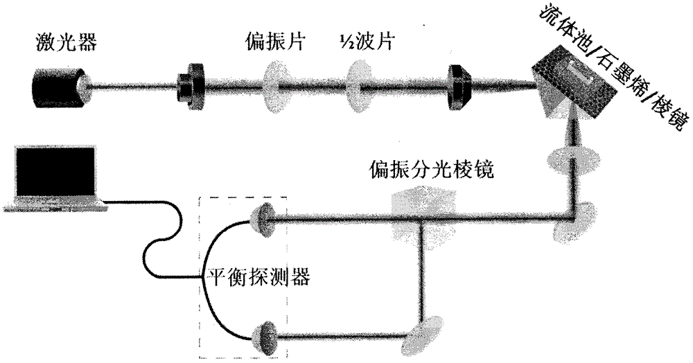
[0052] Graphene polarization-dependent absorption sensor, its specific embodiment is as follows:

[0053] Such as figure 2 As shown, the graphene polarization-dependent absorption biosensor mainly consists of an optical system and a sensing system composed of a sandwich structure of prism-graphene / quartz sheet-fluid pool. Index matching oil was used to bond the graphene / quartz sheet to the prism. The red light emitted by the 633nm laser is adjusted to strictly linearly polarized light by the polarizer, and then focused onto the surface of the prism / graphene through the lens, where total reflection occurs at the graphene interface. A polarization beam splitter divides the reflected light into S light and P light, and a balanced detector is used to detect the separated P light and S light. When the protein to be tested in the solution reacts specifically with the probe protein on the graphene surface, the graphene’s absorption of P light and S light changes due to the polariz...

Embodiment 3

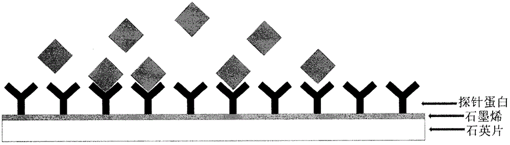
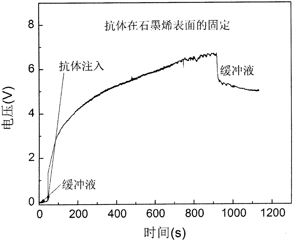
[0055] The fixation of probe protein, its specific technical scheme is as follows:

[0056] First, oxygen plasma is used to treat the graphene surface prepared in Example 1, and the treatment time is 15 seconds, so that the graphene surface layer contains more oxygen-containing functional groups, and secondly, the mixed solution of EDC / NHS is used to activate graphene Oxygen-containing functional groups on the surface, followed by, as image 3 As shown, PBS was used to wash the graphene surface, and then the protein (goat anti-rabbit IgG) used as a probe was injected onto the graphene surface, and the probe protein was fixed to the graphene surface by covalent bonding, and then , PBS was used to flush the fluid cell.


PUM

PropertyMeasurementUnit
wavelengthaaaaaaaaaa
Sensitivityaaaaaaaaaa

Abstract

The invention relates to a preparation method of a graphene biosensor for specific protein detection. Graphene in the specific protein biosensor is prepared by a high-temperature annealing method in an inert atmosphere. A functional graphene film is designed and prepared, and the graphene specific protein biosensor is also manufactured. Tests show that the specific protein biosensor can perform real-time unmarked detection on specific protein and allows influences caused by concentration changes of target protein to be seen clearly. A graphene biochip has a bright application prospect in the aspects of biomolecule sensors and biomolecule detection.

Description

technical field [0001] The invention relates to the technical field of preparation of graphene sensing and biosensors, in particular to a graphene film-specific protein biosensor and a preparation method thereof. Background technique [0002] Proteins are the executors of life functions and contain important biological information. Protein-protein interactions play an important role in the process of biomolecular interactions. The interaction process between drugs and proteins is the main process of disease treatment, and the biological dynamic information at the molecular level is very important for the research of drug screening. Specific protein biosensors use various changes (binding, dissociation, elution) caused by specific reactions between proteins to record and analyze the reactions between the probe protein and the probe protein, and This reaction is converted into detectable light, electricity, force and other signals to complete the detection and monitoring of ...

Claims


Application Information

Patent Timeline
Patent Type & Authority Applications(China)
IPC IPC(8): G01N21/41
CPCG01N21/41
Inventor 刘智波蒋文帅田建国
Owner NANKAI UNIV
Why Eureka
Try Eureka