Preparation method of nano-pearl powder/C-HA composite scaffold
A technology of nano pearl powder and composite scaffold, which is applied in the field of bioengineering, can solve the problems that hydroxyapatite is not easy to degrade, and the degradation speed of tricalcium phosphate cannot be regulated, and achieves the advantages of proliferation, good biological activity and good wettability. Effect
- Summary
- Abstract
- Description
- Claims
- Application Information
AI Technical Summary
Problems solved by technology
Method used
Image
Examples
Embodiment 3
[0053] 1. Reagents, as shown in Table 2
[0054] Table 2
[0055] Reagent
company and model
micron pearl powder
Hainan Nanjing Run Pearl Biotechnology Co., Ltd.
Sodium hyaluronate
H107141, Aladdin
C105803, Aladdin
acetic acid
Analytical pure, Aladdin
[0056] 2. The instrument is shown in Table 3
[0057] table 3
[0058]
[0059]
[0060] 3. Operation steps
[0061] 1) Grind the micron pearl powder into nano pearl powder by mechanical ball milling with a nano ceramic sand mill disperser, dry and sterilize;
[0062] 2) Dissolve 4wt% chitosan and 1wt% hyaluronic acid into 1% acetic acid solution respectively, stir for 5min at a speed of 2000rpm in a Thinky mixer, and place it at room temperature for 24h to obtain chitosan acetic acid solution and hyaluronic acid acid acetic acid solution;
[0063] 3) After 24 hours, mix the chitosan acetic acid solution and the hyaluronic acid acetic acid solution...
Embodiment 1 Embodiment 5
[0070] 2. Experimental reagents and instruments
[0071] The same as the reagent of embodiment and instrument
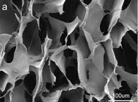
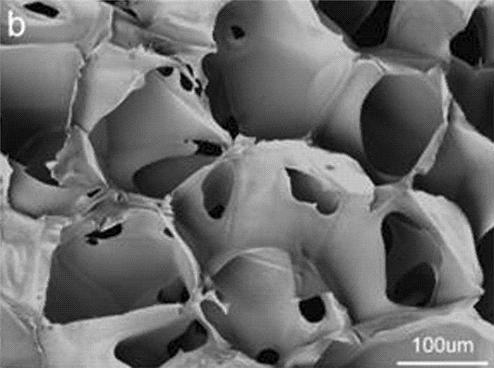
[0072] 1) Experiment 1: Determination of the surface morphology of the composite scaffold
[0073] 1. Observation of the surface morphology of composite scaffolds by scanning electron microscopy
[0074] Cut the sample into thin slices with a width of 3 mm to make the surface of the sample as flat as possible. After drying with a vacuum freeze dryer, stick the samples on the conductive adhesive in turn, mark the upper left corner of the conductive adhesive, and spray gold on the surface of the sample for 60 seconds. Then open the sample chamber of the scanning electron microscope (MIRA3, Czech Republic), fix the sample to be observed, vacuumize, and directly observe the cross-sectional morphology with the scanning electron microscope.
[0075] 2. Conclusion
[0076] like figure 1 As shown, the scaffold is highly porous with uniformly distributed pores and good c...
PUM
| Property | Measurement | Unit |
|---|---|---|
| Diameter | aaaaa | aaaaa |
| Height | aaaaa | aaaaa |
| Diameter | aaaaa | aaaaa |
Abstract
Description
Claims
Application Information
 - R&D
- Intellectual Property
- Life Sciences
- Materials
- Tech Scout
- Unparalleled Data Quality
- Higher Quality Content
- 60% Fewer Hallucinations
Browse by: Latest US Patents, China's latest patents, Technical Efficacy Thesaurus, Application Domain, Technology Topic, Popular Technical Reports.
© 2025 PatSnap. All rights reserved.Legal|Privacy policy|Modern Slavery Act Transparency Statement|Sitemap|About US| Contact US: help@patsnap.com


